Cell Biology - BIOB10 (post midterm)
 Call Kai
 Learn
Learn Practice Test
 Spaced Repetition
 Match
Match Flashcards
 Knowt Play
1/771

There's no tags or description
Looks like no tags are added yet.
Name | Mastery | Learn | Test | Matching | Spaced | Call with Kai | Chat |
|---|
No analytics yet
Send a link to your students to track their progress
772 Terms
Unfolded Protein Response
If there is a large amount of unfolded/misfolded proteins accumulating in the ER, cells have a plan of action called … (3)
Chaperones, proteases
Unfolded Protein Response (UPR):
What 2 things are made more?
Degrade
Unfolded Protein Response (UPR):
More proteases are made to … defective proteins
Decreased
Unfolded Protein Response (UPR):
Translation of new proteins not involved in UPR is …
(increased or decreased)
Chaperones
Unfolded Protein Response (UPR):
Activation on UPR is based on … binding to sensors decreasing
Decreasing
Unfolded Protein Response (UPR):
Activation of UPR is based on chaperones binding to sensors …
(increasing or decreasing)
Increases
Unfolded Protein Response (UPR):
There is less binding of chaperones to sensors because binding to misfolded proteins …
(increases or decreases)
Native form
Some genetic diseases have mutation leading to the expression of a mutant protein, which means they cannot fold properly into … (2)
Destroyed
When a mutant protein is recognized by the quality control pathway in the ER, it is … which loses protein & protein function in the cell
Cystic fibrosis
An example of a ER protein folding disease (2)
Vesicular
Transport of proteins via intermediates
Vesicles
Dynamic organelle involved in selective transport of proteins/materials
Bud
Vesicles … off donor compartment
Coat
Budding vesicles have a … of soluble proteins assembling on the membrane of donor compartment
Cytosolic
Budding vesicles have coat of soluble proteins on donor compartment.
Where are these soluble proteins from? Lumenal or cytosolic
Coat proteins
Proteins that mechanically force the membrane to curve and form a budding vesicle (2)
Cargo
Vesicles:
What protein is needed to go from donor to target?
COP II
Vesicle budding + Coat Proteins:
Vesicles that transport proteins forward from ER to Golgi (anterograde)
COP I
Vesicle budding + Coat Proteins:
Transport proteins backwards from Golgi to ER (retrograde)
Clathrin
Vesicle budding + Coat Proteins:
Any transport from the Golgi onwards is done by …
COP II
Vesicles: COP II, COP I, or clathrin?
Transport from ER to Golgi
COP I
Vesicles: COP II, COP I, or clathrin?
Transport proteins from Golgi to ER
Clathrin
Vesicles: COP II, COP I, or clathrin?
Protein sorting to plasma membrane, lysosomes, endocytosis, etc.
6
Clathrin Coat Proteins (vesicle budding):
Clathrin has # proteins
Triskelion
Clathrin Coat Proteins (vesicle budding):
Clathrin has 6 proteins total that form a … structure.
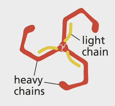
Basket
Clathrin Coat Proteins (vesicle budding):
The Clathrin triskelions come together to form a … around vesicle
Adaptor proteins
Clathrin Coat Proteins (vesicle budding):
What binds to clathrin to form the inner coat between vesicle and clathrin shell? (2)
Inner coat
Clathrin Coat Proteins (vesicle budding):
Adaptor proteins bind to clathrin to form the … (2) between vesicle & clathrin shell
Phosphoinositides, PI
Clathrin Coat Proteins (vesicle budding):
Adaptor proteins form inner coat.
What binds specific adaptor proteins based on phosphorylation of inositol sugar at 3’, 4’, and 5’ position? (1,1)
Phosphorylation, inositol
Clathrin Coat Proteins (vesicle budding):
PI (phosphoinositides) bind specific adaptor proteins based on … of their … sugar at 3’, 4’, 5’ position
Different
Clathrin Coat Proteins (vesicle budding):
Different organelles have … PIPs (phosphoinositidyl phosphates)
(different or same)
Phosphoinositide, cargo receptor
Clathrin Coat Proteins (vesicle budding):
Adaptor protein binds to a … head group and to a … (2) to enhance binding to plasma membrane
Fission
Vesicle budding depends on membrane …
Dimeric membrane bending proteins
Clathrin Coat Proteins (vesicle budding):
Membrane curvature where adaptor proteins assemble (4?)
Fission, dynamin
Clathrin Coat Proteins (vesicle budding):
Severing the vesicle from donor compartment is done by … proteins, specifically …
Dynamin
Clathrin Coat Proteins (vesicle budding):
What is the specific fission protein severing vesicle from membrane?
Neck
Clathrin Coat Proteins (vesicle budding):
The way dynamin severs vesicle from membrane is it creates a collar around the … of budding vesicle
GTP
Clathrin Coat Proteins (vesicle budding):
Dynamin (severs vesicle from membrane) is a … binding protein (acronym)
GTP hydrolysis
Clathrin Coat Proteins (vesicle budding):
Dynamin creates a collar around the neck of the budding vesicle. What is the actual mechanism that does the severing? (2)
Coat recruitment GTPases, Rab proteins, SNAREs
Vesicles are mediated by 3 proteins that control movement & fusion
Coat recruitment GTPases
Targeted vesicle transport (3):
Initiates coated vesicle formation (3)
Rab proteins
Targeted vesicle transport (3):
Directs vesicles
SNAREs
Targeted vesicle transport (3):
Docking & fusion
Pinched
COP II vesicle transport:
Once the entire coat is assembled, the vesicle is … off
Sar1, COP II proteins
COP II vesicle transport:
GTP hydrolysis occurs once vesicle leaves ER which releases 2 things
Cargo receptor
COP II vesicle transport:
Proteins that are fully/partially exposed on the lumenal surface of COP II vesicles
Rab
Rab or SNARE proteins [vesicles]:
Tethering
SNARE
Rab or SNARE proteins [vesicles]:
Docking
SNARE
Rab or SNARE proteins [vesicles]:
Fusion
Rab, GTP
Vesicle tethering is done by … proteins which are small … binding proteins
Lipid
Vesicle tethering: Rab
Associated to membranes by a … anchor
Organelles
Vesicle tethering: Rab
Specific Rab proteins associate with specific …
Cytosol
Vesicle tethering: Rab
Recruit tethering proteins from the …
Positive
Vesicle tethering: Rab
… feedback loop of active Rab5 recruits Rab5 GEFS to make more Rab5s
Fusion
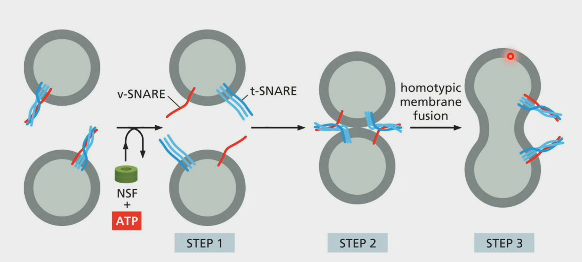
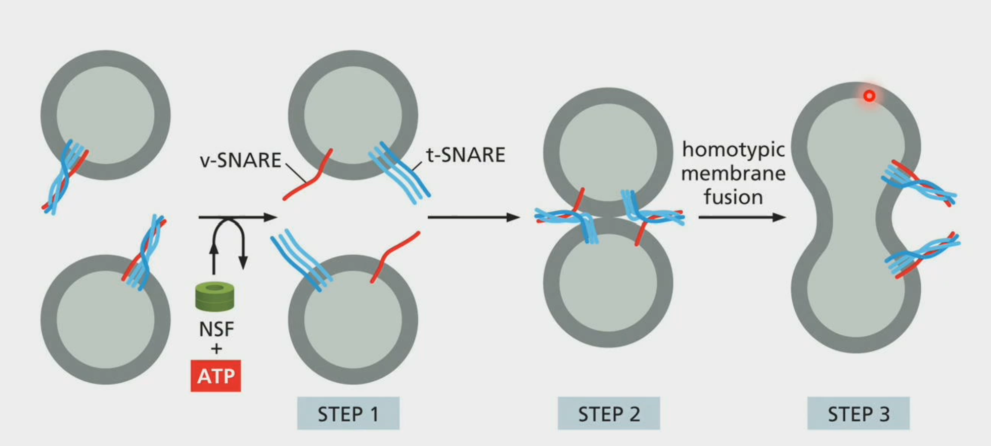
Vesicle docking: SNAREs
Process leads to vesicle …
Vesicle
Vesicle docking: SNAREs
v-SNAREs are on … membrane
v-SNARE
Vesicle docking: SNAREs
SNARE protein on vesicle membrane
Target
Vesicle docking: SNAREs
t-SNARES are on … membrane
t-SNARE
Vesicle docking: SNAREs
SNARE protein on target membrane
Fuse
Vesicle fusion: SNAREs
Interaction between v-SNAREs & t-SNAREs will pull respective membranes together and will … together
Bilayer
Vesicle fusion: SNAREs
SNARE proteins allow for the 2 membranes (vesicle & membrane) will create a new …
NSF
Vesicle fusion:
Once fusion has occurred, unzipping the SNARE proteins require … protein which is ATPase (acronym)
v-SNAREs
Vesicle fusion:
When unzipping SNARE proteins post fusion, … are retrieved and returned to compartment of origin
Fuse, vesicular tubular clusters
After COPII coat is lost, ER derived vesicles … together in … (3)
ER Golgi Intermediate Compartment
ERGIC (expand)
Vesicular tubular clusters
ERGICs are made of … (3)
Retrograde transport vesicles, COPI
If misfolded proteins or ER resident proteins accidentally exit via budding vesicles, they can be sent back via … (3, 1)
Coatomer
Vesicle transport:
COPI coats are recruited as a preassembled complex called …
Signal sequence, C-terminal
How do we know if ER resident protein retrieval is necessary?
Because ER resident proteins have a … (2) on the … of the protein
Nucleus
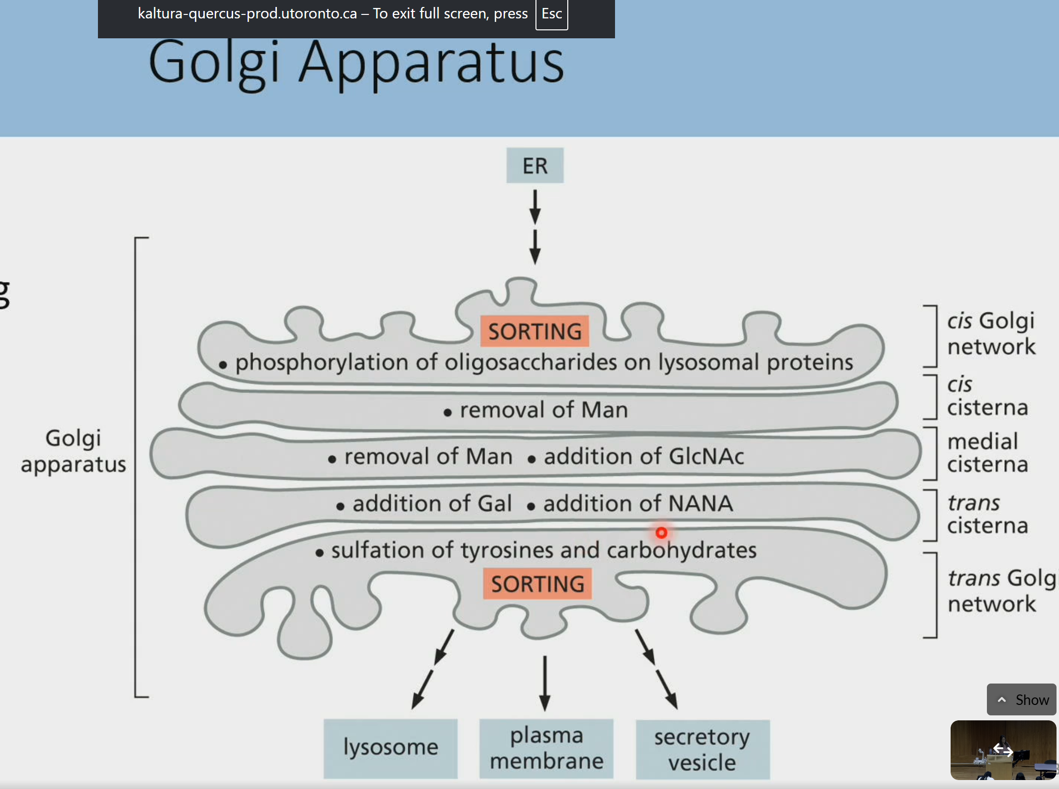
Golgi Apparatus is located adjacent to the … above the rough ER
Cisternae
Golgi apparatus:
Consists of ribbon-like stacks of membranous …
Functions
Golgi apparatus:
Cisternae are distinct which means they can perform several distinct …
Cis
Golgi apparatus:
Cisternae closest to the ER
Medial
Golgi apparatus:
Middle cisternae
Trans
Golgi apparatus:
Cisternae leading to proteins budding off
Cis to medial to trans
Golgi apparatus:
Protein progression through the golgi networks (… to … to …)
No
Golgi apparatus:
Are there discrete boundaries between different cisternae?
Processed, modified, sorted
What 3 important processes are done in the golgi?
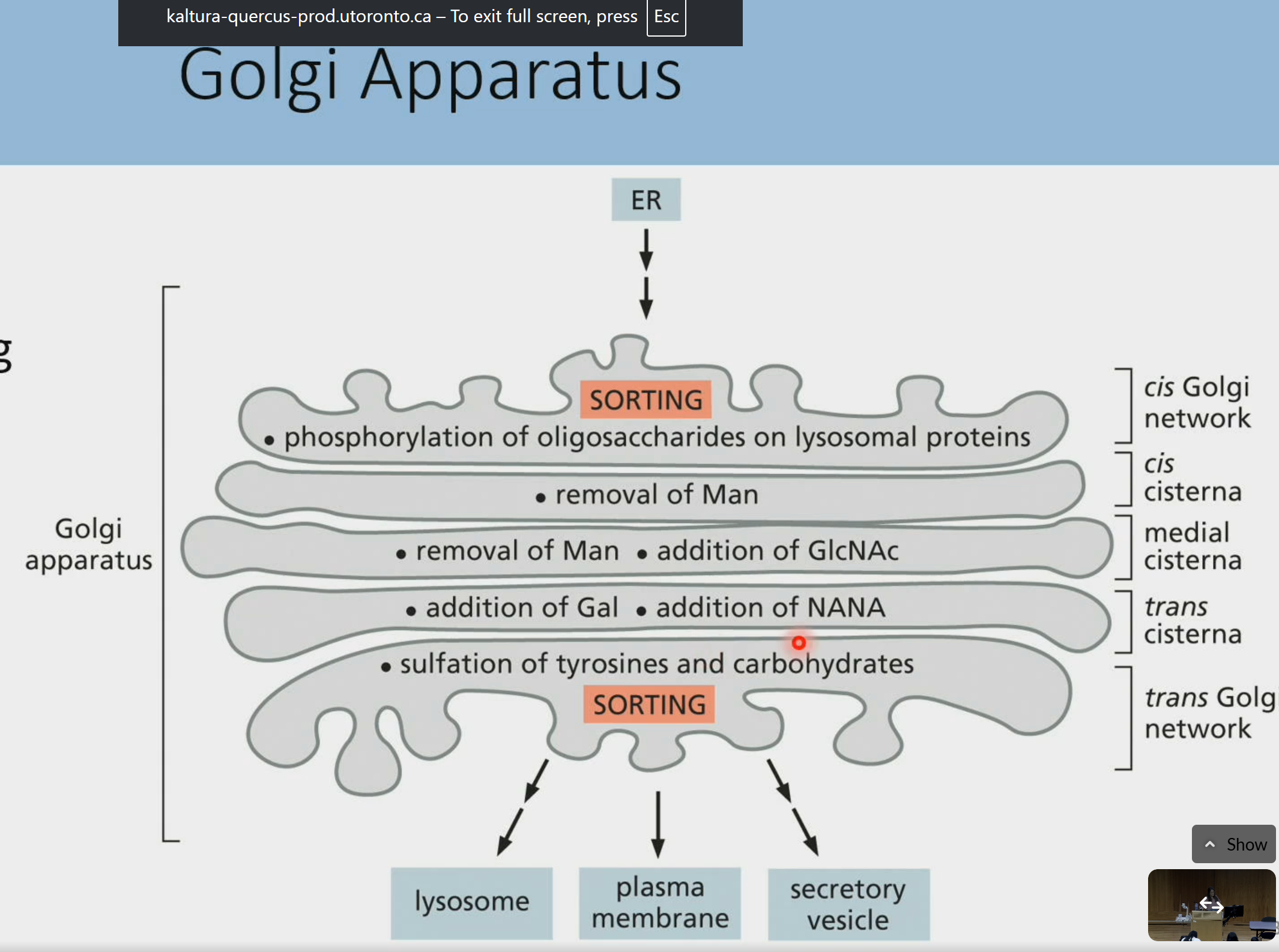
Functional compartmentalization
The fact that proteins are processed, modified, and sorted is seen as … (2) in the golgi
Soluble, aggregates
Golgi:
Proteins are glycosylated for 2 reasons:
Keeps intermediates of protein folding … and prevents …
Glycocode
Golgi:
Proteins are glycosylated for 2 reasons:
Keeps intermediates of protein folding soluble and prevents aggregates
Sequential modification signify progression of protein folding via …
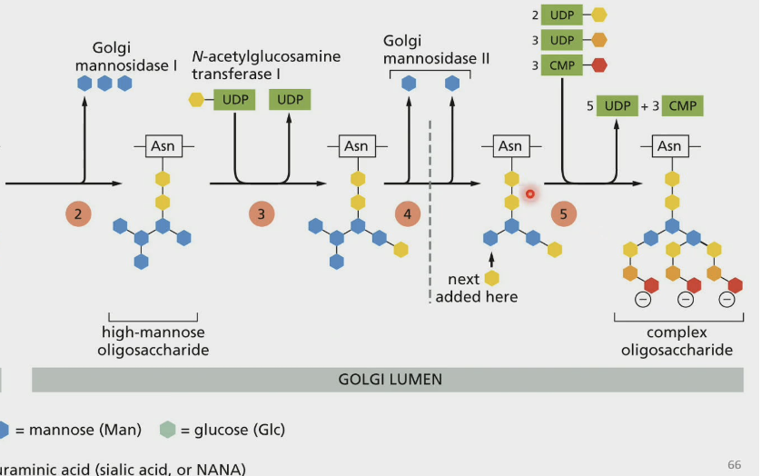
Mannose
N-glycosylation in Golgi:
You will lose an additional 3 … which creates a high-mannose oligosaccharide
GlcNac, mannose
N-glycosylation in Golgi: COMPLEX
Addition of … and trim of 2 …
High mannose oligosaccharide, complex oligosaccharides
Any N-linked glycans going through golgi have 2 fates
O-linked glycosylation
A further Golgi protein modification where sugars are attached to serine or threonine on specific proteins (3)
N-linked, O-linked
Protein modification glycosylation in the golgi (2)
Vesicle transport, stationary cisterna
How does Golgi maintain polarized structure?
Hypothesis 1:
2 names, each cisterna is static so all protein movement through the golgi requires vesicles
Cisternal maturation
How does Golgi maintain polarized structure?
Hypothesis 2: [more accurate, 2]
Cisterna are formed by vesicles from ER
Cis-end to trans-end
How does Golgi maintain polarized structure?
In Hypothesis 2 or Cisternal Maturation Model, cisternal physically move from the (… to …) as they mature
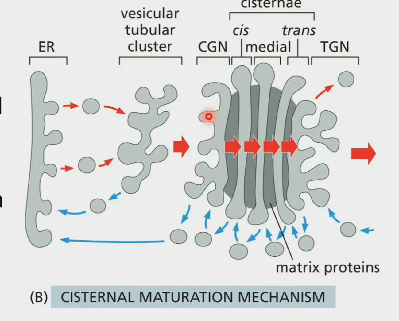
Vesicular tubular cluster
How does Golgi maintain polarized structure?
In hypothesis 2 or Cisternal Maturation Model, it is more accurate because without the … (3), the entire golgi falls apart
Golgins
Cytosolic Golgi proteins forming long tethers to capture vesicles by interacting w/ Rab proteins
Lysosomes
Digestive organelles which break down macromolecules & move products back to cytosol
Acidic
Lysosomes have a highly … environment
Oxidation, lipids
Peroxisomes rely heavily on … to break things down (H2O2) and they break down …
Amino acids, nucleotides
Peroxisomes break down lipids.
Lysosomes break down … (2 things)
Glycosylated
The reason why lysosomes don’t eat themselves is because most proteins are …
Phagocytosis, endocytosis, autophagy, macropinocytosis
Lysosomes get 4 major pathways of delivery
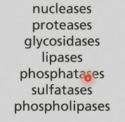
Acid hydrolases
Lysosomes are considered bags of … (2) which function in acidic pHs which can break down entire organelles
Proton pumps
The reason why lysosome is hella acidic is because it has a lot of … (2) on the membrane driving H+ into lumen using ATP (low pH)
Into
The reason why lysosome is hella acidic is because hella proton pumps drive H+ … lumen using ATP
(into or out of)