2.1.1 Cell Structure
1/90
Earn XP
Description and Tags
Completed set
Name | Mastery | Learn | Test | Matching | Spaced |
|---|
No study sessions yet.
91 Terms
How to prepare a light microscope
Staining and sectioning
What does staining do
Allows the specimen to become visible (chemical stains binds to on or in specimen)
E.g
See certain (named) organelles
Improves contrast
What are the different stains that bind to specific cell structures
Acetic orcein stains DNA dark red
Eosin stains cytoplasm pink
Sudan black stains membranes and other lipids black
What does sectioning do
Makes thin sections which allow the light to pass through
What problems can happen because of sectioning
Artefacts
Why do we use light microscopes
To look at whole cells and tissues
What organelles can you see with a light microscope
Nucleus
Mitochondria
Chloroplast
Cell membrane
Cell wall
What does TEM stand for
Transmission Electron Microscope
How does a transmission electron microscope work
Electron beam passed Through the specimen
Why does the specimen need to be thin when using a TEM
Specimen = thin to prevent deflection of electrons
Why do we use TEM’s
To look at organelles in detail
What organelles can you see with an electron microscope
Mitochondria
Golgi apparatus
ER
Lysosomes
Why do some TEM images appear different from each other
The mitochondria have been cut along different planes/ angles
What does SEM stand for
Scanning Electron Microscope
How does a scanning electron microscope work
The beam of electrons scans the surface of the specimen
The reflected beam is detected
The detector and the source are on the same side of the specimen
Why do we use SEM’s
To look at the cell surface
How to remember Laser Scanning Confocal microscope
Liesel Sees Colour
How does the Laser Scanning Confocal microscope work
Specimen treated with fluorescent dye
The laser focuses on this causing the dye to be seen
Facts about Laser Scanning Confocal Microscope
Very thin specimen needed
High resolution
Used to find eye problems
Why do we use Laser Scanning Confocal microscope
To look at an object at a certain depth within the cell
(e.g cytoskeleton in the cell)
Magnification definition
The number of times larger the image is in comparison to the object
(20x means 20 times larger)
Resolution definition
The ability to distinguish between two points as sperate entities
The ability to distinguish between very small structures that are close together in detail
Light microscope magnification and resolution
Up to x1500 (magnification)
50-200 nm (max resolving power)
TEM magnification and resolution
up to 500,000 (magnification)
0.05-1nm (max resolving power)
SEM magnification and resolution
Up to 100,000 (magnification)
0.20nm (max resolving power)
How to go from cm to mm to μm to nm
cm x10 → mm x1000 → μm x1000 → nm
How to go from cm to mm to μm to nm image

μm meaning
Micrometers
What two pieces of equipment do you need to calibrate a microscope
Eyepiece graticule
Stage micrometer
calibrating video
…
Marks for drawing from a microscope image
Use up to half the page
Clear and continuous lines (not ragged and broken) and no shading
Proportions correct
Label
Rule the label lines in pencil and don’t cross them
Label lines touch part that you’re labeling (no arrow heads)
Annotations
Scale
Title
What is the magnification formula
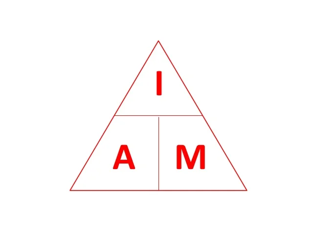
Magnification = Image over actual
(IAM)
Hard IAM question
If you have a scale bar and you need to find the actual size of the specimen
Measure length with ruler for Image size
Then measure length of scale bar of and convert to nano/ micrometers then sub into the IAM to find the magnification which is the length of scale bar measured with a ruler divided by the actual scale bar, then that’s the magnification
What is a nucleus
Nucleus controls nearly all the activities of the cell.
Contains DNA (genetic info) which controls the genetic code for proteins
What’s the difference between the nucleus and the nucleolus
Nucleolus has dense DNA
Nucleus is whole thing (Chromatin, nuclear envelope and pores)
What do the nuclear pores do
Allow substances in and out the nucleus, like mRNA leaving
What is the Rough Endoplasmic Reticulum (structure)
Flattened membrane bound sacs called cisternae.
Continuous with the nuclear envelope
RER has ribosomes on it
What happened at the rough ER
Protein synthesis
What is the Smooth Endoplasmic Reticulum (structure)
Flattened membrane bound sacs called cisternae.
Continuous with the nuclear envelope
Smooth ER has NO ribosomes
What happened at the smooth ER
Lipid and hormone production
What is the Golgi apparatus
A stack of membrane-bound flattened sacs
What happens at the Golgi apparatus
It modifies and packages proteins into vesicles
Some of these vesicles may be secreted out of the cell
What is the Ribosome
Tiny organelles, (in cytoplasm and on RER)
What happens at the Ribosomes
Site of protein synthesis
Where mRNA assembles proteins from amino acids
What is the mitochondria structure
Have 2 membranes separated by a fluid filled space.
The membrane is highly folded cristae
Central part = matrix
What happens at the mitochondria
ATP (adenosine triphosphate) is made during aerobic respiration
What is the lysosome structure
Spherical sacs surrounded by membrane
What happens at the lysosome
Contains powerful digestive enzymes
(break down materials)(used a lot in wbc)
Where is the chloroplast and its structure
Found only in plant cells
2 membranes separated by a fluid filled space
Inner membrane = continuous with a network of flattened disks called thylakoid (where chlorophyll is found)
A stack of thylakoids = a granum (plural grana)
What happens at the chloroplast
The site of photosynthesis, which is driven by light energy
What is the plasma/ cell surface membrane made from
Made out of phospholipid bilayer
What happens at the plasma/ cell surface membrane
Controls what goes in and out of the cell
What are centrioles
Small tubes of protein fibers, a pair of then found next to nucleus in an animal cells
What do centrioles do
Take part in mitosis to form spindle fibres
What is the cell wall
Plant cells = made of cellulose
Bacteria cells = made of peptidoglycan
What is the qualities of the cell wall
Provides high tensile strength
Insoluble
Inert
What is the flagella structure & where
Found in procaryotes
Have 9 + 2 arrangement
What does the flagella do
Causes whole cell to move
Powered by chemiosmosis
What is the cilia
Finger like appendages on ciliated epithelial cells
What happens at the cilia
They move substances along
(mucus in the trachea)
What is the vacuole structure
Membrane bound organelle found in plant cells
Membrane surrounding vacuole = tonoplast
Filled with water and enzymes
What happens at the vacuole
Removes unwanted substances from the cell and alters the cell shape by changing the amount of water in the vacuole (turgid or flaccid)
What is the vesicle
A membranes bound organelle
What does the vesicle do
Transports substances
What is found in a plant cell but not an animal cell
Centrioles
Glycogen granules
(Lysosomes)
(Cilia)
(Flagella)
What is the interrelationship between the organelles
To produce a protein (enzyme..) or hormone in that cell
(simple) Protein synthesis
Nucleus produces mRNA
mRNA leaves the nuclus through the nuclear pore
mRNA attaches to a ribosome
Either free in the cytoplasm or bound to an RER
Once protein made through protein synthesis, it goes ito a vesicle an dis transported to the golgi apparatus.
At the Golgi apparatus/ body, the protein is modified (adding carbohydrates..) and packded into a vesicle again
Vesicle moved to cell surface membrane of a eukaryotic cell = exocytosis occurs
mitochondira - produces atp = needed for contractile filimaents in the cytoskeleton
Cytoskeleton - made of contractile filaments which need atp to move vesicles from the golgi body, to the cell surface membrane to allow exocytosis to occor
What are the 3 main components of the cytoskeleton
Microfilaments
Microtubules
Intermediate filaments
Importance of the cytoskeleton
Whole cell support
Movements of cilia and flagella
Changing cell shape
Moving organelles (like a vesicle)
Movement of chromosomes
What does the cytoskeleton need
Cytoskeleton needs ATP - made through aerobic respiration in mitochondria
Because they are contractile filaments
What is a microfilament
6nm
Made of actin
Contract and are used in cytoskeleton
Importance of microfilaments
Changes in cell shape
Microtubules
25nm
Made of globular tublin proteins form tubes
Importance of microtubules
Moves chromosomes in mitosis (forming spindle)
Acts as tracks for organelles to move along
Moves organelles around the cell e.g vesicles
Intermediate filaments
10nm
Actin and microtubules
Importance of Intermediate filaments
Gives mechanical strength to cells, whole support
Importance of the flagella
(only in prokaryotes)
Movement of whole organism
By using ATP
using a motor
Importance of the cilia
Found in Eukaryotic cells
(esophagus, nasal cavity)
Movement of substances
Difference between prokaryotic and eukaryotic cells
Eukaryotic - membrane bound organelles and nucleus
PRo - no membrane bound organells an dno nucleus
Cell wall (capsule) of a Prokaryotic cell
Made of murein or peptidoglycan
High tensile strength
Cell surafce or plalsma membrane of a Prokaryotic cell
Allows substances in and out of the cell
Only membrane
Cytoplasm of a Prokaryotic cell
Site of chemcial reactions
Ribosomes of a Prokaryotic cell
Site of protein synthesis
70s
Size of ribosome in a eukaryotic cell
80s
Plasmids in a Prokaryotic cell
Circular DNA
Can be passed to other bacteria to exchange genetic info
(through pili to other surrounding bacteria - if pili tehere)
Pili in a Prokaryotic cell
Used for lateral flow of DNA to other bacteria
Nucleioud in a Prokaryotic cell
Circular DNA (eukaryotes have linear)
Genetic info
(not all have this)
Bacterial flagellum in a Prokaryotic cell
Used to move the cell
Needs ATP
Where is DNA found in a Prokaryotic cell
Plasmids or in Nucleoid - no nucleus
Where is ATP made in a Prokaryotic cell
Mesosome - in folded regions of the cell membrane
Prokaryotes and Eukaryotes link
Chloroplasts and mitochondria have evolved from prokaryotes
internal structure = similar
DNA found is circular and smaller ribosomes (70s not 80s)