L5 - Benign Bone Tumors
1/71
There's no tags or description
Looks like no tags are added yet.
Name | Mastery | Learn | Test | Matching | Spaced |
|---|
No study sessions yet.
72 Terms
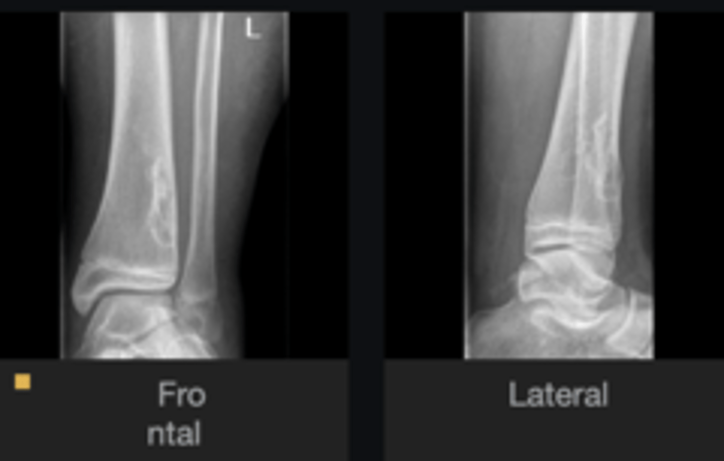
Lateral proximal tibia
Bone

- Longitudinal: epiphysis/metaphysis (up to joint surface)
- Eccentricity: eccentric
- Transverse: medullary
Location

Monostotic
Mono/polyostotic

>1 cm
Size

- Lytic
- Geographic
- Short zone of transition
Behavior

- Cortical thinning
- Cortical expansion
Cortex
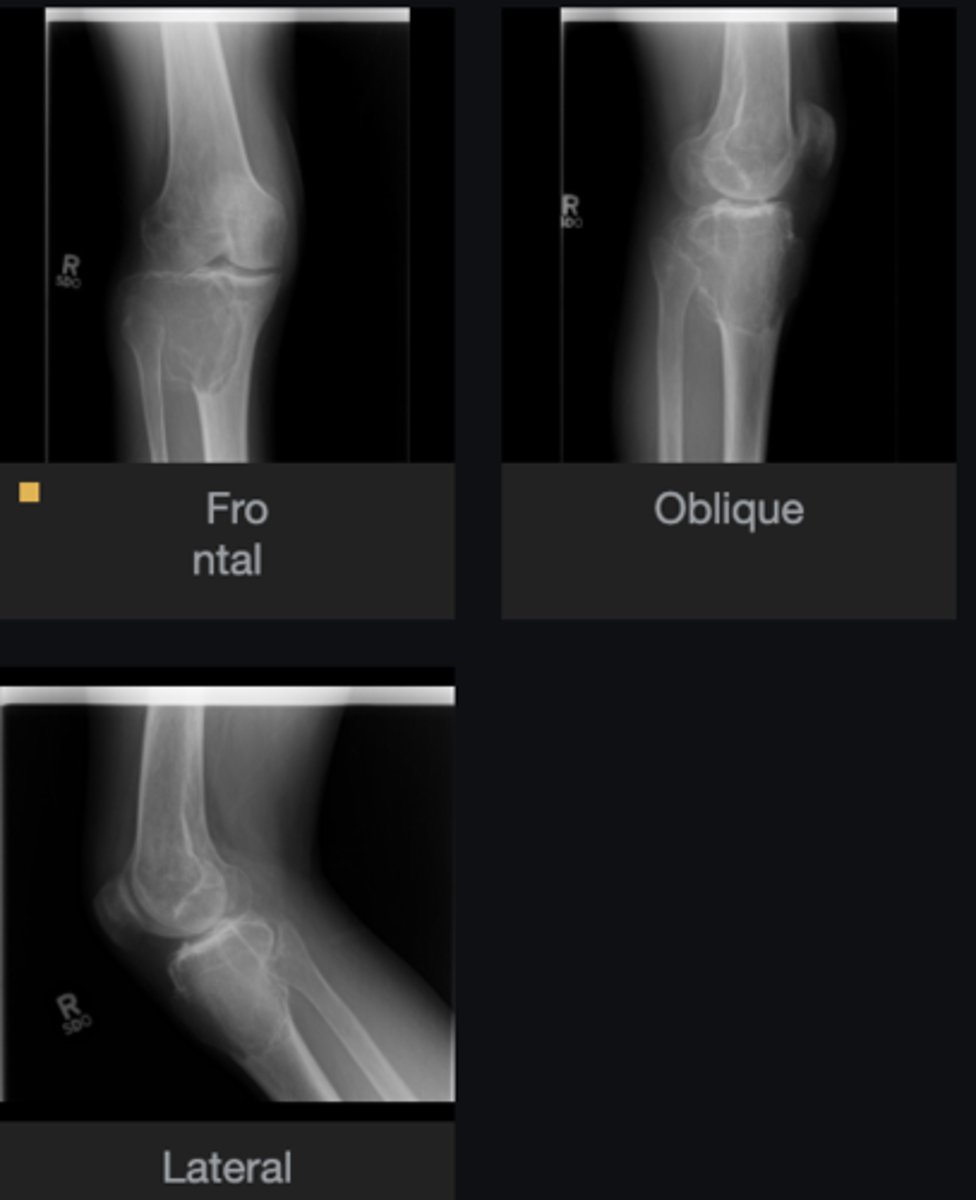
Nothing visible
Matrix

None
Periosteal reaction

Soft tissue swelling
Soft tissue involvement

Joint surface involved
Joint involvement

Giant cell tumor
Most likely diagnosis?

Refer to orthopedist or oncologist
Next step?

Could be aggressive (20%)
Concerns/complications?

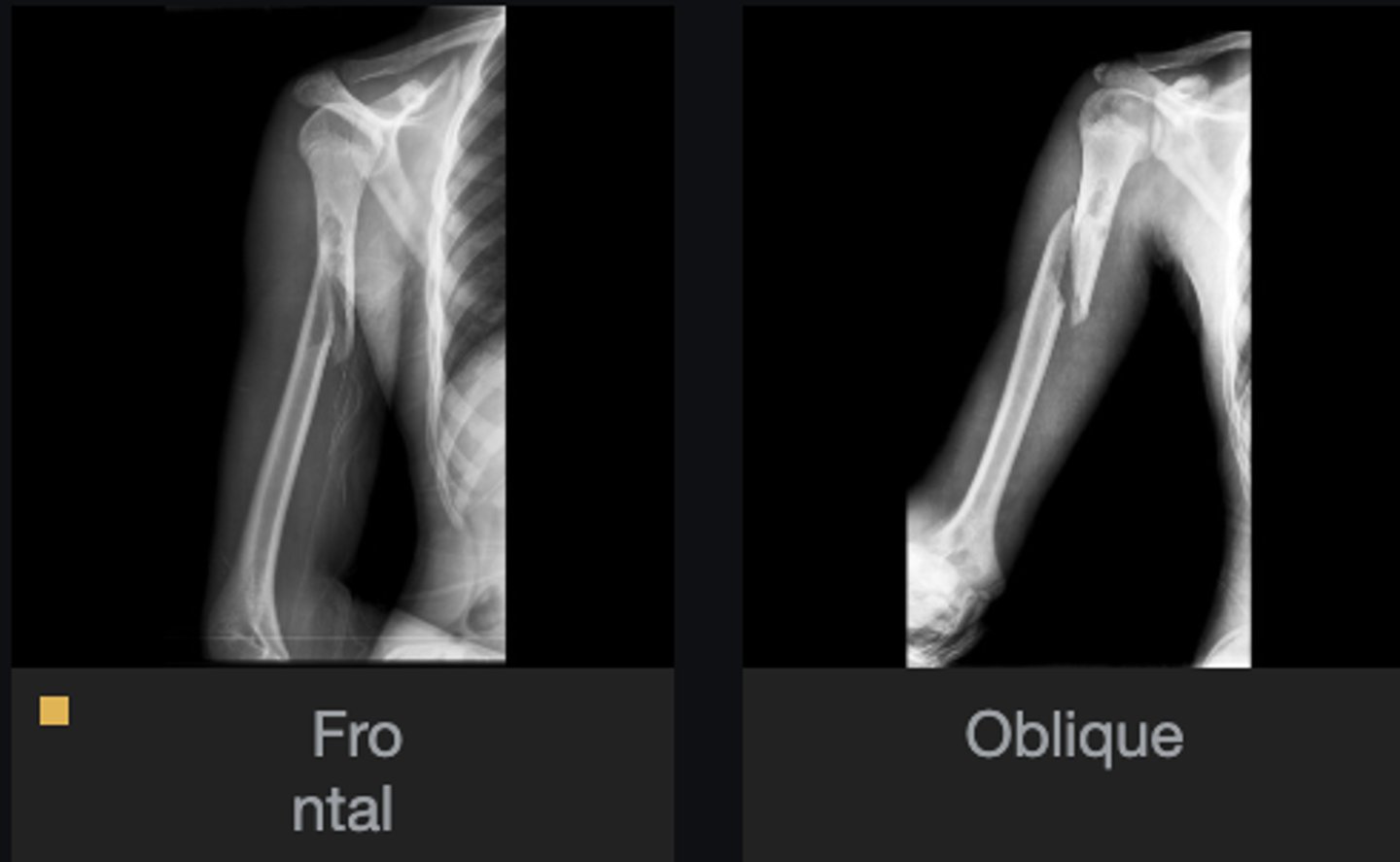
- Distal femur involved
- Elongated stalk
- Cortex and medulla are continuous
- Pointing away from joint
- Narrow base
Describe the bone lesion
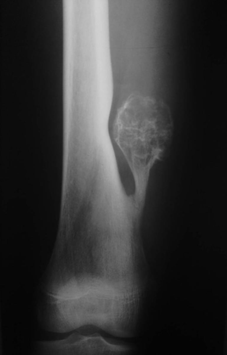
Pedunculated solitary osteochondroma
Most likely diagnosis?

Refer to orthopedist
Next step?

- Fracture
- Malignant transformation (chondrosarcoma)
- Bursitis
- Neurologic injury
Concerns/complications?

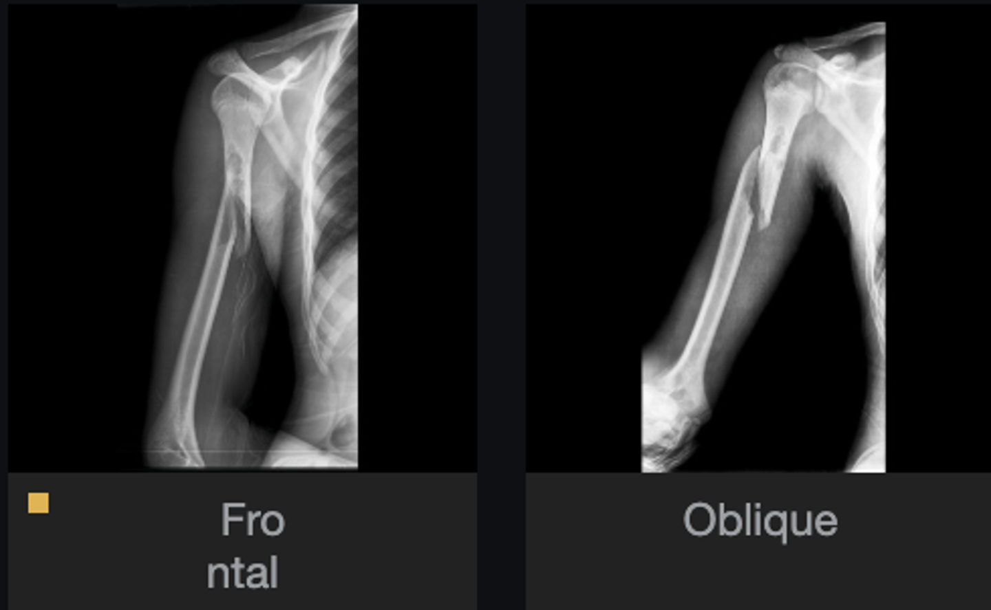
- Distal femur
- Metaphysis
- Anterolateral
- Broad base
- Cortex and medulla continuous
Describe the bone lesion
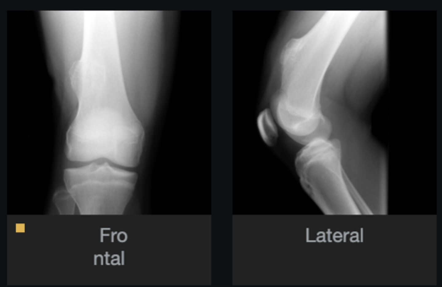
Sessile solitary osteochondroma
Most likely diagnosis?

Hereditary multiple exostoses
Most likely diagnosis?
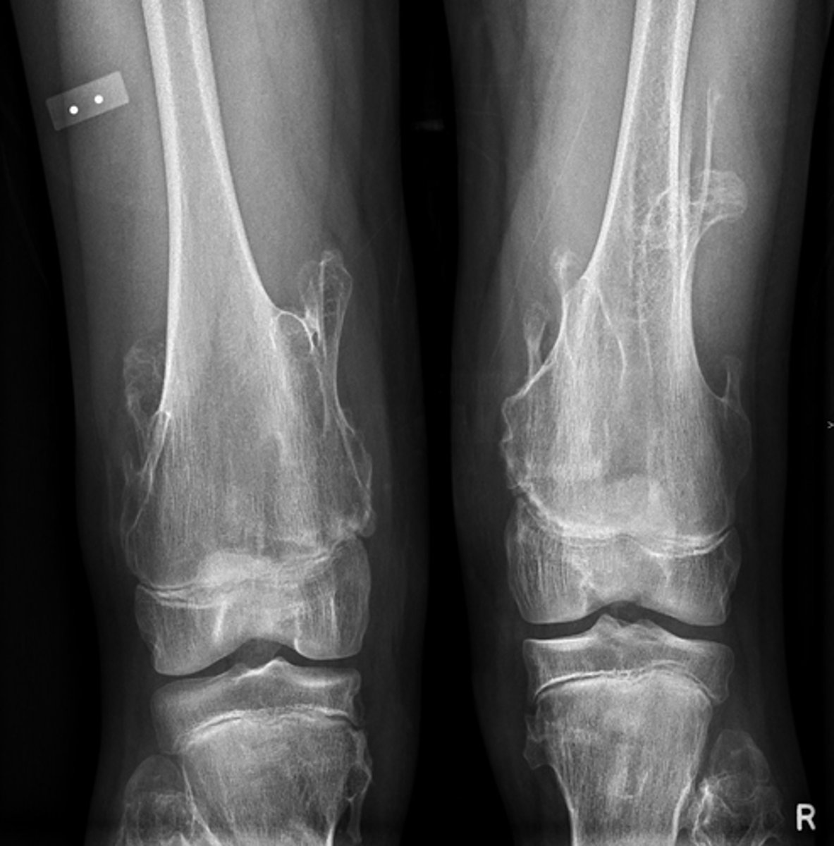
Malignant transformation (5-25%)
Concerns/complications?

Inherited metaphyseal overgrowth
How do you get this?

- Superior aspect of sacrum on right side
- Lucent/opaque brush-border oval
Describe the lesion
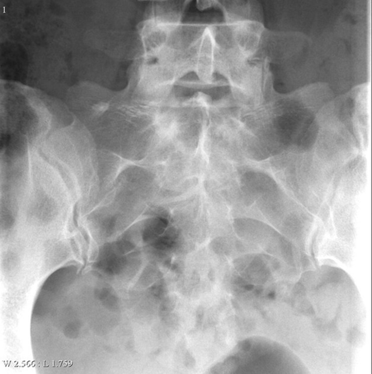
Bone island
Most likely diagnosis?

Nothing
Next step?

Osteochondroma
Concerns/complications?

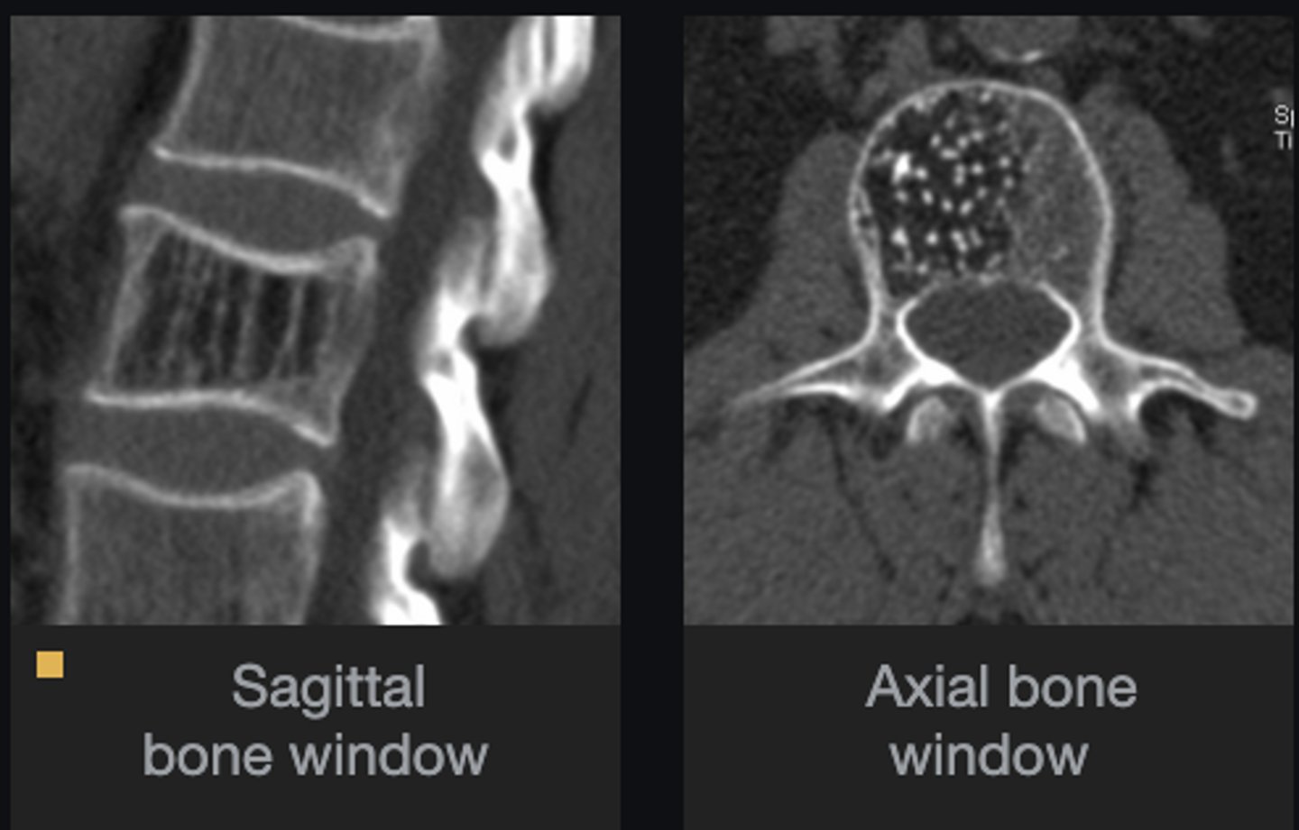
- Ulna
- Diaphysis
- Sclerotic
- Small, lucent nidus (< 1cm) in the cortex
- Geographic)
- Cortical thickening
Describe the lesion
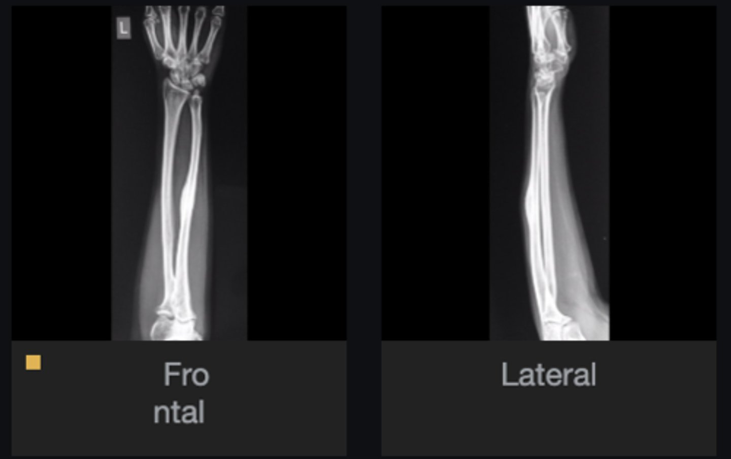
Osteoid osteoma
Most likely diagnosis?

Severe pain (worse at night, relieved by aspirin)
What is the hallmark history of a patient with this lesion?

- Refer to orthopedist
- Adjust spine
Next step?

None
Concerns/complications?

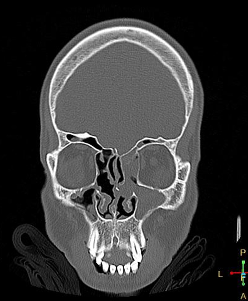
Paranasal sinus
What is the location of the lesion?

Osteoma
Most likely diagnosis?

Headaches
Concerns/complications?

Refer to EENT
What is your next step?
L1
What vertebral level is involved?
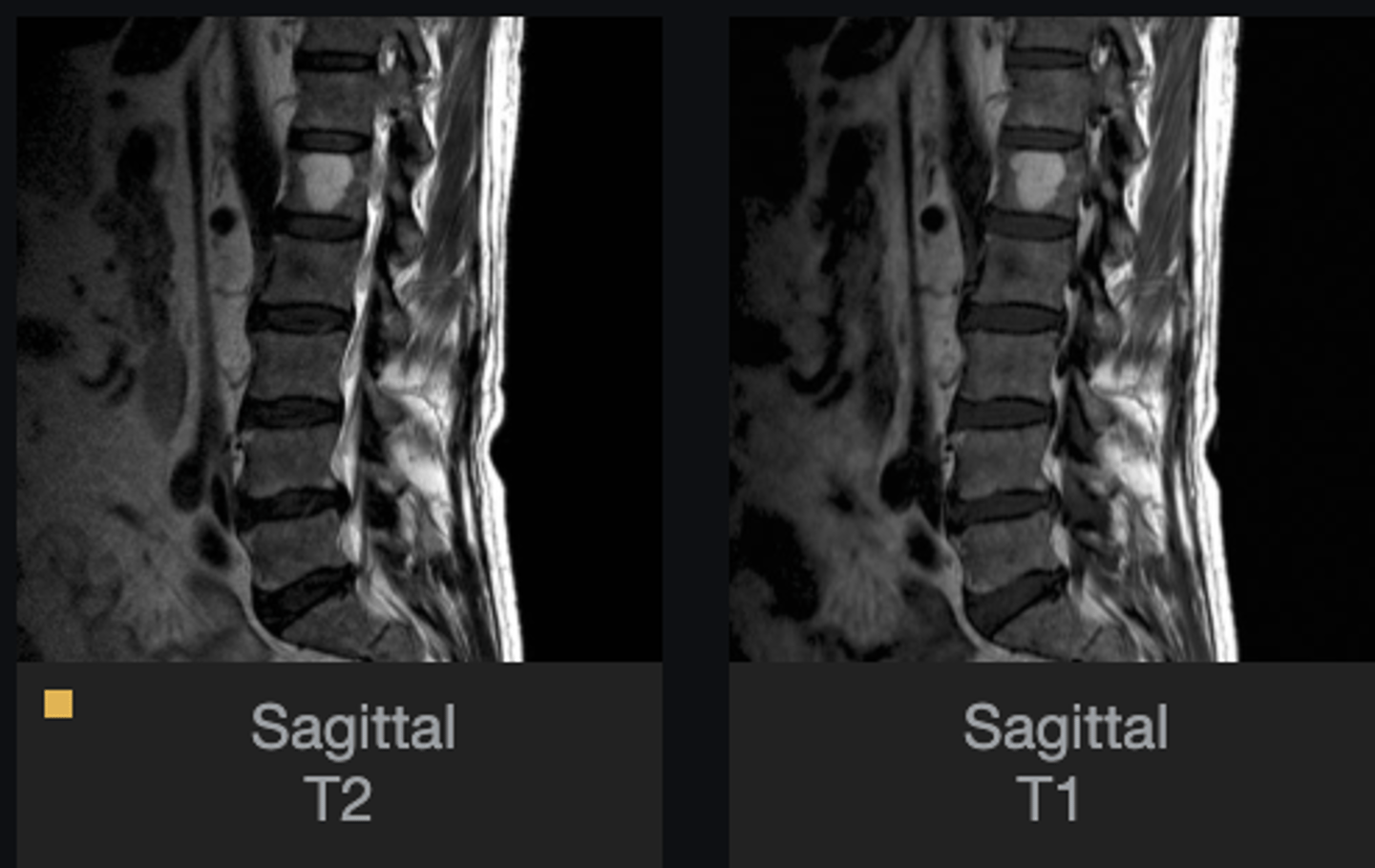
- Solitary vascular neoplasm
- Slow-growing
- Vertical striations (corduroy cloth)
- Fatty lesion
Describe the bone lesion

Vertebral hemangioma
Most likely diagnosis?

- None with this patient
- Expansion (rare) may result in neurologic findings
Concerns/complications?
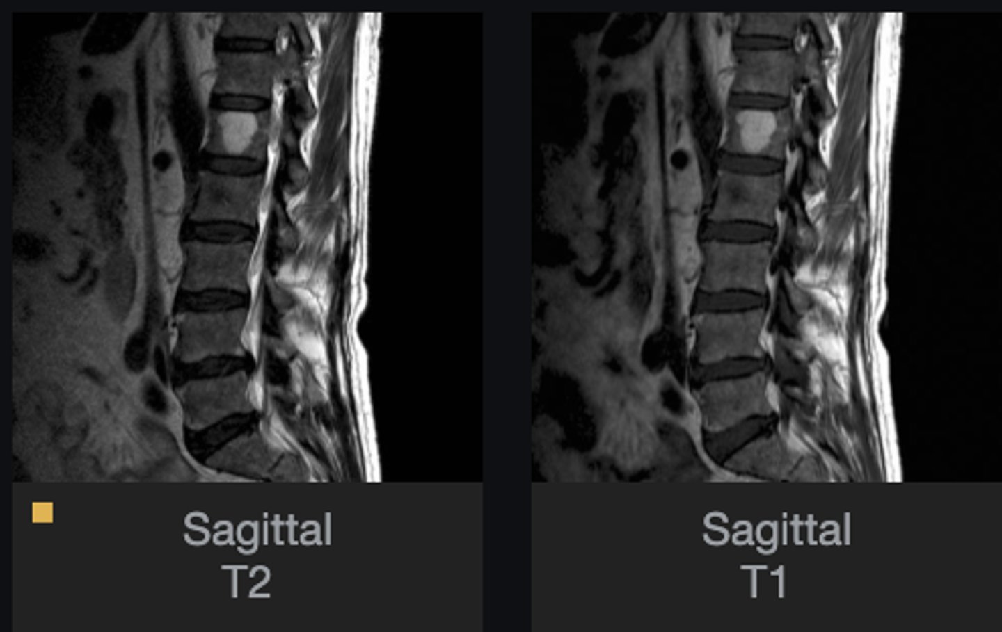
Lumbar
What spinal anatomy is involved?
- Fat body
- Mammillary process
- Posteromedial facets
Are there any distinguishing features?

Vertebral hemangioma
Most likely diagnosis?

Adjust
Next step?

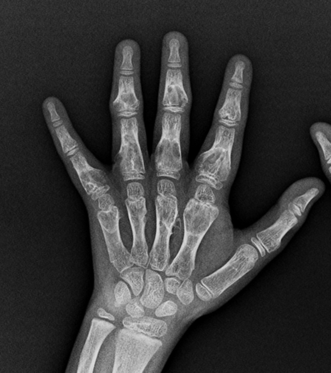
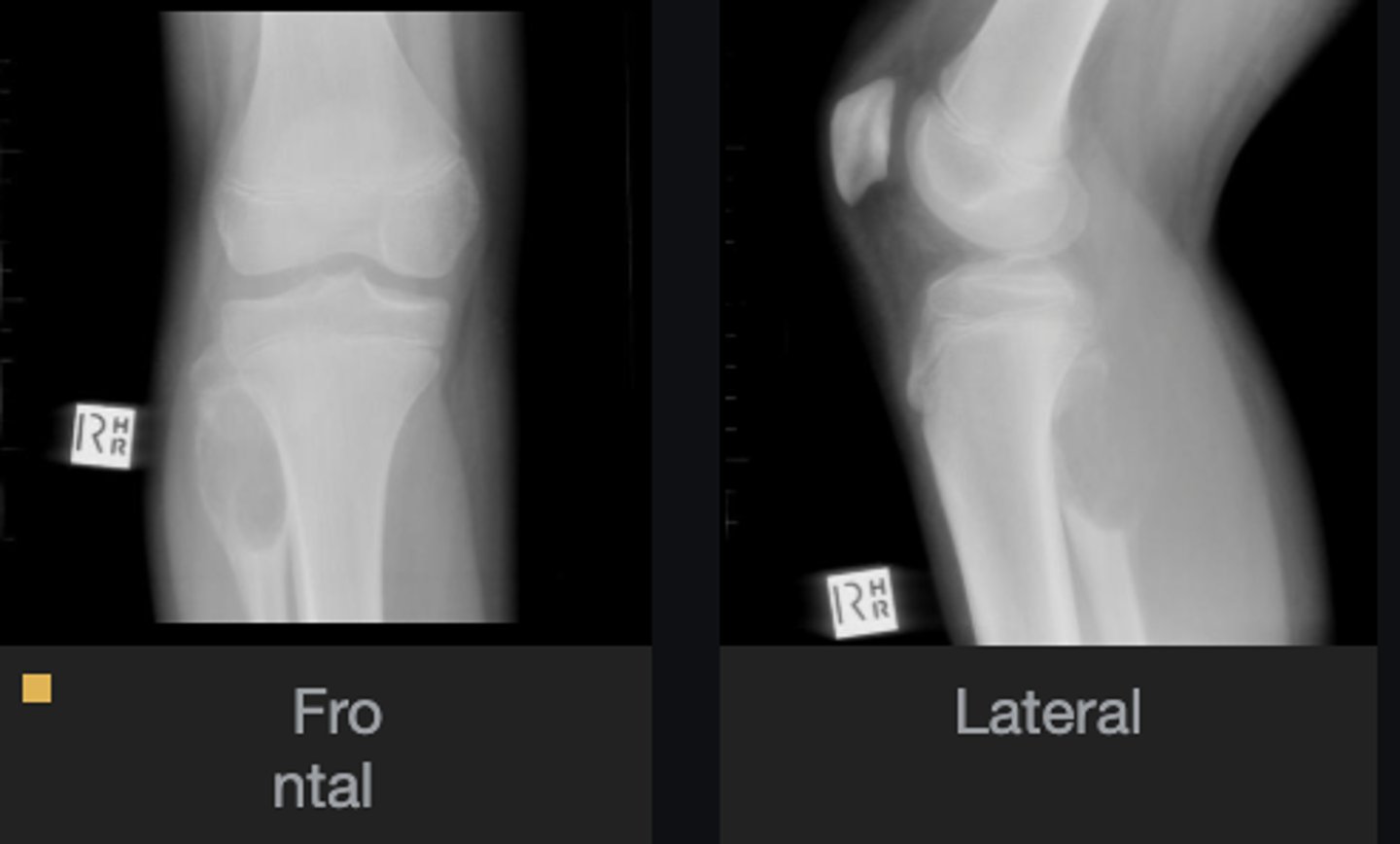
- Head of 5th metacarpal
• Cortical thinning
• Lucency in head
• Pathologic fracture
• Stippled calcification
• Cartilaginous matrix
• Geographic
• Short zone of transition
Describe the lesion
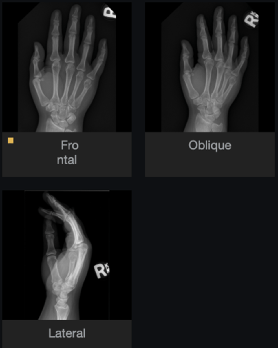
Solitary enchondroma
Most likely diagnosis?

Fracture
What complication has occurred?
Refer to orthopedist
Next step?

Multiple enchondromas (Ollier's disease)
Diagnosis?

- Malignant transformation (10-50%)
- Fracture
Concerns/complications?
- Flocculent calcification
- Metaphysis/diaphysis
- Enchondroma
- Cartilaginous matrix
Describe the lesion

Enchondroma
Most likely diagnosis?

Malignant transformation (rare)
Concerns/complications?
Refer to orthopedist
Next step?

- Solitary
- Eccentric
- Geographic
- Multiloculated
- Fibrous matrix
- Small
- Cortical thinning
- Sclerotic border
Describe the lesion

Non-Ossifying Fibroma
Diagnosis?
Nothing
Next step?

- Anterolateral distal tibia
- Metaphysis
- Septation
- Cortical thinning
- Geographic
- Sclerotic border
- Fibrous matrix
Describe the lesion
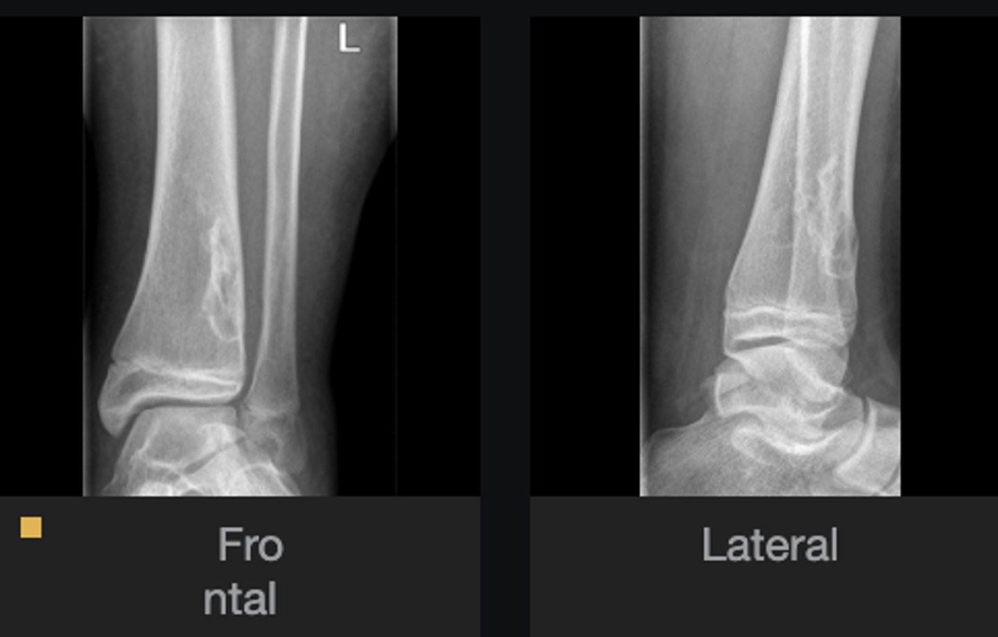
Non-Ossifying Fibroma
Diagnosis?
- Look for fracture
- Refer to orthopedist
Next step?

- Humerus
- Central
- Diaphysis
- Geographic
- Cortical thinning
Describe the lesion
Pathologic fracture
What complication has occurred?

Simple bone cyst
Most likely diagnosis?

Refer to orthopedist
Next step?
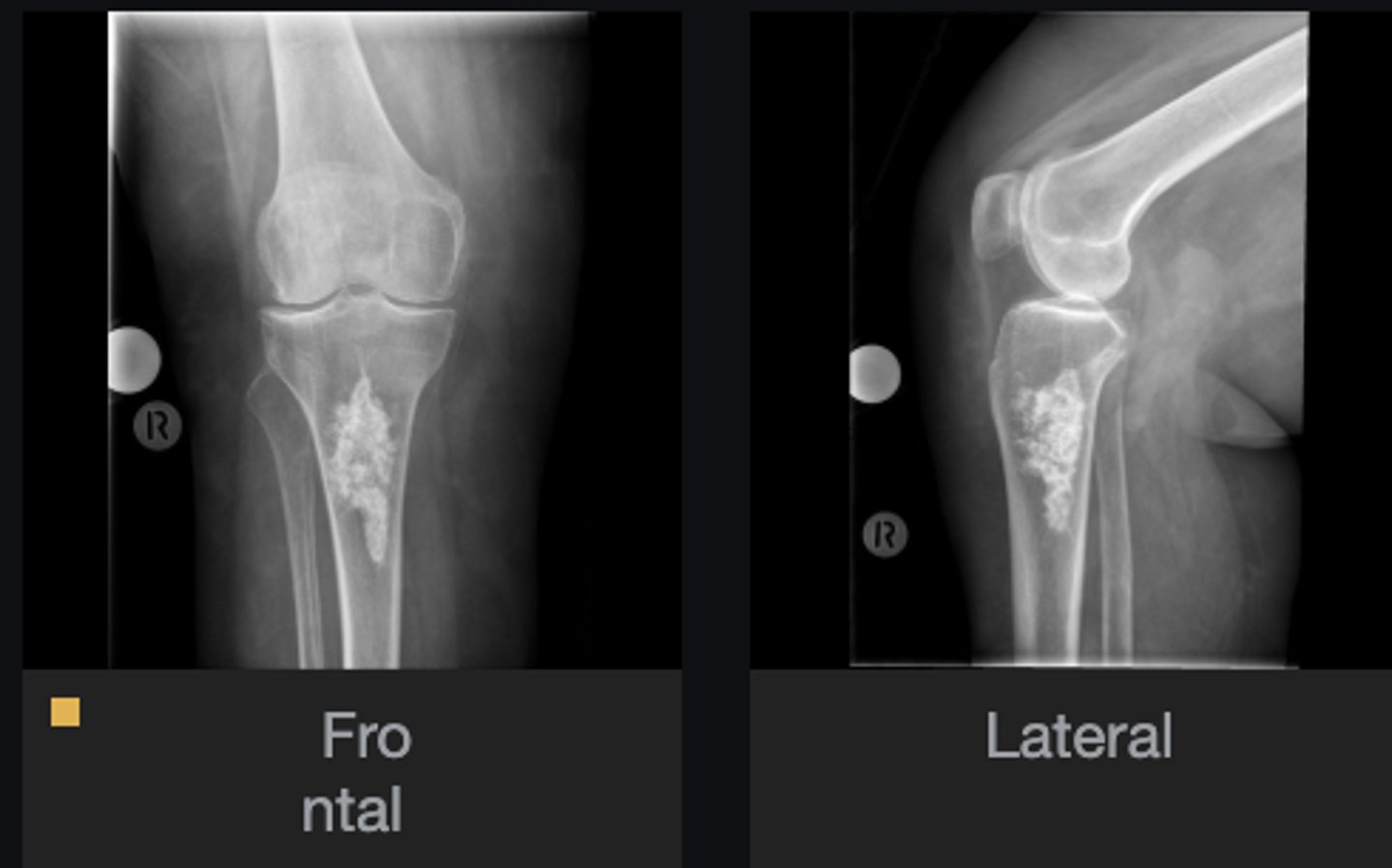
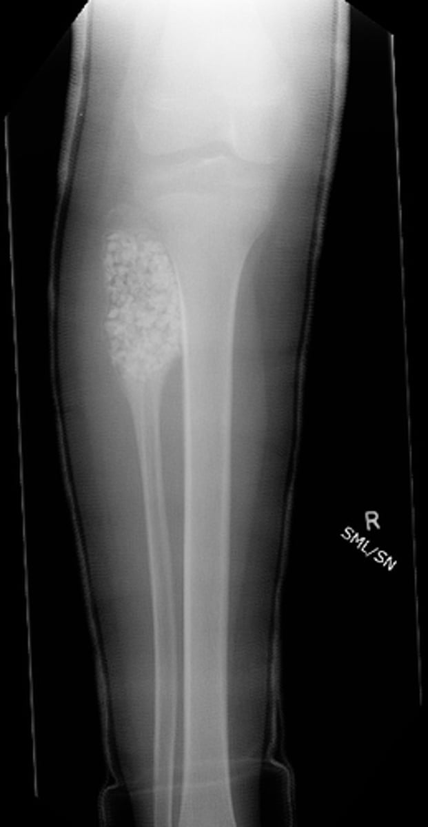
- Proximal fibula
- Metaphysis/diaphysis
- Eccentric
- Saccular ballooning of cortex
- Periosteal buttressing
Describe the lesion

Aneurysmal bone cyst
Most likely diagnosis?

Refer to orthopedist
Next step?
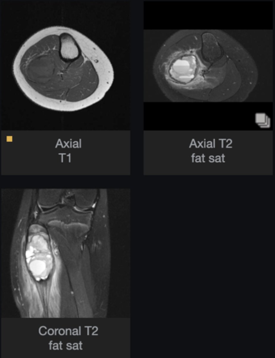
- Fluid-Fluid Levels
- Aneurysmal bone cyst
- Are there any features that help you diagnose this lesion?
- What is the most likely diagnosis?
Bone chips
What has occurred?
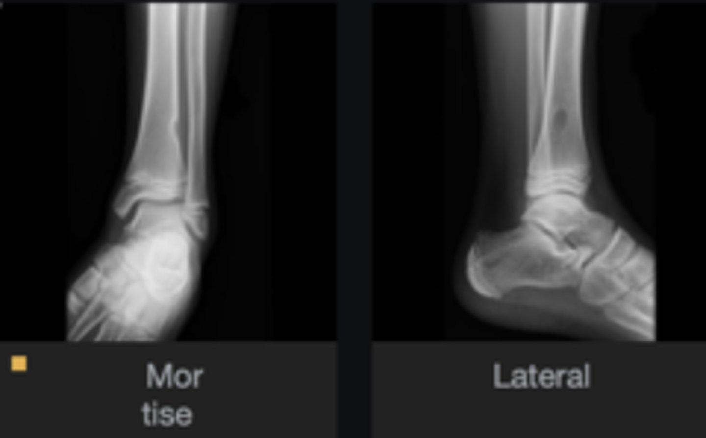
- Body of calcaneus
- Geographic
- Lucent
- Central target sequestrum
Describe the lesion

Intraosseous lipoma
Most likely diagnosis?

Refer to orthopedist or specialist
Next step?
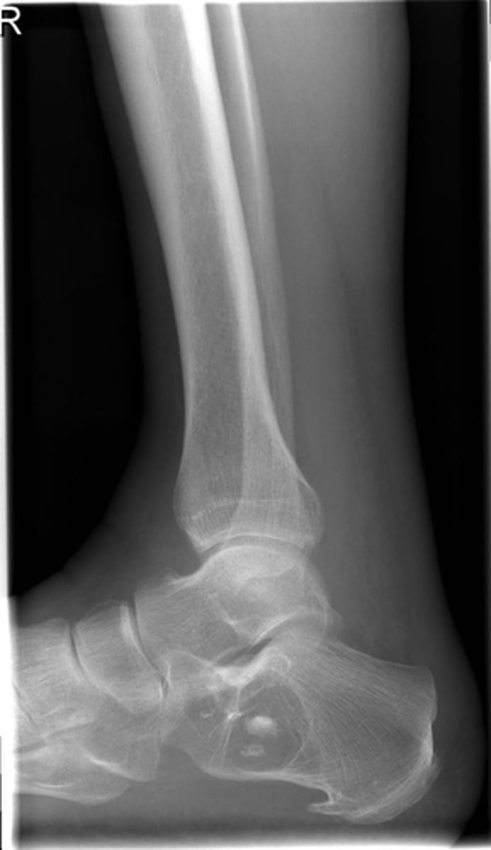
Heel spur
What is going on with the posteroinferior calcaneus?
