Neurobiology Exam 3
1/60
Earn XP
Description and Tags
Chapters 8, 10, 15
Name | Mastery | Learn | Test | Matching | Spaced |
|---|
No study sessions yet.
61 Terms
Motor Systems
Performs so many different motor tasks with speed and accuracy: reflex, rhythmic, and voluntary
What processes give motor systems the ability to perform activities?
1. commands to motor neurons and muscles are distributed in hierarchically interconnected areas of the spinal cord, brain stem, and forebrain.
2. sensory information relating to movement is processed in different systems that operate in parallel.
Movement
A single relocation of a body part, usually resulting from a brief muscle contraction
Reflex
A simple, stereotyped, and unlearned response to particular stimulus
Acts
Complex, sequential behaviors
Motor Plan (Program)
A set of muscle commands that is established before the action occurs
Electromyography (EMG)
Records the electrical activity of muscles
Tendons
Connect muscle to bone in a reciprocal fashion
Antagonistic Muscle
When one muscle group contracts and it stretches the other group
Synergist Muscle
Muscles that act together to move a limb
Skeletal Muscles
Used for movement of the skeleton
What are Skeletal Muscles made of?
Striate muscle
Striate muscle
Overlapping layers of proteins myosin and actin give a striped appearance
Muscle Anatomy: Muscle Belly / Body is…
Whole muscle
Muscle Anatomy: The muscle is made up of…
Muscle Fibers
Muscle Anatomy: Muscle fibers contain…
Myofibrils
Muscle Anatomy: Myofibrils contain…
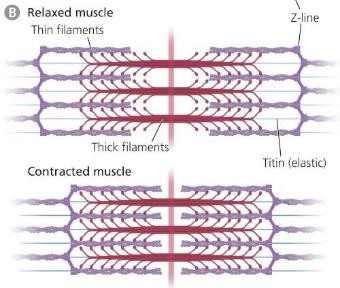
Sarcomeres (Actin and Myosin)

Action of the Muscle: Sliding Filament Model
Contraction of the muscle increases the overlap of actin and myosin filaments with muscle fibers
As they slide past each other, the muscle shortens
Distance between the z-lines shorten
What makes actin and myosin interact?
ATP, which generates contractile muscle force
Neuromuscular Junction (NMJ) Process
Acetylcholine (ACh) is released at the NMJ
ACh reaches receptors on the membranes of muscle fibers
Na++ channels open and sodium ions enter the cytoplasm of the muscle fiber
The sodium influx also causes the release of stored calcium ions into the muscle
The calcium ions cause the actin and myosin to slide along one along another to produce a contraction
For the muscle to relax the calcium lvls must decrease a bit
Tetanus
If motor action potentials occur very rapidly (high frequency over short time) there is no time to reduce calcium, which causes uncontrollable sustained muscle contractions.
Cause of Tetanus:
Caused by tetanus toxin (bacteria) which blocks GABA release from inhibitory neurons, so there is an overexcitation of motor neurons and powerful contractions
Rigor Mortis
2-3 hours after death, all stored calcium is released and triggers muscle contractions
Muscle stay contracted until they start to disintegrate
Myasthenia Gravis
Autonimmune disorder where the body produces antibodies that break down ACh receptors.
Muscle contractions become weak if not treated, they may stop breathing
Treatment:
Immunosuppressants to decrease antibodies production
Administer an enzyme that breaks down acetylcholinesterase (which is a ACh inhibitor)
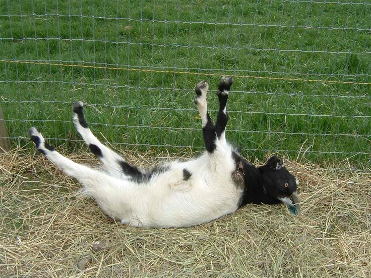
Myotonia
Neuromuscular condition in which the relaxation of a muscle is impaired
Ex.) Myotonic “fainting” goats
Motor Unit
Motoneuron’s axon and all of its target fibers
Motor units very in what?
In hoe forcefully they twitch and how quickly they fatigue
When muscle contractions get stronger…
Larger motor units are recruited
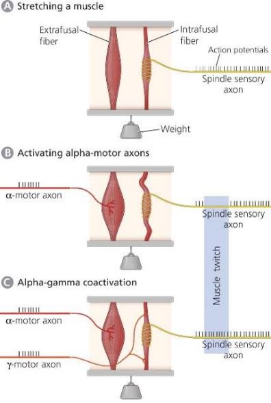
What do spiraling sensory and γ-motor axons innervate?
The intrafusal fibers in muscle spindles
Alpha motor neurons function
Contract muscles
Gamma motor neurons function
Provide proprioceptive info to muscle stretch
Coactivation of Alpha-Gamma motor axons…
Allows muscle spindles to sense muscle stretch even during a contraction
Cardiac Muscle
Cardiac muscle fibers are electrically coupled to one another (use gap junctions)
Rhythmic (no neural input requested)
Neural input regulates Heart Rate (HR)
Norepinephrine (NE) increased HR
Ach decrease HR
Cardiac Muscle: ACh Receptors…
In the heart are metabotropic (muscarinic)
At the NMJ are ionotropic (nicotinic)
Endocrine Glands
Secrete hormones into the blood
Magnocellular neurons in the hypothalamus release oxytocin or vasopressin posterior pituitary gland
Oxytocin release is involved in the ejection of mother’s milk and uterine contractions
Vasopressin (ADH) helps the kidney retain water to regulate BP
Cells in the anterior pituitary gland secrete hormones that control other glands
Portal Veins Function:
Carry releasing factors to the anterior pituitary, where they stimulate the release of pituitary hormones
What causes milk ejection from mammary glands?
Burst of action potentials in supraoptic neurons
Eacj burst causes a pulse of oxytocin release
HPA Axis:
Releasing Hormone: CRH (corticotropin releasing hormone)
Pituitary Hormone: ACTH (adrenocorticotropic hormone, aka corticotropin)
Principle End Organ: Cortex of the adrenal gland
End Organ Hormones: Cortisol
HPG Axis:
Releasing Hormone: GnRH (Gonadotropin releasing hormone)
Pituitary Hormone: LH and FSH (luteinizing hormone and follicle stimulating hormone)
Principle End Organ: Gonads (testes or ovaries)
End Organ Hormones: Testosterone or Estradiol
HPT Axis:
Releasing Hormone: TRH (Thyrotropin releasing hormone)
Pituitary Hormone: TSH (Thyroid stimulating hormone, aka Thyrotropin)
Principle End Organ: Thyroid
End Organ Hormones: Thyroxin and other thyroid hormones
What does hippocampus lesion affect?
Spatial memory recall in part through increased stress hormone release
What does early exposure to the estrogen mimic, DES, do?
Causes adult obesity
DES: Diethylstilbestrol (
Reflex
An involuntary, stereotyped response to stimulus
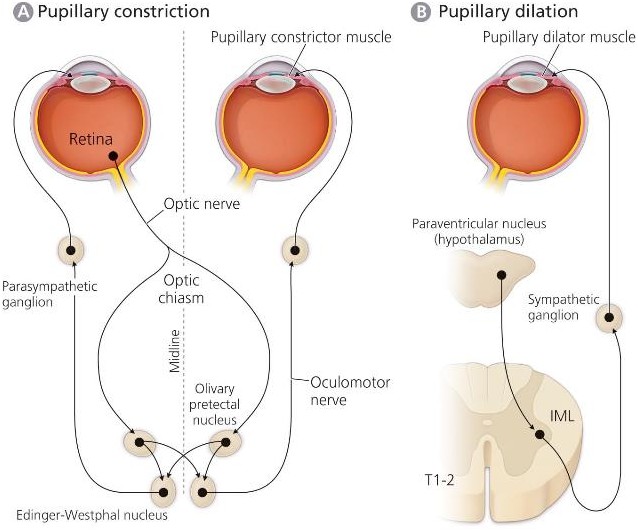
Light-Induced Pupillary Constriction
A reflex that occurs against a background of sympathetic activation promoting pupillary dilation
The default state of pupil size is Pupil Dilation
Light is modulation stimulus
Is a reflex that antagonizes pupillary dilation
Pupillary Constriction Pathway (Response to light)
From Retina
Through Optic nerve
Integrated through Olivary Pretectal Nucleus to Edinger-Westphal Nucleus
Through Oculomotor nerve
Through Parasympathetic Ganglia
To Pupillary constrictor muscle

Pupillary Dilation Pathway (Tonically active)
From Hypothalamus
Through IML
Through Parasympathetic ganglia
To Pupillary dilator muscle
Ways reflexes protect us from harm:
Tears
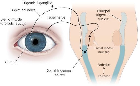
Blinking from corneal stim involves a circuit through spinal trigeminal and facial motor nuclei
Withdraw from pain causes muscle flexors to contract and muscle extensors to relax. Goes through the spinal cord and includes a set of inhibitory interneurons
The corneal eye blink reflex involves:
Three sets of neurons
Two cranial nerves: Trigeminal Nerve, Facial Nerve
The cornea is innervated by the trigeminal nerve that projects to the spinal trigeminal nucleus
Neurons in the spinal trigeminal nucleus project to neurons in the facial motor nucleus that innervates the eyelid closing muscles (Orbicularis oculi)
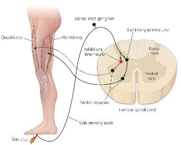
How the leg withdrawal reflex moves the leg out of harm’s way:
Reflex: Step on tack and withdraw your foot
Activates the mechanosensitive neurons (foot) that projects into the spinal cord
Synapse on excitatory interneurons that project to hamstring motor neurons
Sensory neurons also synapse on the inhibitory interneurons (red) that project to motor neurons innervating the quadriceps
Knee jerk response is a stabilizing stretch reflex…
Reflex: Tapping the patellar tendon results in your leg straightening
Stretched tendon —> stretches the quadriceps
Activates quadriceps muscle spindle receptor —> spinal cord motor neurons —> quadricep contract
Also inhibit interneurons that project to motor neurons that innervate hamstring
Reciprocal Innervation
Sending opposing commands to antagonistic muscles
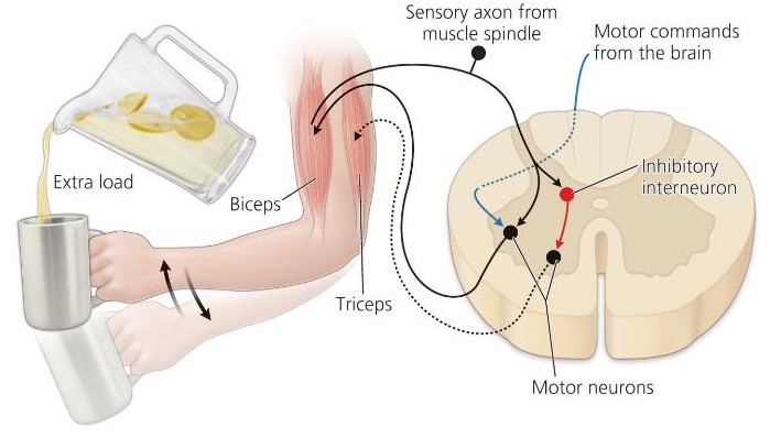
When does the bicep stretch reflex stabilize the hand?
When it experiences a change in load
Motor commands from the brain can adjust the “set point” for limb position
Eye Movements
Oculomotor — 3rd cranial nerve (inward)
Trochlear — 4th cranial nerve (downward)
Abducens — 6th cranial nerve (outward)
Optokinetic Reflex
Slow tracking of moving objects. Eyes reset position to keep images stable on retina. Needs visual input (not in dark)
Eye Stabilization: Optokinetic Reflex Circuitry
Retina: Photoreceptors — bipolar — ganglion cells (optic nerve)
Some ganglion cells send projections to the nucleus of the optic tract — then on to the vestibular complex for each eye
Involves reciprocal innervation for both eyes to look in the same direction
Vestibular Ocular Reflex
Stabilizes gaze by countering head movements
Head moves side to side while eyes stayed focused, therefore eyes are moving in the opposite direction
Turn head LEFT Semicircular canal input to vestibular complex to turn eyes to the RIGHT
So abducens nucleus to turn Right E (R) and oculomotor nucleus to turn Left E (R)