Human Physiology
0.0(0)
0.0(0)
Card Sorting
1/86
Earn XP
Description and Tags
Midterm #2 Up to slide 81/137
Study Analytics
Name | Mastery | Learn | Test | Matching | Spaced | Call with Kai |
|---|
No study sessions yet.
87 Terms
1
New cards
What are the three different type of muscle tissue?
- Skeletal Muscle
- Cardiac Muscle
- Smooth Muscle
- Cardiac Muscle
- Smooth Muscle
2
New cards
What is the primary function of Muscle Tissue?
To Generate Force to Muscle Tension.
(creates movement, maintains posture, stabilizes joints, generates heat, regulates flow of materials through hollow organs, guards body entrances and exits.
(creates movement, maintains posture, stabilizes joints, generates heat, regulates flow of materials through hollow organs, guards body entrances and exits.
3
New cards
What is the Structure of Skeletal Muscle Tissue?
Long, cylindrical striated muscle fibres; cells are MULTINUCLEATED.
4
New cards
Where is Skeletal Muscle Tissue located?
Mostly attaches to Skeleton; contraction can produce movement of a body part.
5
New cards
Is skeletal muscles voluntary or involuntary?
Voluntary (controlled by conscious thought
6
New cards
What is the function of skeletal muscle tissue
Produces movement of the body.
7
New cards
What is the structure of cardiac muscle tissue?
Short, wide, branching striated cardia muscle cells with intercalated discs; cells have a single nucleus or two nuclei.
8
New cards
Where are cardiac muscles located?
The heart
9
New cards
Are cardiac muscle voluntary or involuntary?
Involuntary (not controlled by conscious thought)
10
New cards
What are the functions of cardiac muscle tissues?
Produces beating of the heart
11
New cards
How are skeletal muscles and cardiac muscles similar?
They are both striated muscle cells.
12
New cards
What is the structure of smooth muscle tissue?
Thin, smooth muscle cells, generally joined by gap junctions; cells have a single nucleus.
13
New cards
Where is smooth muscle tissue found?
Walls of hollow organs, as well as in the skin, and the eyes.
14
New cards
Are smooth muscles voluntary or involuntary?
Involuntary
15
New cards
What is the function of smooth muscle tissue?
- Changes diameter of hollow organs
- Causes hairs to stand erect
- Adjust the shape of the lens and the size of the pupil of the eye.
- Causes hairs to stand erect
- Adjust the shape of the lens and the size of the pupil of the eye.
16
New cards
What is smooth muscle tissue linked together by?
Gap junctions which allows for synchronized contractions.
17
New cards
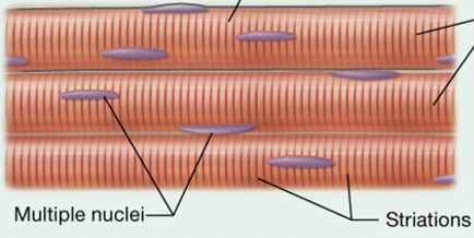
What kind of muscle tissue is this?
Skeletal Muscle Tissue
18
New cards
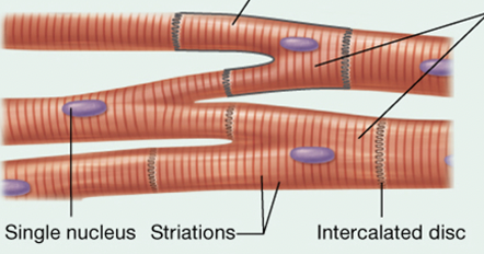
What kind of muscle tissue is this?
Cardiac Muscle Tissue
19
New cards
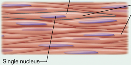
What kind of muscle tissue is this?
Smooth Muscle Tissue.
20
New cards
What are the 5 important properties of muscle cells?
1. Contractility
2. Excitability
3. Conductivity
4. Extensibility
5. Elasticity
2. Excitability
3. Conductivity
4. Extensibility
5. Elasticity
21
New cards
What is contractility?
Gives the muscle the ability to contract; proteins in the cell draw closer together.
22
New cards
What is excitability?
The ability for a muscle to respond to a stimulus (e.g., skeletal muscle fibres respond to electrical signals from a motor neuron)
23
New cards
What is conductivity?
The ability to conduct electrical signals across the entire plasma membrane
24
New cards
What is extensibility?
The ability to be stretched (up to 3 times resting length) without rupturing
25
New cards
What is elasticity?
The ability to return to original length after stretching
26
New cards
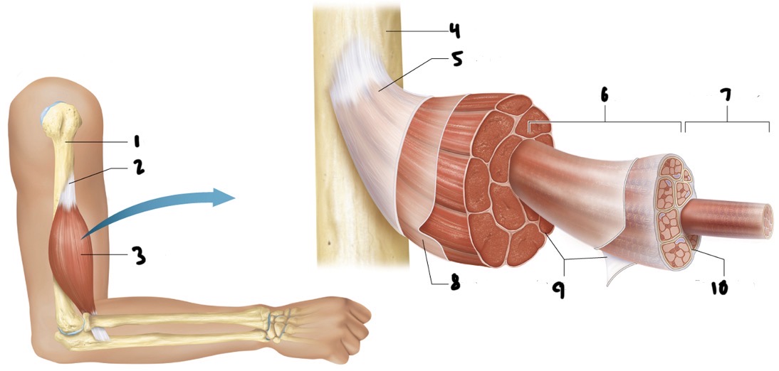
Label the following correctly:
- Fascicle
- Epimysium
- Endomysium
- Muscle cell fibre
- Bone
- Tendon
- Skeletal muscle
- Bone
- Muscle fascia
- Perimysium
- Fascicle
- Epimysium
- Endomysium
- Muscle cell fibre
- Bone
- Tendon
- Skeletal muscle
- Bone
- Muscle fascia
- Perimysium
1. Bone
2. Tendon
3. Skeletal muscle: wrapped by the epimysium
4. Bone
5. Muscle fascia
6. Fascicle: a bundle of muscle cell fibres, they vary from muscle to muscle (arrangement). Wrapped by perimysium
7. Muscle cell fibre: wrapped by endomysium
8. Epimysium
9. Perimysium
10. Endomysium
2. Tendon
3. Skeletal muscle: wrapped by the epimysium
4. Bone
5. Muscle fascia
6. Fascicle: a bundle of muscle cell fibres, they vary from muscle to muscle (arrangement). Wrapped by perimysium
7. Muscle cell fibre: wrapped by endomysium
8. Epimysium
9. Perimysium
10. Endomysium
27
New cards
What is the structure of the skeletal muscle fibre?
Skeletal muscle tissue consists of many fibres and surrounding the edomysium (extracellular matrix).
- Fibres are thin cylinders; lengths up to 30 centimetres and thickens up to 100 micrometers.
- Fibres are thin cylinders; lengths up to 30 centimetres and thickens up to 100 micrometers.
28
New cards
Where is calcium stored and what part of the muscle fibre releases calcium ions (required for contraction).
It is stored within the sarcoplasmic reticulum.
29
New cards
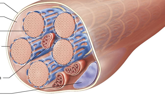
Label to following muscle cell (myocyte) from top to bottom.
- Sarcoplasmic reticulum
- Myofibril
- Mitochondrion
- Sarcoplasm
- Nucleus
- Sarcolemma
- Sarcoplasmic reticulum
- Myofibril
- Mitochondrion
- Sarcoplasm
- Nucleus
- Sarcolemma
1. Sarcolemma
2. Sarcoplasm
3. Myofibril
4. Sarcoplasmic reticulum
5. Mitochondrion
6. Nucleus
2. Sarcoplasm
3. Myofibril
4. Sarcoplasmic reticulum
5. Mitochondrion
6. Nucleus
30
New cards
Where does extracellular fluid enter through?
It goes into the opening of the T-tubule (transverse tubules). T-tubules are extensions of the sarcolemma that surround each myofibril.
31
New cards
What makes up the triad?
The sarcoplasmic reticulum and the sarcolemma together create the triad.
32
New cards
What are the three types of myofilaments?
Thick filaments: bundles of contractile protein myosin
Thin filaments: proteins actin, tropomyosin, and troponin
Elastic filaments: Single massive, spring-like structural protein (titin); Stabilizes myofibril structure; resists structure; resists excessive stretching.
Thin filaments: proteins actin, tropomyosin, and troponin
Elastic filaments: Single massive, spring-like structural protein (titin); Stabilizes myofibril structure; resists structure; resists excessive stretching.
33
New cards
What is myosin?
Myosin consists of globular heads at each end linked by intertwining tails. Heads are connected to tails by hinge-like neck. Each head has active site that binds with actin. Myosin tails from opposite sides are anchored together at the M-line
34
New cards
What is actin?
Multiple actin subunits string together; form two intertwining strands in functional thin filament. Each bead-shaped actin has active site; binds with myosin heads.
35
New cards
What is tropomyosin?
Long, rope-like regulatory protein; twists around actin, covering up active sites
36
New cards
What is troponin?
Small globular regular protein; holds tropomyosin in place; assists with turning contractions on and off.
37
New cards
What kind of filaments are found within the I band?
Only thin filaments.
38
New cards
Where is the Z disc and what does it do?
The Z disc is in the middle of the I band and it is composed of structural proteins that anchor thin filaments in place and to one another, Serve as attachment points for elastic filaments, attach sarcomeres to one another across entire diameter of muscle fiber.
39
New cards
Where is the A band and what important structures can you find within it?
The A band is in-between both of the I bands. Within the A band is the zone of overlap with both thick and thin filaments that generate tension during contraction. The H zone is also in the A band where only thick filaments exists. The M Line is in the middle of the A band and it is a dark line in the middle. These structural proteins hold thick filaments in place; serve as anchoring point for elastic filaments.
40
New cards
What is the functional unit of contraction. Hint: extends from one Z-disc to the next.
A sarcomere.
41
New cards
What are the 3 stages within the sliding filament mechanism?
Myosin heads attach to actin
Pull thin filaments toward M-line
Brings Z-discs closer together (shorten sarcomere).
Pull thin filaments toward M-line
Brings Z-discs closer together (shorten sarcomere).
42
New cards
What happens to I band, H zone, and A band during the sliding filament mechanism?
Both the I band and the H zone narrow, and the A band is unchanged.
43
New cards
What is cross-bridge formation?
When myosin and actin are bound to each other.
44
New cards
What is cross-bridge cycling?
The on and off interaction between myosin and actin that pulls thin filaments toward the M-line.
45
New cards
Put the following in order to match the steps of the cross-bridge cycle:
- Power stroke: ADP and Pi are released from myosin. this causes myosin to pull actin toward the M-line as it pivots to a relaxed position. Cross-bridge is at 45 degree angle relative to thick filament.
- Myosin Head becomes Cocked once ATP is bound and energy is gathered through ATP hydrolysis
- Cross-bridge formation: Myosin binds to actin at a 90 degree angle relative to the thick filament
- Another ATP binds to myosin and this breaks the attachment to actin.
1. Myosin Head becomes Cocked once ATP is bound and energy is gathered through ATP hydrolysis
- Cross-bridge formation: Myosin binds to actin at a 90 degree angle relative to the thick filament
2. Cross-bridge formation: Myosin binds to actin at a 90 degree angle relative to the thick filament
3. Power stroke: ADP and Pi are released from myosin. this causes myosin to pull actin toward the M-line as it pivots to a relaxed position. Cross-bridge is at 45 degree angle relative to thick filament.
4. Another ATP binds to myosin and this breaks the attachment to actin.
46
New cards
What needs to happen in order for the contraction cycle to repeat?
The stimulus to contract continues and ATP is available.
47
New cards
How many times does the contraction cycle repeat for an average contraction?
20-40 times for each myosin head in each sarcomere.
48
New cards
A property of excitable cells is that they produce________.
Action potentials
49
New cards
True or false. Muscle fibers are excitable cells.
True.
50
New cards
What are excitable cells defined by?
A rapid and temporary change in membrane potential. (a large increase to positive mV values followed by a quick return to negative mV values
51
New cards
What are excitable cells generated by?
The opening and closing of Na+ and K+ channels.
52
New cards
True or false.
The membrane is permeable to Na+ and K+ without the presence of channels and pumps to allow them to pass across.
The membrane is permeable to Na+ and K+ without the presence of channels and pumps to allow them to pass across.
False, The membrane would be impermeable.
53
New cards
How are the Na+/K+ concentration gradients maintained?
They are maintained by the Na+/K+ pump.
54
New cards
True or false.
ATP is not required in order to pump these ions (Na+ and K+) against their gradients.
ATP is not required in order to pump these ions (Na+ and K+) against their gradients.
False, ATP is required.
55
New cards
Between the Cytosol side and the extracellular fluid side which has high/lo concentrations of Na+/K+?
The extracellular fluid side (ECF) has low K+ and high Na+ concentrations.
The cytosol side has High K+ and low Na+ concentrations.
The cytosol side has High K+ and low Na+ concentrations.
56
New cards
What is a channel that is always open called?
Leak channels; they are like pores in the membrane. Substances that can pass will flow down their gradient.
57
New cards
What are gated channels and what are the three types?
Gated channels can be chemically electrically or mechanically gated.
58
New cards
Explain a chemically gated channel.
Gating controlled by a molecule (ligand) that binds to the protein receptor
59
New cards
Explain an electrically gated channel.
Gating controlled by voltage differences across the plasma membrane.
60
New cards
Explain a mechanically gated channel.
Physical forces put pressure on the membrane and the channel pops open.
61
New cards
What is the potential difference across a cells plasma membrane called?
Membrane potential.
62
New cards
What is the electrical potential difference across the plasma membrane measured in?
Millivolts
63
New cards
True or False.
Membrane potential is different for every cell type in the human body but it is always negative. when the cell is at rest.
Membrane potential is different for every cell type in the human body but it is always negative. when the cell is at rest.
True. (E.g., -90mV)
64
New cards
How is Membrane potential primarily maintained?
It is maintained by Na+ and K+ concentration differences across the plasma membrane
65
New cards
Match the following to its correct function:
1. concentration gradients
2. electric gradients
- Favor Na+ flowing into the cell and K+ flowing out of the cell
- Favor flow into the cell (due to the negative resting membrane)
1. concentration gradients
2. electric gradients
- Favor Na+ flowing into the cell and K+ flowing out of the cell
- Favor flow into the cell (due to the negative resting membrane)
1. Favor Na+ flowing into the cell and K+ flowing out of the cell
2. Favor flow into the cell (due to the negative resting membrane)
2. Favor flow into the cell (due to the negative resting membrane)
66
New cards
What is the electrochemical gradient?
The sum of both chemical and electrical forces. Each ion has its own electrochemical gradient.
67
New cards
What is the approximate value of the resting membrane potential across the sarcolemma of skeletal muscle fibers?
-90mV
68
New cards
What is the depolarization stage?
When membrane potential becomes less negative.
In response to a stimulus; voltage-gated Na+ channels open and Na+ enter the cell, making the membrane potential less negative.
In response to a stimulus; voltage-gated Na+ channels open and Na+ enter the cell, making the membrane potential less negative.
69
New cards
What is the repolarization stage?
Membrane potential returns to more negative state.
Na+ channels close while voltage-gated K+ channels open and K+ leave the cell, making the membrane potential more negative again.
Na+ channels close while voltage-gated K+ channels open and K+ leave the cell, making the membrane potential more negative again.
70
New cards
What stage comes before the action potential is triggered?
The resting stage; before a stimulus arrives, the membrane is at the resting membrane potential (RMP) and voltage-gated Na+ and K+ channels are closed.
71
New cards
What kind of channels are in the sarcolemma?
Voltage-gated Na+ and K+ channels, these are electrically gated channels, meaning they will open in response to a change in voltage across the sarcolemma.
72
New cards
What are the 4 steps of skeletal muscle contraction?
1. Excitation phase
2. Excitation-contraction coupling
3. Contraction phase
4. Muscle relaxation
2. Excitation-contraction coupling
3. Contraction phase
4. Muscle relaxation
73
New cards
What is a skeletal muscle fibre innervated by?
A motor neuron, in order to contract, a skeleton muscle fibre must receive a stimulus from the motor neuron.
74
New cards
True or false.
1 motor neuron communicates with multiple muscle fibres.
1 motor neuron communicates with multiple muscle fibres.
True.
75
New cards
What is a motor unit compare to a motor neuron?
A motor unit consists of 1 motor neuron and all the muscle fibres it innervates.
76
New cards
What is the neuromuscular junction?
The connection point between the motor neuron and a muscular fibre. This is where a nerve impulse (action potential) is transmitted to the sarcolemma of the muscle fibre.
77
New cards
What are the three components of the neuromuscular junction?
1. Axon terminal: End of the axon where the electrical signal is transmitted to a chemical signal. Contains voltage-gated Ca2+ ion channels in the membrane and synaptic vesicles filled with a neurotransmitter; Acetylcholine (ACh)
2. Synaptic cleft: The space between the axon terminal and muscle fibre
3. Motor end plate: Specizlized region of the muscle fibre sarcolemma. Folded surface with a high density of ligand-gated Na+ channels.
2. Synaptic cleft: The space between the axon terminal and muscle fibre
3. Motor end plate: Specizlized region of the muscle fibre sarcolemma. Folded surface with a high density of ligand-gated Na+ channels.
78
New cards
What is a neurotransmitter?
A chemical that transmits a signal from a neuron --> triggers changes in a target tissue.
79
New cards
What occurs in the excitation phase: Events at the neuromuscular junction.
1. An action potential arrives at the axon terminal and triggers Ca+ channels in the axon terminal to open.
2. Ca2+ entry triggers exocytosis of synaptic vesicles
3. Synaptic vesicles release ACh into the synaptic cleft
4. ACh binds to ligand gated ion channels (chemically gated ion channels in the motor end plate
5. Ion channels open and Na+ enter the muscle fibre
6. Entry of Na+ depolarizes the sarcolemma (membrane) locally, producing an end-plate potential
80
New cards
Put the following in the correct order for skeletal muscle relaxation:
- Active sites on actin become blocked again
- Sarcolemma returns to resting membrane potential due
to K+ exit through voltage-gated K+channels
- Acetylcholinesterase* breaks down ACh
- Ca2+ pumped back into the SR
- Action potentials down motor neuron start
- Active sites on actin become blocked again
- Sarcolemma returns to resting membrane potential due
to K+ exit through voltage-gated K+channels
- Acetylcholinesterase* breaks down ACh
- Ca2+ pumped back into the SR
- Action potentials down motor neuron start
1. Action potentials down motor neuron start
2. Acetylcholinesterase* breaks down ACh
3. Sarcolemma returns to resting membrane potential due
to K+ exit through voltage-gated K+channels
4. Ca2+ pumped back into the SR
5. Active sites on actin become blocked again
2. Acetylcholinesterase* breaks down ACh
3. Sarcolemma returns to resting membrane potential due
to K+ exit through voltage-gated K+channels
4. Ca2+ pumped back into the SR
5. Active sites on actin become blocked again
81
New cards
Why do need ATP for skeletal muscle function?
- Contraction and relaxation
- To pump Ca2+ back into the SR
- To power the Na+/K+ active transport pump in order to maintain ion gradients
- To pump Ca2+ back into the SR
- To power the Na+/K+ active transport pump in order to maintain ion gradients
82
New cards
How can muscle fibres generate ATP?
1. Immediately via a reaction with creatine phosphate in the cytosol
2. Rapidly via anaerobic metabolism in the cytosol (no oxygen required) --> Glycolysis
3. Sustained via Aerobic metabolism in the mitochondria (oxygen required) --> Oxidative metabolism
ATP is released by the mitochondria
2. Rapidly via anaerobic metabolism in the cytosol (no oxygen required) --> Glycolysis
3. Sustained via Aerobic metabolism in the mitochondria (oxygen required) --> Oxidative metabolism
ATP is released by the mitochondria
83
New cards
What are sources of fuel for STP production (skeletal muscle) ?
- Glucose (preferred source)
- Fatty acids
- Amino acids (less)
- Fatty acids
- Amino acids (less)
84
New cards
What is muscle fatigue?
- The inability to maintain a given level of intensity during activity
85
New cards
What are causes of muscle fatigue?
- Depletion of fuel sources: (creatine phosphate, glycogen, and glucose) involved in ATP production
- Imbalance of oxygen supply and demand (increase demand during exercise, depleted myoglobin-bound oxygen and inadequate oxygen intake in lungs to meet the demands of working muscle). Relies more on glycolysis for ATP production which results in lactic acid (lactate) build up.
- Build up of other chemicals and end-products of metabolism
- Environmental conditions, particularly extreme heat; sweating may also cause electrolyte disturbances
86
New cards
What is oxygen debt; Excess post- exercise oxygen consumption (EPOC)?
This is the amount of oxygen required to restore normal, pre-exercise state.
It takes time after exercising to return to pre-exercise state; termed recovery period.
It takes time after exercising to return to pre-exercise state; termed recovery period.
87
New cards
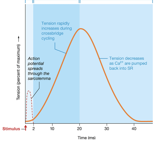
Label the three periods from left to right:
- Relaxation
- Latent
- Contraction
- Relaxation
- Latent
- Contraction
1. Latent Period
2. Contraction Period
3. Relaxation Period
2. Contraction Period
3. Relaxation Period