Anatomy 2300 Unit 2: Joints of the Pelvic Gridle & Lower Limb (Personal List)
 Call Kai
 Learn
Learn Practice Test
 Spaced Repetition
 Match
Match Flashcards
 Knowt Play
1/36

There's no tags or description
Looks like no tags are added yet.
Name | Mastery | Learn | Test | Matching | Spaced | Call with Kai | Chat |
|---|
No analytics yet
Send a link to your students to track their progress
37 Terms
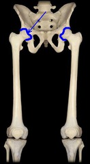
Hip (Coxal) Joint:
(1) Articulating Bones
(2) Structural Type
(3) Functional Type &
Movements Allowed
Definition: The joint where the femur (thigh bone) connects with the pelvis, allowing the leg to move in many directions.
(1) Articulating Bones:
- Hip bone and femur
(2) Structural Type:
- Synovial; ball-and-socket
(3) Functional Type & Movements Allowed:
- Diarthrotic; multiaxial; flexion, extension, abduction, adduction, rotation, circumduction of thigh
Knee (Tibiofemoral) Joint:
(1) Articulating Bones
(2) Structural Type
(3) Functional Type &
Movements Allowed
Definition: The joint where the femur (thigh bone) connects with the tibia (shin bone), allowing the leg to bend and straighten.
(1) Articulating Bones:
- Femur and tibia
(2) Structural Type:
- Synovial; modified hinge (contains articular discs)
(3) Functional Type & Movements Allowed:
- Diarthrotic; biaxial; flexion, extension of leg, some rotation allowed

Knee (Femoropatellar) Joint:
(1) Articulating Bones
(2) Structural Type
(3) Functional Type &
Movements Allowed
Definition: The joint where the femur connects with the patella (kneecap), allowing the patella to move as the knee bends and straightens.
(1) Articulating Bones:
- Femur and patella
(2) Structural Type:
- Synovial; plane
(3) Functional Type & Movements Allowed:
- Diarthrotic; gliding of patella

Ankle Joint:
(1) Articulating Bones
(2) Structural Type
(3) Functional Type &
Movements Allowed
Definition: The joint where the tibia and fibula connect with the talus, allowing the foot to move up and down.
(1) Articulating Bones:
- Tibia and fibula with talus
(2) Structural Type:
- Synovial; hinge
(3) Functional Type & Movements Allowed:
- Diarthrotic; uniaxial; dorsiflexion and plantar flexion of the foot

Toe (Interphalangeal) Joint:
(1) Articulating Bones
(2) Structural Type
(3) Functional Type &
Movements Allowed
Definition: The joints between the phalanges (toe bones) that allow the toes to bend and straighten.
(1) Articulating Bones:
- Adjacent phalanges
(2) Structural Type:
- Synovial; hinge
(3) Functional Type &
Movements
- Diarthrotic; uniaxial; flexion; extension of toes

Medial Patellar Retinaculum
A band of connective tissue on the inner side of the patella that helps hold the kneecap in place and prevent excessive movement.

Lateral Patellar Retinaculum
A band of connective tissue on the outer side of the patella that helps hold the kneecap in place and stabilize it during knee movement.

Extracapsular Ligament
A ligament located outside the joint capsule that helps stabilize and strengthen the joint.
Intracapsular Ligament
A ligament located within the joint capsule that helps stabilize the joint and limit excessive movement.
Patellar Ligament
A strong band of connective tissue that connects the patella (kneecap) to the tibial tuberosity, helping stabilize the knee and transmit the force of the quadriceps to straighten the leg.

tendon of the quadriceps femoris muscle
A strong tendon that connects the quadriceps femoris muscles to the patella, helping straighten (extend) the knee.

Medial (Tibial) Collateral Ligament (MCL):
(1) Ligament Type?
Definition: A strong ligament on the inner side of the knee that connects the medial epicondyle of the femur to the medial condyle of the tibia, helping stabilize the knee and prevent the knee from bending inward too far.
(1) Ligament Type?
- Extracapsular

Lateral (Fibular) Collateral Ligament (LCL):
(1) Ligament Type?
Definition: An extracapsular ligament on the outer side of the knee that connects the lateral epicondyle of the femur to the head of the fibula, helping stabilize the knee and prevent excessive side-to-side movement.
(1) Ligament Type?
- Extracapsular

Anterior Cruciate Ligament (ACL):
(1) Ligament Type?
(2) Movement it Prevents?
(3) How does it Rupture?
Definition: An intracapsular ligament inside the knee that connects the femur to the tibia and helps stabilize the knee by preventing the tibia from sliding too far forward.
(1) Ligament Type?
- Intracapsular
(2) Movement it Prevents?
- Prevents anterior translation of the tibia in relation to the femur
(3) How does it Rupture?
- ruptures from hyperextension

Posterior Cruciate Ligament (PCL):
(1) Ligament Type?
(2) Movement it Prevents?
(3) How does it Rupture?
Definition: An intracapsular ligament inside the knee that connects the femur to the tibia and helps stabilize the knee by preventing the tibia from sliding too far backward
(1) Ligament Type?
- Intracapsular
(2) Movement it Prevents?
- Prevents posterior translation of the tibia in relation to the femur
(3) How does it Rupture?
- ruptures from landing on tibial tuberosity with flexed knee
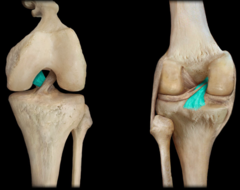
Cruciate
Cross-shaped ligaments (like the ACL and PCL) providing anteroposterior stability to the knee.

Medial Meniscus
C-shaped piece of fibrocartilage on the inner (medial) side of the knee that sits between the femur and tibia and helps absorb shock and stabilize the knee.
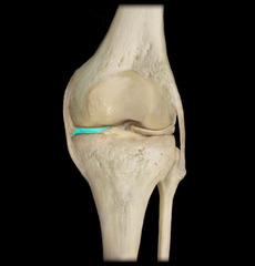
Lateral Meniscus
A C-shaped piece of fibrocartilage on the outer (lateral) side of the knee that sits between the femur and tibia and helps absorb shock and stabilize the knee.

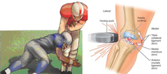
Unhappy Triad
A serious knee injury where three structures are damaged at the same time:
the ACL, MCL, and medial meniscus.
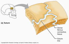
Fibrous Joint
Bones joined by fibrous tissue; no joint cavity; mostly immovable. (Synarthrosis)
Cartilaginous Joint
Bones united by cartilage; no joint cavity; slightly movable. (Amphiarthrosis)

Synovial Joint
Bones separated by a fluid-containing joint cavity; freely movable. (Diarthrosis)

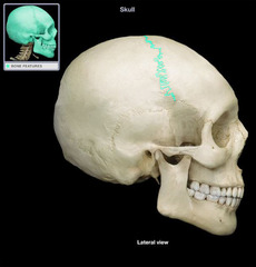
Synarthrosis
An immovable joint.
Amphiarthrosis
A slightly movable joint.

Diarthrosis
A freely movable joint.

Ball-and-Socket Joint
Multiaxial synovial joint allowing movement in all directions.

Plane Joint
Nonaxial synovial joint allowing gliding movements.

Hinge Joint
Uniaxial synovial joint allowing flexion and extension.

Flexion
Movement that decreases the angle of a joint.

Extension
Movement that increases the angle of a joint.

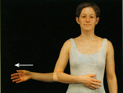
Abduction
Movement of a limb away from the midline.

Adduction
Movement of a limb toward the midline.

Circumduction
Movement describing a cone-like shape in space.

Rotation
Turning of a bone around its own long axis.
Opposition
Movement of the thumb touching the tips of other fingers.

Dorsiflexion
Lifting the foot so its superior surface approaches the shin.

Plantar Flexion
Depressing the foot or pointing the toes downward.
