Cardiovascular pathology 4
1/55
There's no tags or description
Looks like no tags are added yet.
Name | Mastery | Learn | Test | Matching | Spaced | Call with Kai |
|---|
No study sessions yet.
56 Terms
What are the layers of the elastic arteries?
Tunica intima —> Endothelial cell layer over a basal lamina and subendothelial connective tissue
Tunica media (thickest layer) —> Elastic laminae interposed with smooth muscle cells (expand and recoil to accept high volume of blood)
Tunica adventitia —> Collagen, elastic fibres and connective tissue with blood vessels (Vasa vasorum —> vessels that supply large vessels)
How do the muscular arteries differ to elastic arteries?
Fewer layers of smooth muscle cells without elastic laminae
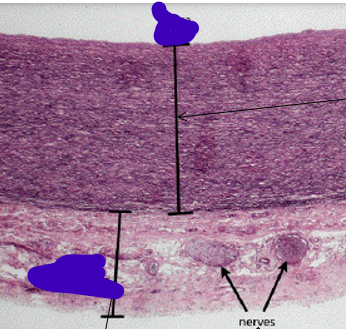
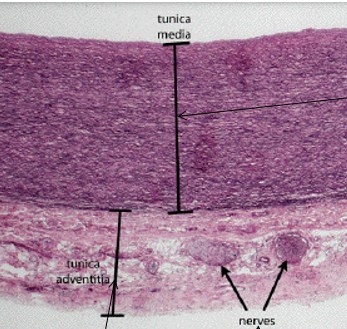
Label the following artery
Describe capillaries
5-10mm diameter
Continuous epithelium
Fenestrated —> endocrine glands, renal glomeruli, small intestine
Discountinous —> liver, BM, spleen
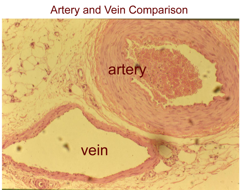
Describe veins vs arteries
Veins = thinner walls (less pressure), valves to prevent retrograde blood flow, wider lumen
Arteries = thick wall, narrow lumen
What is the thickest layer in veins?
Tunica adventitia
What is an aneurysm?
Localized dilation of a thinned and weakened portion of a vessel.
Usually arteries affected, but can occur in veins
Clinically silent until rupture → fatal consequences
What are the different causes of aneurysms?
Copper deficiency in pigs —> Copper neccesary for development of elastic tissue.
Parasitic infestations —> Spirocerca lupi in dogs/strongylus vulgaris in horses.
Disecting aneurysms in birds —> disruption of intima → entry of blood into media dissecting along the wall
What can cause rupture of vessels in horses?
Sudden rupture of ascending aorta due to trauma to ventral thorax from fall (haemothorax or hydropericardium)
Rupture of internal carotid artery into adjacent guttural pouch (esp. In mycosis) —> epistaxis
What can cause vessel rupture in cows?
Rupture of middle uterine artery may occur during parturition
Due to uterine torsion or prolapse
What can cause arterial hypertrophy?
Sustained increase in pressure or volume loads
Mainly affected in muscular arteries with hypertrophy of smooth muscle of tunica media
When does arterial hypertrophy occur in different species?
Cats —> Pulmonary arteries —> parasitic infections (Aelurostrongylus abstrusus, Toxocara sp, Dirofilaria immitis.
Cows —> Pulmonary arteries —> hypoxia induced pulmonary arterial vasoconstriction and pulmonary hypertension from exposure to high altitude (Brisket disease).
All species —> Cardiovascular anomalies that shunt blood left to right lead to pulmonary hypertension and hypertrophy
Describe what is being shown here?
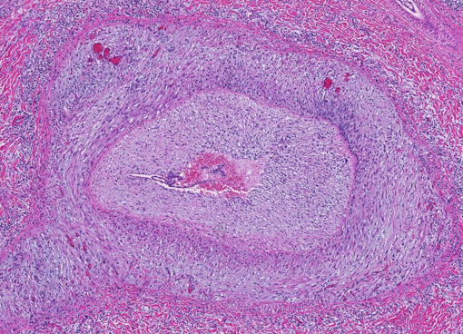
Arterial hypertrophy
Marked thickening of the tunica media
What can cause arterial medial calcification?
Same as endocardial mineralisation
Calcinogenic plant toxicosis
Vitamin D toxicosis (→ hypercalcaemia = Ca deposited in tissues)
Renal insufficiency
Johne’s disease
How does arterial medial calcification present grossly?
Solid, dense, pipe-like structures with raised, solid, white, intimal plaques
How does arterial medial calcification present histologically?
Prominent basophilic, granular mineral deposits in lumen occasionally admixed with iron

What type of finding is arterial intimal calcification?
Normal finding in subendothelium of muscular arteries & arterioles of horses —> incidental & insignificant finding
What is fibrinoid necrosis?
Deposits of an amorphous, homogeneous, eosinophilic PAS+ protein material composed of serum proteins and fibrinogen
No differentiation between tunica media and adventitia
Lost structure of tunica media → replaced by amophous band of eosinophilic material
What is the pathogenesis of fibrinoid necrosis?
Endothelial and muscular damage of the arterial wall with extravasation of proteins and deposition in the vessel wall
What are the causes of fibrinoid necrosis in different species?
Pigs —> Selenium/vit E deficiency or Oedema disease (E. coli with shiga-like toxin)
Dogs —> Uraemia
What are the predisposing factors for thrombosis?
Endothelial damage
Turbulence or stasis
Hypercoagulative states
What does this image show?
“Saddle thrombosis” in cat —> occlusion of vessel
What does this image show?
“Saddle thrombosis” in dog —> thrombi get longer & extend down iliac arteries
What does this image show?
“Saddle thrombosis” in horse —> becomes nidus for further development
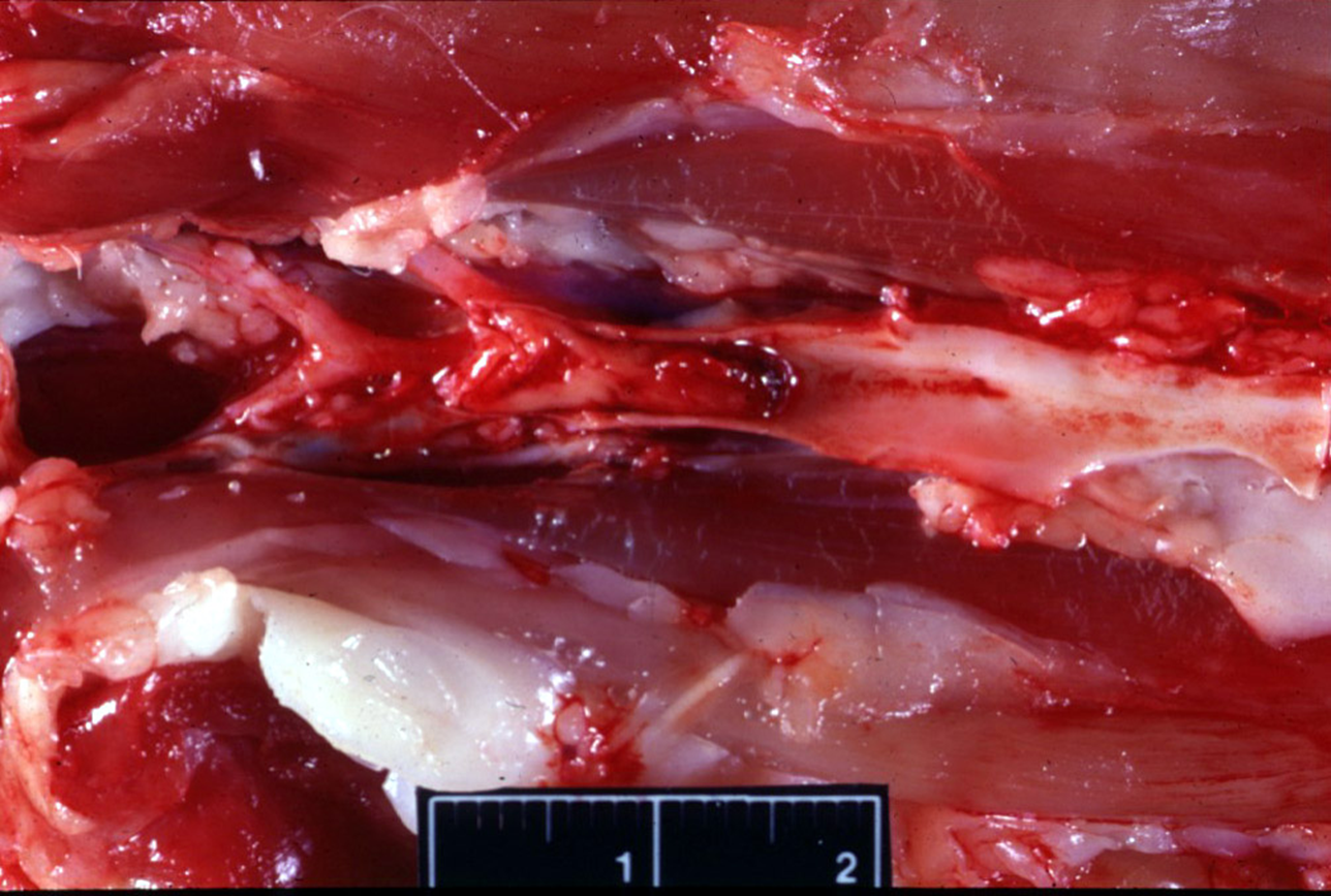
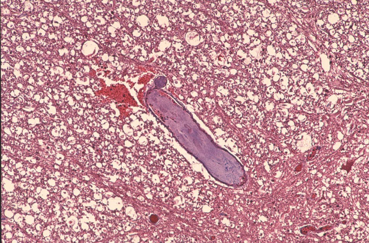
What does this image show?
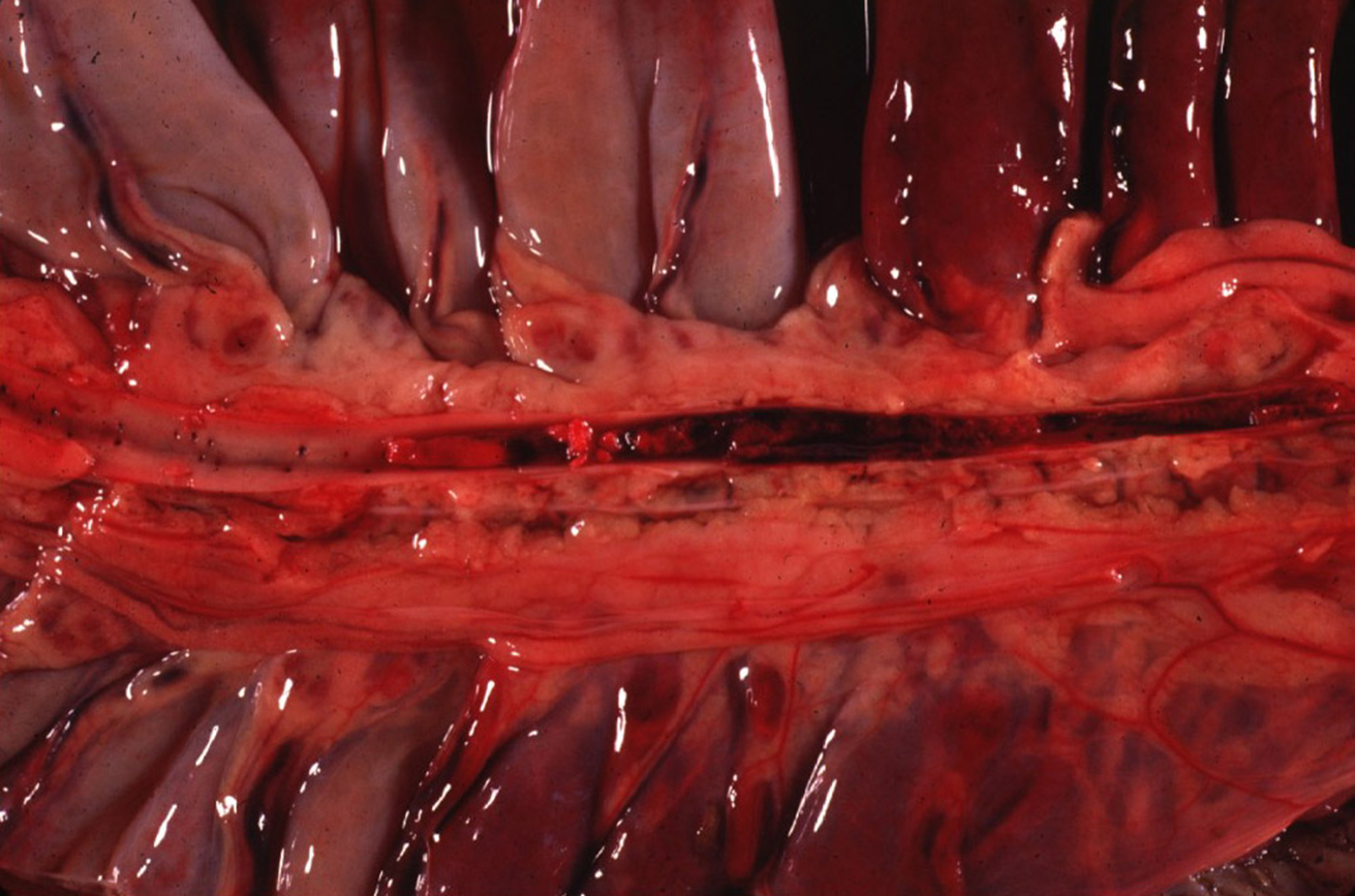
Thrombosis of cranial mesenteric artery in horse caused by strongylosis —> L4 creates endothelial damage, arteritis & thrombus
What is the difference in size between dirofilariosis and strongylosis?
Dirofilariosis much bigger than strongylosiss —> vascular oclusion
What can cause DIC?
Severe end of disease
Endotoxaemia
Viraemia (FIP and canine infectious hepatitis)
Dirofilariasis
Tumours (hemangiosarcoma and leukemia)
Shock, haemolysis, extensive necrosis (burns)
What is DIC?
Clotting phenomenon due to endothelial damage with exposure of subendothelial collagen & subsequent platelet aggregation & intravascular activation of coagulation process
Extensive clotting depletes coagulation factors, resulting in widespread haemorrhages
What is an emboli?
Oclussion of arteries by lodgement of foreign materials such as disrupted fragments of thrombi, neoplastic cells, bacteria, parasites, fat etc.
Describe septic emboli
Originate from lesions of vegetative endocarditis in lung (R side) or myocardium, kidneys, spleen, joints, leptomeninges (L side)

Describe parasitic emboli
Fragments of dead intravascular parasites, such as dirofilaria, into pulmonary circulation of carnivores following administration of adulticidal drugs
What does a fat embolism occur secondary to?
Bone fractures (fat cells in bone marrow enter blood)
BM has central core of adipose tissue w/ peripheral rim of haematopoietic cells
What is fibrocartilaginous embolism?
Fibrocartilagenous fragments from the spinal cord gets into spinal vasculature
Leads to infarction of the spinal cord and paralysis of the hindlimbs
Where does a thromboembolism often occur?
Pulmonary arterial tree in dogs & cats
What is the difference grossly between thrombi and post-mortem clots?
Thrombi = adhered to wall
Post-mortem clots = able to pull away from wall
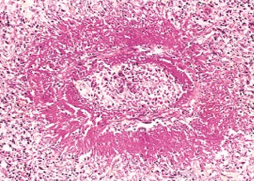
Describe what is circled in this image
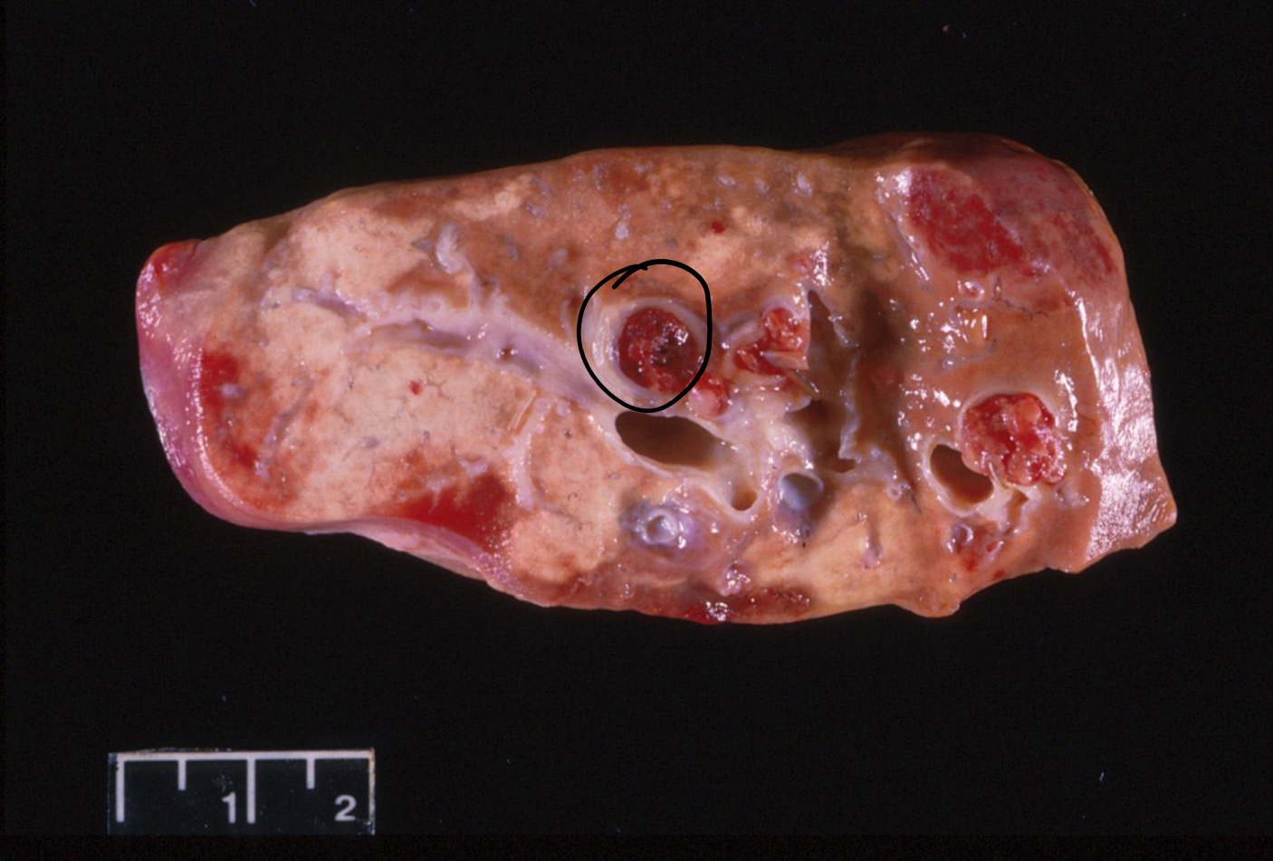
Artery occluded by thrombus in lung
Thrombi adherred to wall of vessel
Endothelial damage, layer of platelet anchor clot
What can cause vasculitis?
Haematogenous dissemination
Local extension of suppurative-inflammatory processes,
Immune-mediated processes
Parasitic infections
How does vasculitis appear grossly?
Medium-sized arteries appear thick and tortuous
Associated haemorrhages, aneurysms and thrombosis (causing infarction)
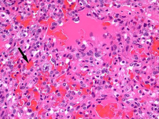
How does vasculitis appear histologically?
Fibrinoid necrosis (indicated vascular damage)
Inflammation of the intima and media
Leukocytes present within and surrounding walls
Endothelial damage causes thrombosis → infarction
What are the different viral causes of vasculitis and what do they cause?
Bluetongue of sheep (orbivirus) —> haemorrhage at the base of the pulmonary artery
Equine viral arteritis (arterivirus) —> oedema and petichia
Feline Infectious Peritonitis (coronavirus) —> Pyogranulomatous vasculitis
Malignant Catarrhal Fever (MCF) of cattle (gamma-herpesvirus) —> Polyarteritis and periarteritis
Equine Infectious Anemia (retrovirus) —> Polyarteritis and periarteritis
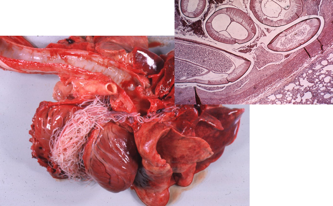
What does Dirofilaria immitis cause in the dog?
Microfilaria are found throughout the circulation.
Villous pulmonary endoarteritis (inflammation of the intima) and obstruction and narrowing of the lumina
Increased pulmonary resistance causing right ventricular hypertrophy which may progress to right heart failure
What does Strongylus vulgaris cause in the horse?
4th stage larvae in mesenteric arteries:
Causes intense focal inflammatory reaction in the walls of the larger arteries
Aneurysmal dilation,
Thrombosis,
Infarction distal to involved site (thromboembolic colic of horses)
Which species is polyarteritis nodosa common in?

Rats (idiopathic) —> lesion of mesenteric arteries
What does Idiopathic necrotising polyarteritis cause?
Cervical pain with still gait and stiff neck with a hunched body posture in beagles
(affects vessels in cervical spine —> painful)

What is the meaning of phlebitis?
Inflammation of veins
What are the different causes of phlebitis?
Systemic infections (vasculitis) —> Salmonellosis, colibacilosis, FIP.
Local infections —> Metritis (inflam of the uterus), hepatic abscesses.
Intravenous injection sites
What is Omphalophlebitis?
Inflammation of the umbilical vein —> naval ill in neonatal farm animals → septicaemia, suppurative arthritis, hepatic & umbilical abscesses
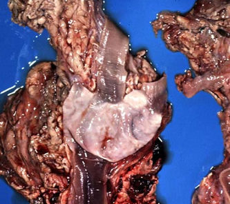
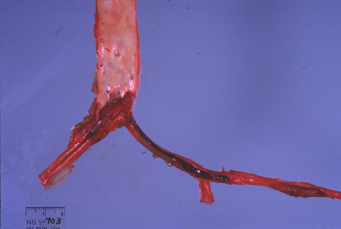
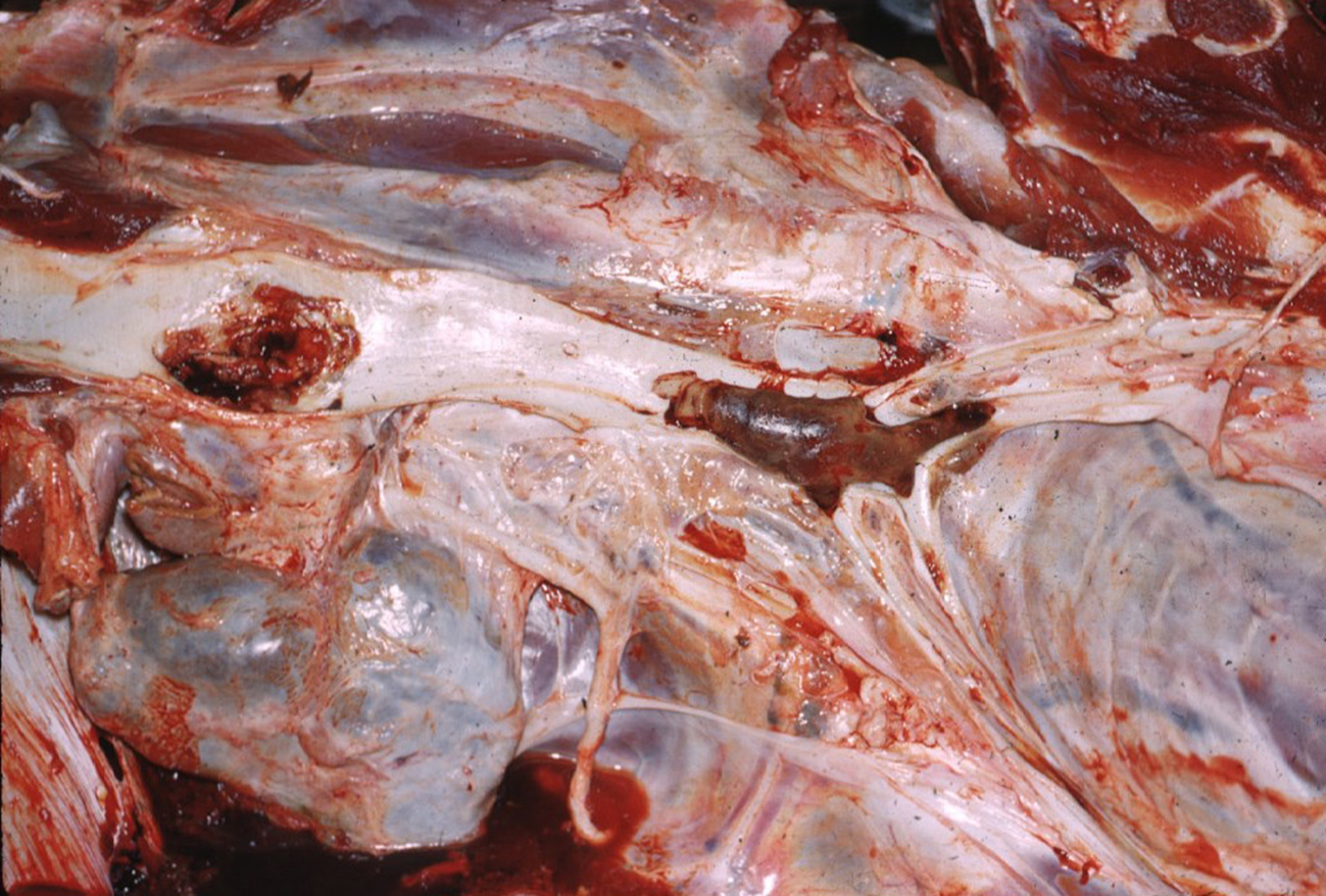
What is being shown here? What caused this?
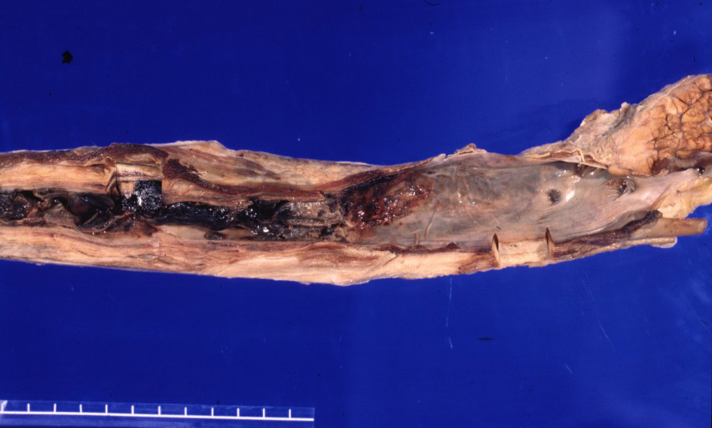
Vascular damage & development of phlebitis & thrombosis jugular vein (horse) —> IV injections of irritant solutions or intimal trauma produced by IV catheters
What is being shown here?
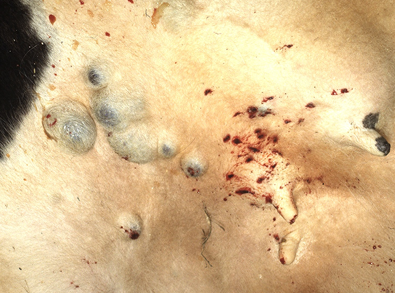
Haemangioma in vessels in the skin
Describe haemangioma histologically?
Variably sized vascular spaces filled with erythrocytes and lined by a single layer of uniform endothelial cells
Fine capillary-like structures lined by layer of endothelial cells
Benign form —> endothelial cells normal size & shape but proliferate abnormally
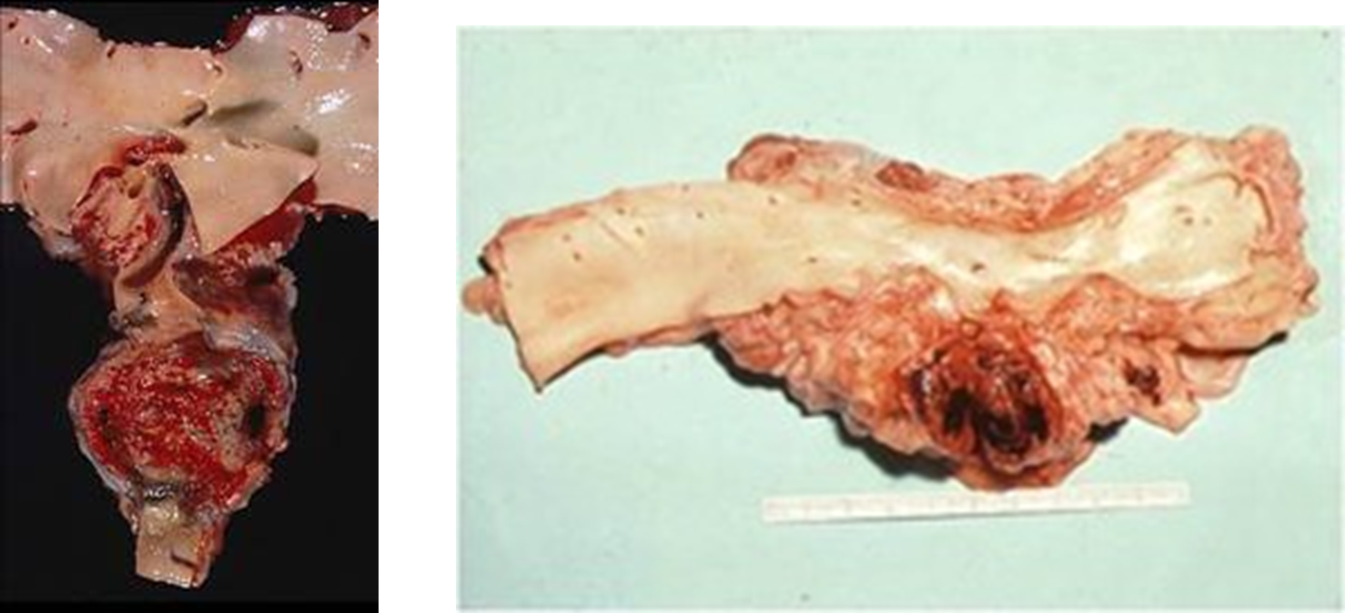
What has happened to the spleen in this image?
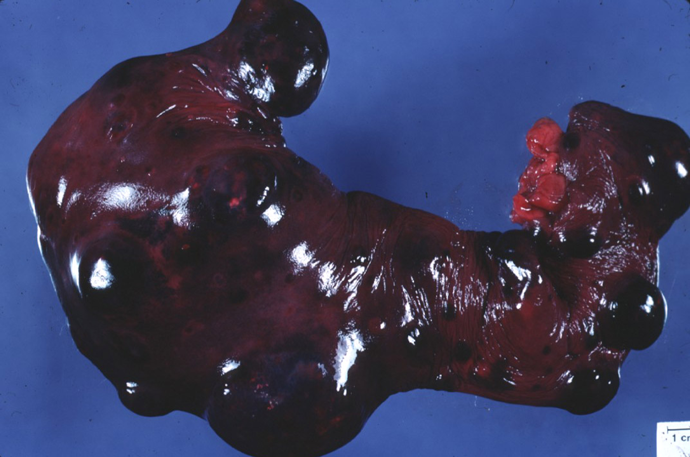
Haemangiosarcoma —> vascular tumour (malignant neoplasm of endothelium)
Where are the primary tumours and metastasis of Haemangiosarcomas often located?
Primary tumours —> any location, but typically right atrium, spleen, skin
Metastasize —> commonly to lungs, liver, other sites possible
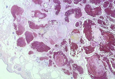
How do haemangiosarcomas present grossly?
Large red (haemorrhagic) nodular masses
How do haemangiosarcomas present histologically?
Irregular vascular clefts, channels, atypical cells, high mitotic rate, usual signs of malignancy
Vascular spaces more disorganised
Solid pattern, plump cells, large nuclei
What do haemangiopericytomas mean?
What are the features of them?
How is distant metastases prevented?
Tumour in the wall of a vessel
Occurring exclusively in dogs, solitary, multilobulated masses occurring around the joints of limbs. White and firm
Low grade malignancy —> excision to prevent distant metastases but must remove completely

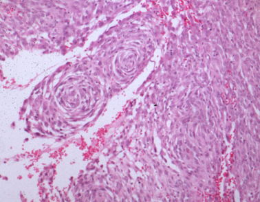
How do haemangiopericytomas present histologically?
Perivascular whorls of fusiform cells
Low metastatic potential
Centre of whorls = vessel