Dental Embryology - Basic Science Core
 Call Kai
 Learn
Learn Practice Test
 Spaced Repetition
 Match
Match Flashcards
 Knowt Play
1/36

Earn XP
Description and Tags
Embryology Exam 1
Name | Mastery | Learn | Test | Matching | Spaced | Call with Kai | Chat |
|---|
No analytics yet
Send a link to your students to track their progress
37 Terms
ODONTOGENESIS
Development results in a complex and intricate cascade of gene expression that takes place to self- organize,
Expand in number, and differentiate along the proper developmental trajectory (teeth).
Odontogenesis Genes (5)
1) Bone morphogenetic protein BMP
2) Sonic Hedgehog (shh)
3) Ectodysplasin A (Eda)
4) Wingless-related integration site(wnt)
5) Fibroblast growth factor(FGF)
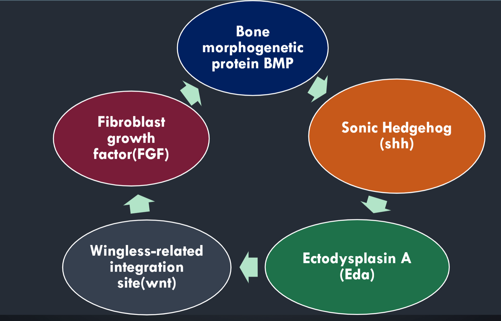
DIFFERENTIATION (MOLECULAR SIGNALING IN TOOTH)
The epithelium derived from the first pharyngeal arch is essential for tooth development, and over 90 genes, including Sonic Hedghog (Shh) are expressed in cells of the developing oral epithelium and mesenchyme
STAGES OF TOOTH BUD DEVELOPMENT (5)
1) Initiation
• Phase of deciduous tooth 5th month in utero
• Phase of permanent tooth – 6 th month
Phase of accessional tooth 4th month in utero until 4 to 5 years
2) Proliferation
3) Histodifferentiation
4) Morpho differentiation
5) Apposition
Primary epithelial band.
The maxillary and mandibular processes have a thickened epithelium called the primary epithelial band.
The epithelium rests on a basal lamina.
The underlying embryonic connective tissue is called ectomesenchyme because neural crest cells from the ectoderm migrate into the mesenchyme.
Dental Lamina
Parts of the dental lamina will develop into teeth.
The dental lamina is surrounded by a condensation of ectomesenchymal cells (MC) that will become the dental papilla and dental follicle
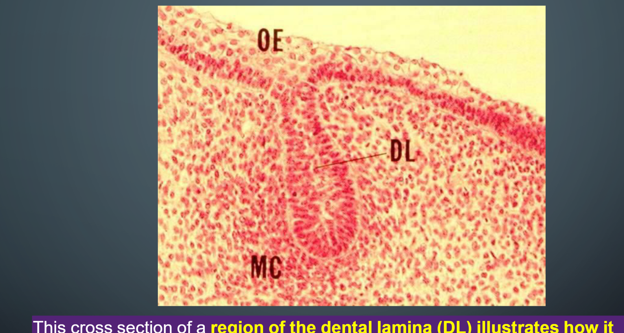
At about 6-7 weeks of development, the primary epithelial band grows into … (2)
1) vestibular lamina
2) dental lamina
The Vestibular Lamina
The vestibular lamina will become the space between the cheek and gingiva.
PLACODE
Dental placodes are believe to initiate the various tooth families
Placode is a determinant in tooth formation. Smaller-than the normal
placodes lead to missing and smaller teeth , whereas larger placodes
induce supernumerary and larger teeth
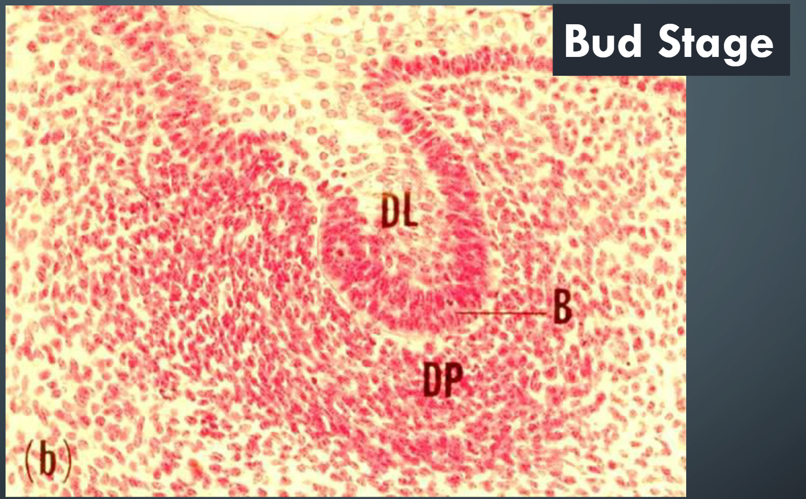
dental papilla (DP)
The dental papilla (DP) is a crescent-shaped Condensation of ectomesenchymal cells that will develop into cells that will produce the dentin, cementum and pulp of a tooth.
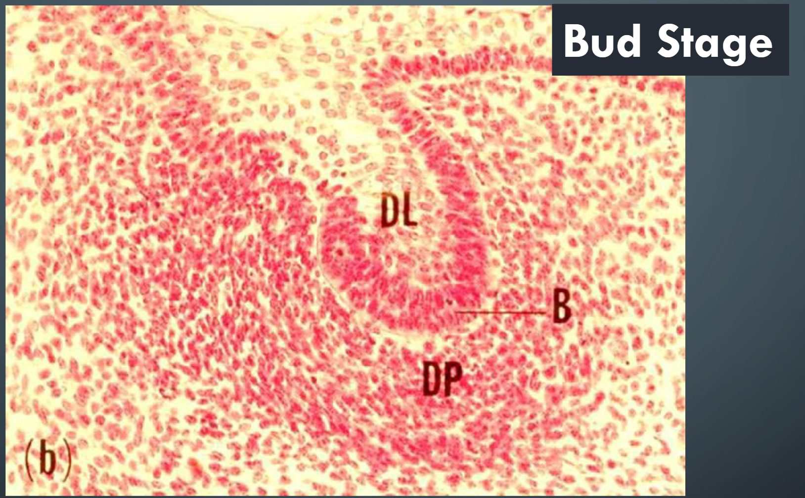
A Bud in tooth development
At about 8 weeks of development, along the dental lamina (DL) at
each site where a tooth will develop, the epithelium thickens to form a
bud (B).
Some cells in each bud will develop into ameloblasts that will
produce the enamel crown for one tooth.
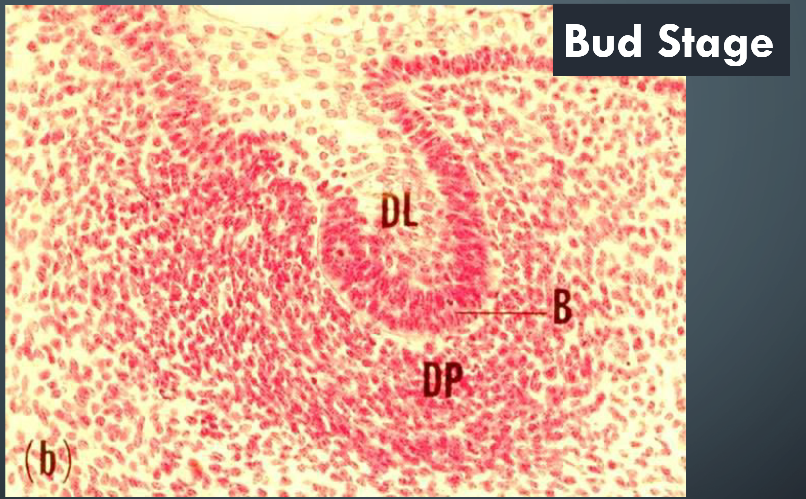
8 Weeks in development is also known as the ______ Stage
The Bud Stage
In this stage bud stage of tooth development as the dental laminae fold inward over the developing maxilla and mandible.
10 Weeks in development is also known as the ______ Stage
Cap Stage
CAP STAGE AND THE ENAMEL ORGAN
At about 10 weeks, the bud develops into the cap
shaped enamel organ
Since the cells are interconnected with desmosomes, they become star-shaped (i.e. stellate) as they are forced apart by the increasing intercellular fluid volume.
The enamel organ layers of cells (3)
1) the outer enamel epithelium
(OEE),
2) the inner enamel epithelium (IEE)
3) the stellate reticulum (SR).
Inner enamel epithelium Cells
Cells of the inner enamel epithelium will differentiate into enamel-producing Ameloblasts
The IEE begins to enclose the ectomesenchymal cells that form the
dental papilla (DP).
The stellate reticulum (SR)
The SR cells secrete glycosaminoglycans that
draw water into the enamel organ and cause it
to swell.
I am on Slide 15, I need to go back, I will start agian on slide 28
The Formation of the Permanent Dentition
The successional permanent teeth develop between week 20 of fetal development and 10 months after birth.
The permanent molars start to develop between week 20 of fetal development (first permanent molar) and age 5 years (third molar). The permanent molars have no deciduous precursors. Once the body of the mandible grows long enough, the dental lamina grows posteriorly into the ectomesenchyme and forms tooth buds for the first, second and third molars. This is why 3rd molars are located far back in the mandibular ramus.
DENTAL HARD TISSUE FORMATION
Dental Hard Tissues result from the
histodifferentiation of
• Enamel from Ameloblasts
• Dentin from Odontoblasts
• Cementum from Cementoblasts
Ameloblasts →
Become Enamel
Odontoblasts →
Become Dentin
Cementoblasts →
• Become Cementum
Amelogenesis
At the junction between the enamel organ and the dental papilla, the Ameloblasts differentiate first, and then odontoblasts.
Odontoblasts secrete pre-dentin first which then stimulates Ameloblasts to produce enamel facing the pre-dentin.
Ameloblasts mature through several morphologically distinct functional stages during enamel production.
The enamel matrix is deposited by ameloblasts directly on the surface of the previously formed pre-dentin
Ameloblasts and Enamel Formation
Ameloblasts continue to produce enamel matrix until the full
thickness of the enamel is achieved
Maturation of the enamel matrix
Maturation of the enamel matrix involves removal of organic material (proteins) and water and continued mineralization from influx of calcium and phosphate into the maturing enamel.
Active ameloblasts have many mitochondria to fuel the transport of organic material out and mineral into the enamel.
AMELOBLAST FUNCTIONAL STAGES (3) (Do not need to know)
1) Presecretory Stage
2) Secretory Stage
3) Post- secretory or Maturation Stage
Presecretory Stage
Golgi and rough
endoplasmic reticulum of ameloblasts produce
vesicles of proteins:
1. Morphogenic Stage
2.Histodifferentiation Stage
Secretory Stage
ameloblasts deposit the proteins
and minerals of the enamel onto predentin:
1. Initial Secretory Stage (no
Tome’s Process)
4. Secretory Stage (with Tome’s Process)
Secretory Stage ameloblasts release
secretory granules of enamel matrix
proteins and hydroxyapatite from Tome’s
process
Post- secretory or Maturation Stage
ameloblasts alternate between two morphologies
that remove protein and transport mineral ions that
harden the enamel:
5. Ruffle-ended Ameloblast
6. Smooth-ended Ameloblast
7. Protective Stage
8. Desmolytic stage
Secretory Stage ameloblasts release
secretory granules of enamel matrix
proteins and hydroxyapatite from Tome’s
process
Enamel cannot exist without
Dentin, as dentin is a cushion for the enamel
REMOVE unsupported enamel in dentistry
Ruffle-ended maturation stage
Ruffle-ended maturation stage ameloblasts secrete enzymes to
remove proteins and additional mineral ions to harden the enamel.
Smooth-ended maturation stage
Smooth-ended maturation stage ameloblasts absorb water and
degraded proteins from the enamel matrix.
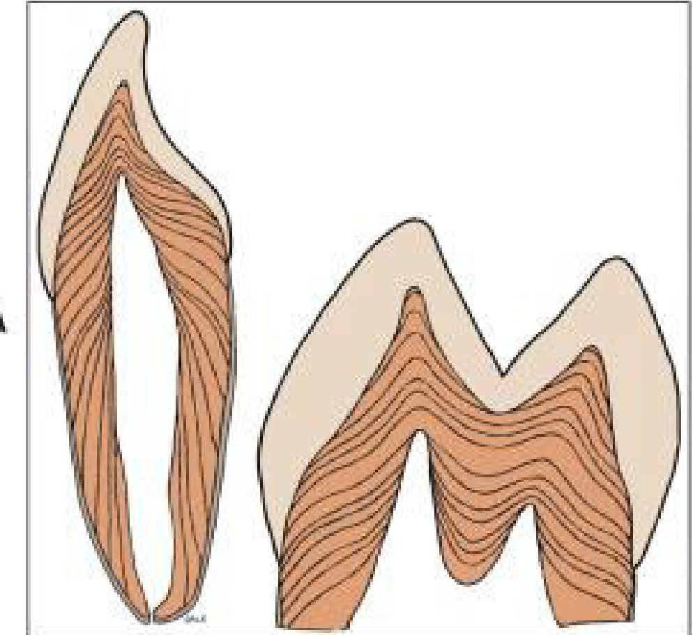
DEVELOPMENT OF DENTIN
As dentin is produced, it creates a pattern of incremental lines. In a
tooth section of a person who received tetracycline intermittently, the
drug has been incorporated at successive dentin-forming fronts,
highlighting the incremental line patterns.
Hertwig's Epithelial Root Sheath (HERS)
of the dental follicle.
cementoblasts