anat 2
1/29
There's no tags or description
Looks like no tags are added yet.
Name | Mastery | Learn | Test | Matching | Spaced |
|---|
No study sessions yet.
30 Terms
Which of the following lymph nodes do we find in the brachial plexus?
A) Mandibular lymph nodes
B) Tracheobronchial lymph nodes
C) Superficial cervical lymph nodes
D) Axillary lymph nodes
D
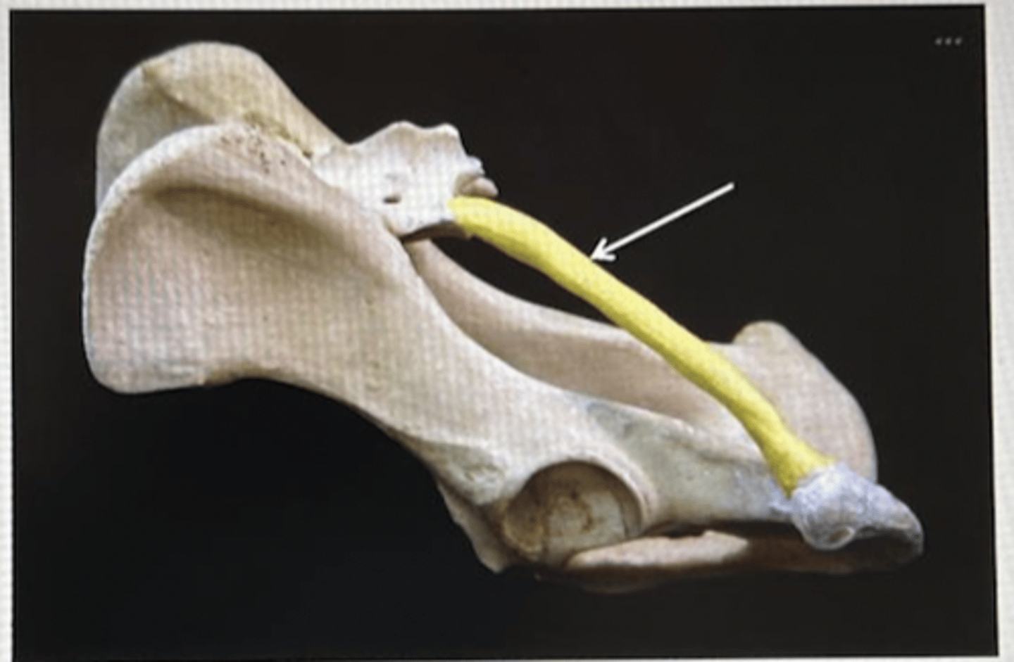
What joint is the following picture showing? And which ligament is absent?
A) Scapulohumeral joint. Collateral ligaments
B) Humero radial joint. Lateral and medial glenohumeral ligaments
C) Scapulohumeral joint. Cruciate ligaments
D) Humero radial joint. Transverse humeral ligaments
A
Which of the following arteries do we find running together with the femoral vein and saphenous nerve in the femoral canal and we would normally use to measure the pulse rate in the pelvic limb?
A) Saphenous artery
B) Femoral artery
C) Popliteal artery
D) External iliac artery
B
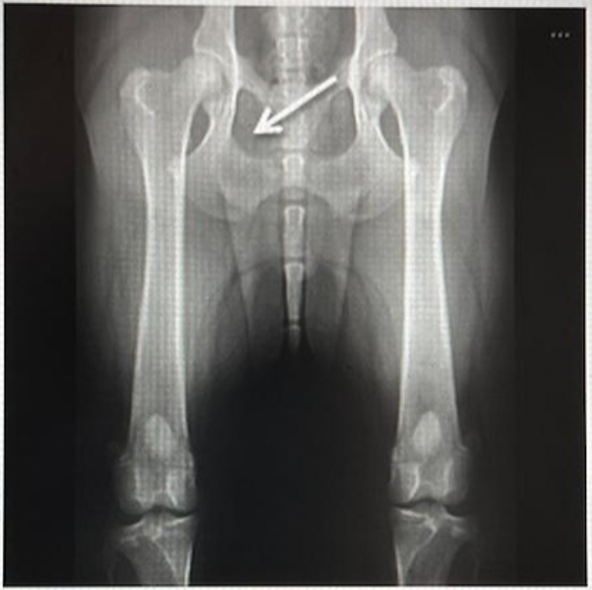
Identify the anatomic structure the arrow is pointing at in the following radiograph cranio-caudal view of the femur.
A) Obturator foramen
B) Ischial arch
C) Pubis
D) Ischial tuberosity
A
Which of the following muscles does not fix the scapula to the trunk?
A) Trapezius muscle
B) Cranial dorsal serrate muscle
C) Omotransverse muscle
D) Ventral serrate muscle of the neck
B
Which muscles are involved on the formation of the common calcanean tendon also known as the Achilles tendon?
A) Gastrocnemius muscle, gracilis muscle, biceps muscle of the thigh, semitendinous muscle, and superficial digital flexor muscle
B) Gastrocnemius muscle, gracilis muscle, semimembranous muscle and superficial digital extensor muscle
C) Gastrocnemius muscle, gracilis muscle, biceps muscle of the thigh, semimembranous muscle and straight muscle of the thigh
D) Gastrocnemius muscle, gracilis muscle, biceps muscle of the thigh, semitendinous muscle and supinator muscle
A
Identify the structure the arrow is pointing at and which of the following anatomic structures joins here?
A) Calcaneus, Common calcaneal tendon.
B) Talus. Lateral collateral ligament
C) Calcaneus. Medial collateral ligament
D) Talus. Common calcanean tendon
A
Which of the following species lacks the acromial portion of the deltoid muscle?
A) Carnivore
B) Equine
C) Small ruminant
D) Large ruminant
B
Which of these is located at the fetlock joint?
A) navicular bone
B) coffin bone
C) Proximal sesamoid bones
D) Short pastern bone
C
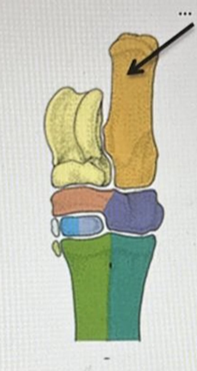
Identify the anatomic structure pointed at by an arrow and the specie it belongs to.
A) Cannon bone. Ruminant
B) Rudimentary 2nd metacarpal bone. Equine
C) Rudimentary 4th metacarpal bone. Equine
D) Pisiform carpal bone. Ruminant
B
Choose the correct answer regarding the quadriceps muscle of the thigh.
A) It has five different heads
B) it is innervated by the femoral nerve
C) All the heads of this muscle insert at the patella and tibial tuberosity
D) Answers B and C are correct
D
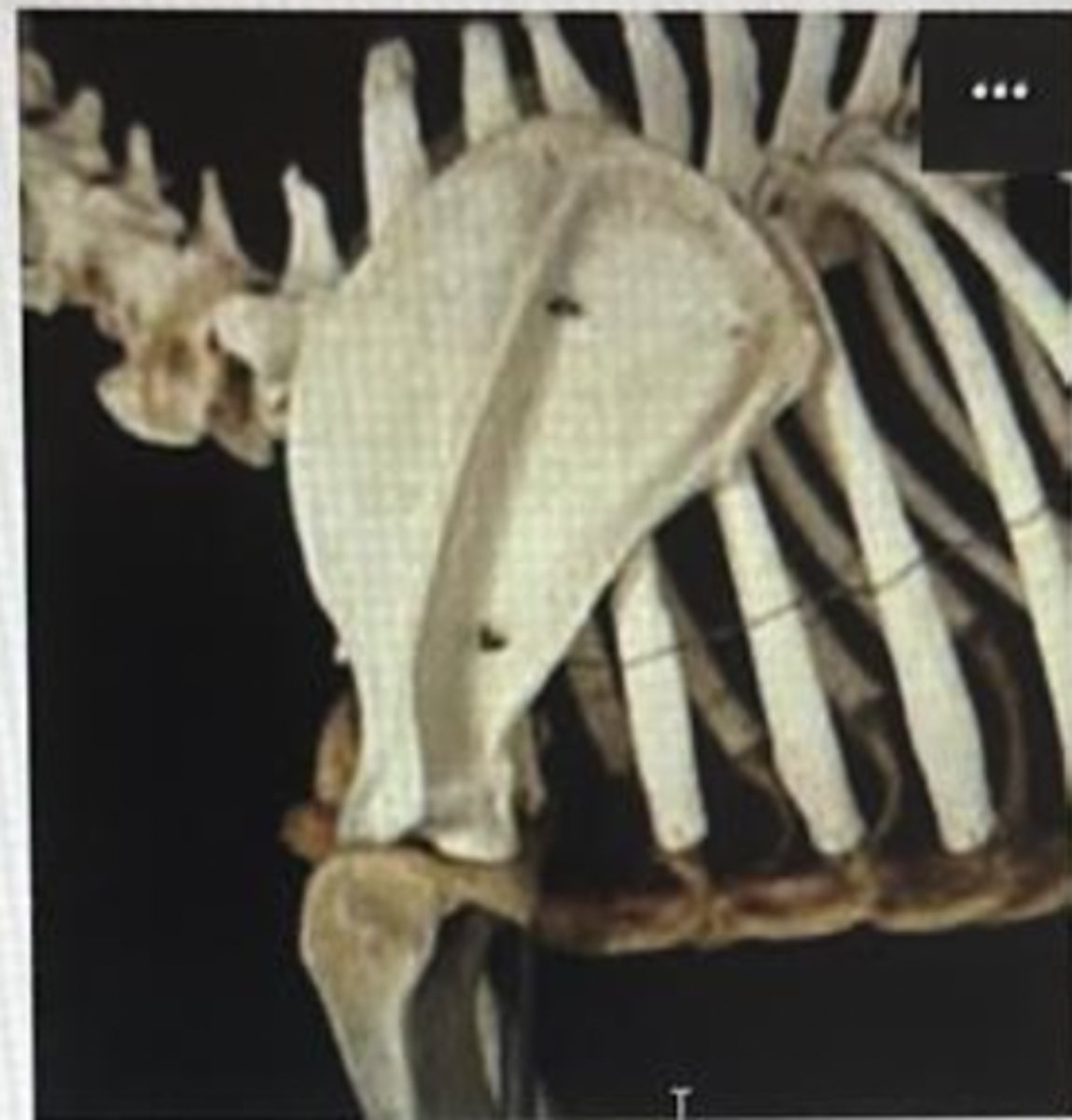
Identify the anatomic structure the arrow is pointing at and its extension in carnivores.
A) Sacroiliac ligament. From sacrum to iliac crest
B) Sacrotuberous tendon. From sacrum to ischial tuberosity
C) Sacroiliac tendon. From sacrum to iliac crest
D) Sacrotuberous ligament. From sacrum to ischial tuberosity
D
What kind of movements are proximal and distal radioulnar joints going to allow? In what species do we find these joints?
A) Flexion and extension. Ruminants and carnivores
B) Flexion and extension. Every specie
C) Pronation and supination. Ruminants and carnivores
D) Pronation and supination. Carnivores
D
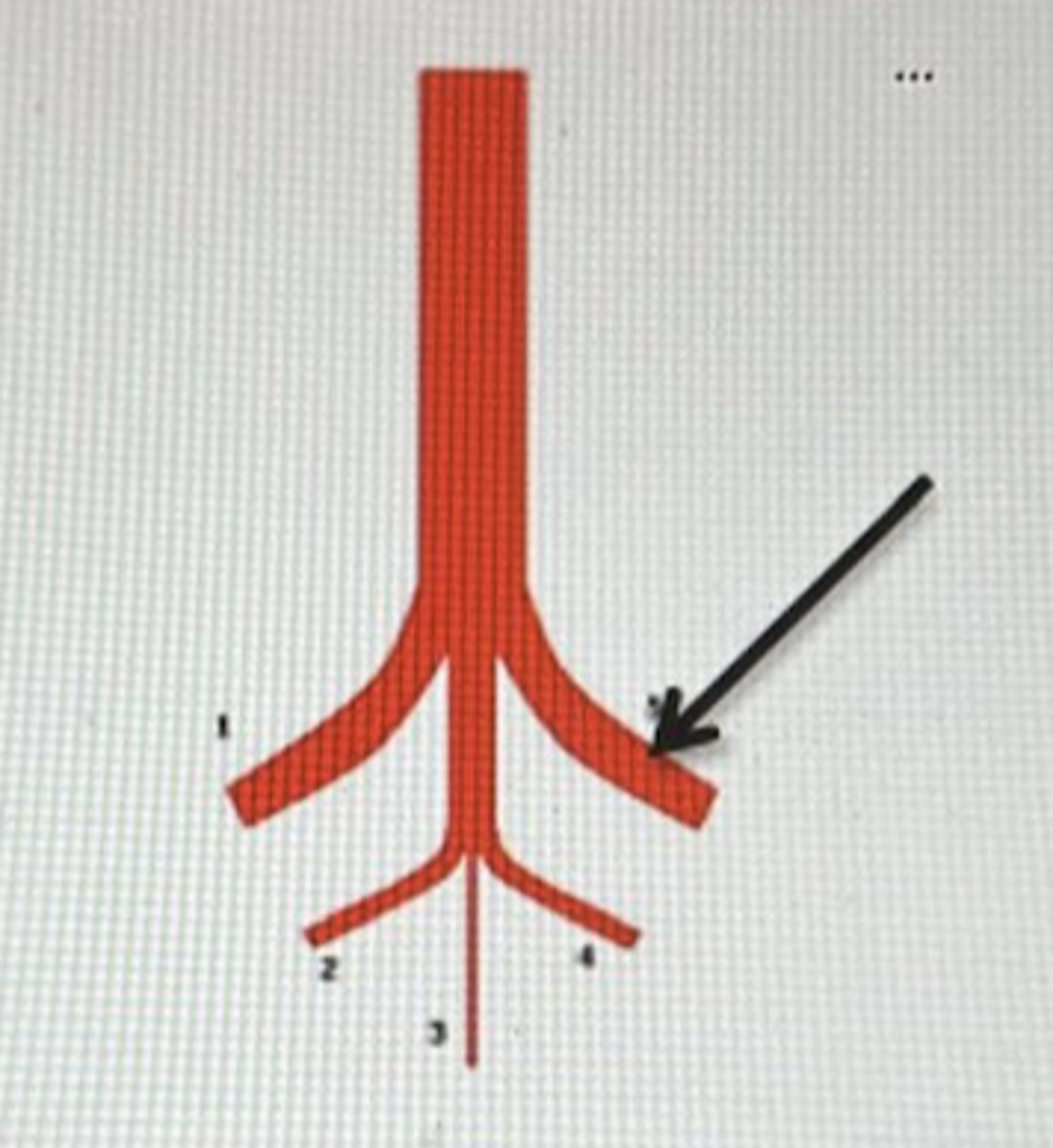
The following picture shows the last branches of the abdominal aorta. What branch is the arrow pointing at which is going to vascularize most of the pelvic limb?
A) Internal iliac artery
B) Median sacral artery
C) External iliac artery
D) Deep circumflex iliac artery
C
Which of the following joints of the pelvic limb are synovial?
A) Coxofemoral joint
B) Sacroiliac joint
C) Tarsal joints
D) All the above answers are correct
D
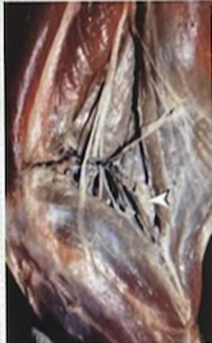
Which of the following lymph nodes found caudally to the knee joint and that should always be explored by veterinarians is the arrow pointing at?
A) Inguinal lymph node
B) Popliteal lymph node
C) Axillary lymph node
D) Aortic lymph node
B
Which muscles are considered to be the main anteversor and retroversor muscles of the thoracic limb?
A) Anteversor: Brachiocephalic muscle. Retroversor: Broadest muscle of the back
B) Anteversor: Brachiocephalic muscle. Retroversor: Ventral serrate muscle of the thorax
C) Anteversor: Omotransverse muscle. Retroversor: Broadest muscle of the back
D) Anteversor: Omotransverse muscle. Retroversor: Ventral serrate muscle of the thorax
A
If we need to place an intravenous catheter on the thoracic limb, which superficial vein would you use?
A) Saphenous vein
B) External Jugular vein
C) Maxillary vein
D) Cephalic vein
D
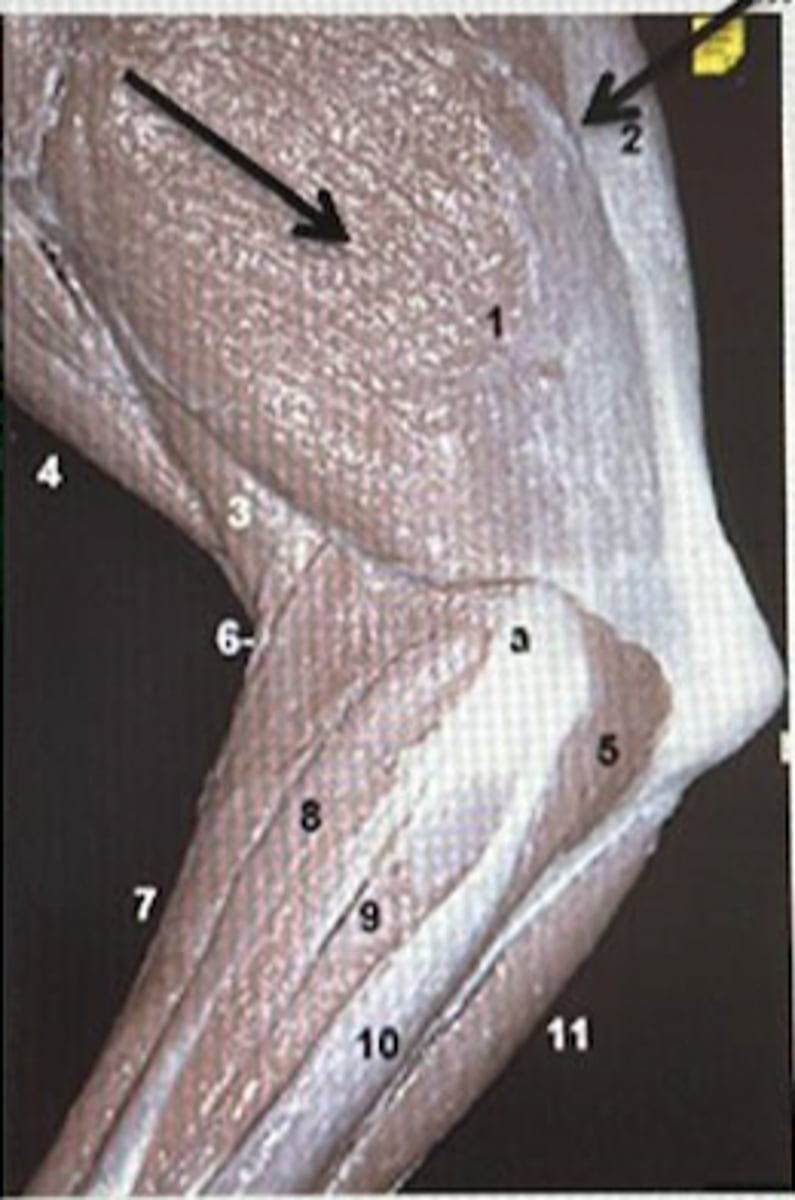
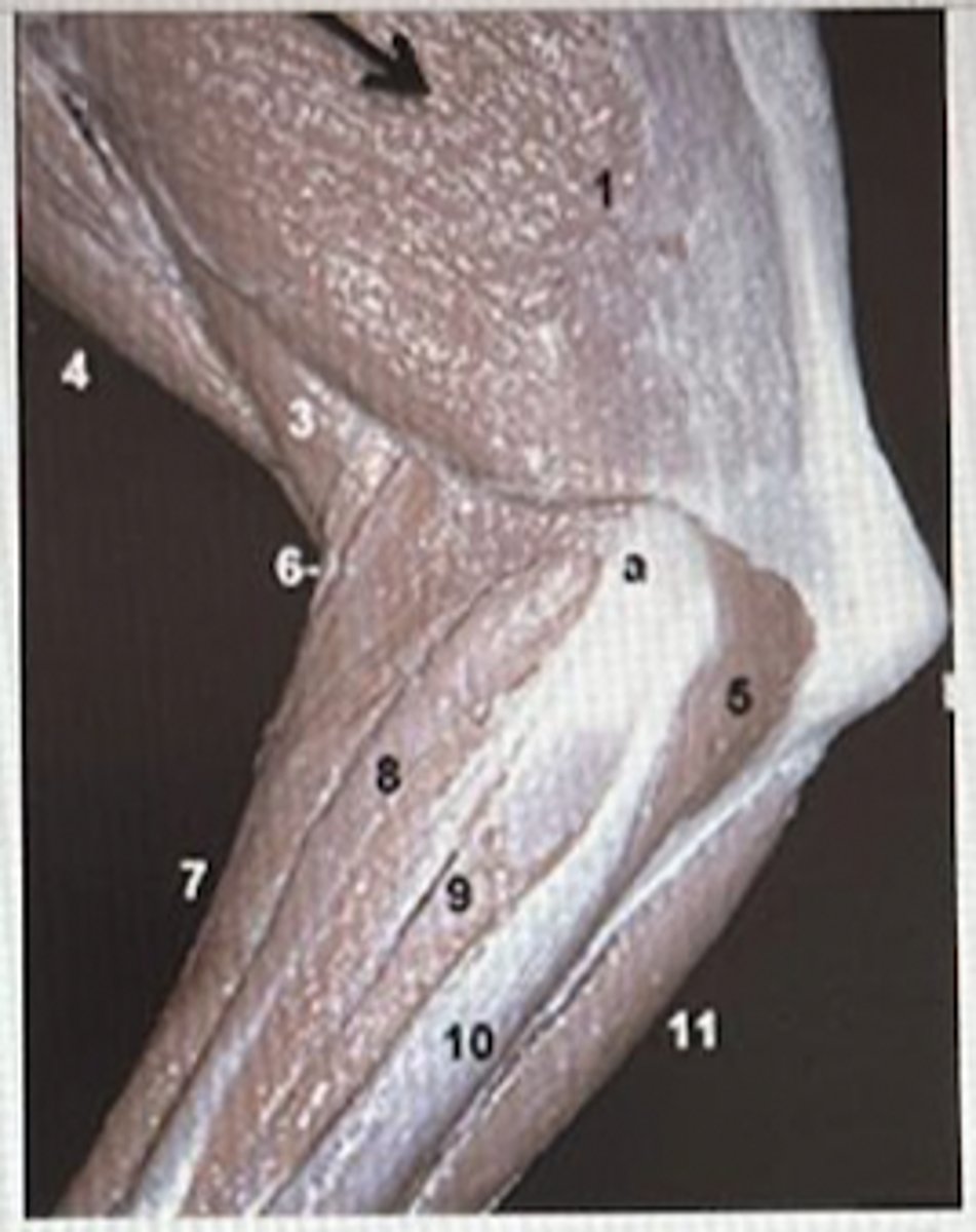
Which muscles are the arrows pointing at and what function are they going to carry out?
A) Lateral and long heads of the triceps muscle of the forearm. Flexion of the elbow joint.
B) Medial and accessory heads of the triceps of the forearm. Flexion of the elbow joint
C) Lateral and long heads of the triceps muscle of the forearm. Extension of the elbow joint.
D) Medial and accessory heads of the triceps of the forearm. Extension of the elbow joint
C
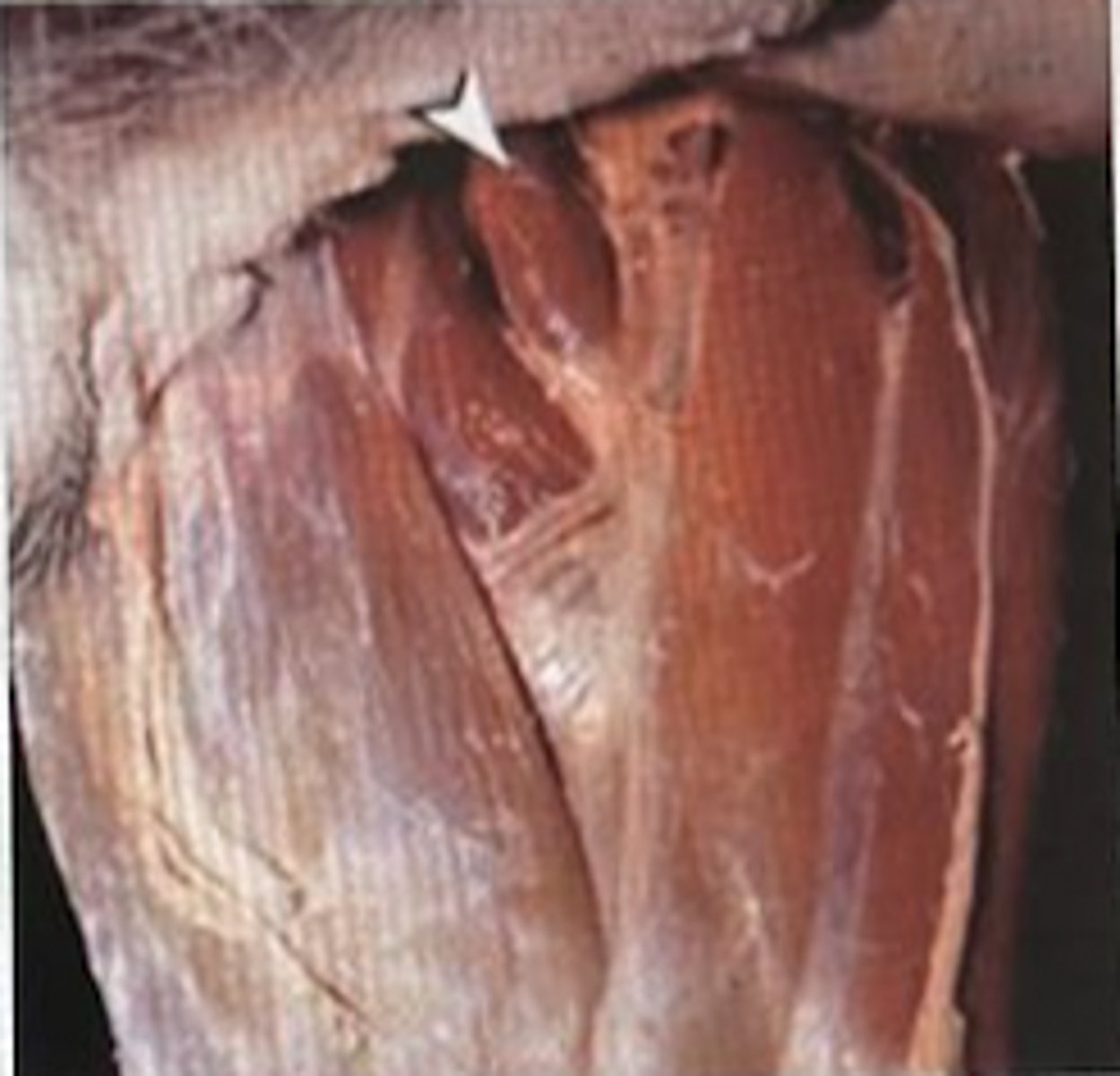
Identify the muscle shown on the following picture which extends from the humerus neck to the proximal cranial surface of the radius and ulna.
A) Biceps muscle of the forearm
B) Brachial muscle
C) Triceps muscle of the forearm
D) Tensor muscle of the antebrachial fascia
B
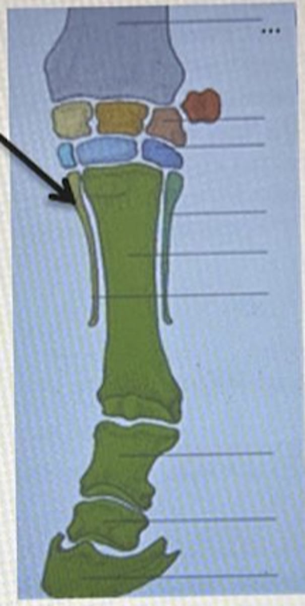
Choose the correct answer regarding the articular surfaces of the fetlock joint in equine.
A) 2nd phalanx. 3rd phanx and navicular bone
B) 3rd metacarpal bone, 1st phalanx and navicular bone
C) 3rd metacarpal bone, 1st phalanx and proximal sesamoid bones
D) 1st phalanx and 2nd phalanx
C
Choose the correct answer regarding innervation of the pelvic limb.
A) The most important muscle innervated by the femoral nerve is the biceps muscle of the thigh
B) The cranial and caudal gluteal nerves are very superficial nerves
C) The brachial plexus is incharged of the innervation of the pelvic limb
D) The sciatic nerve is the largest nerve in the body and is covered the the biceps muscle of the tigh
D
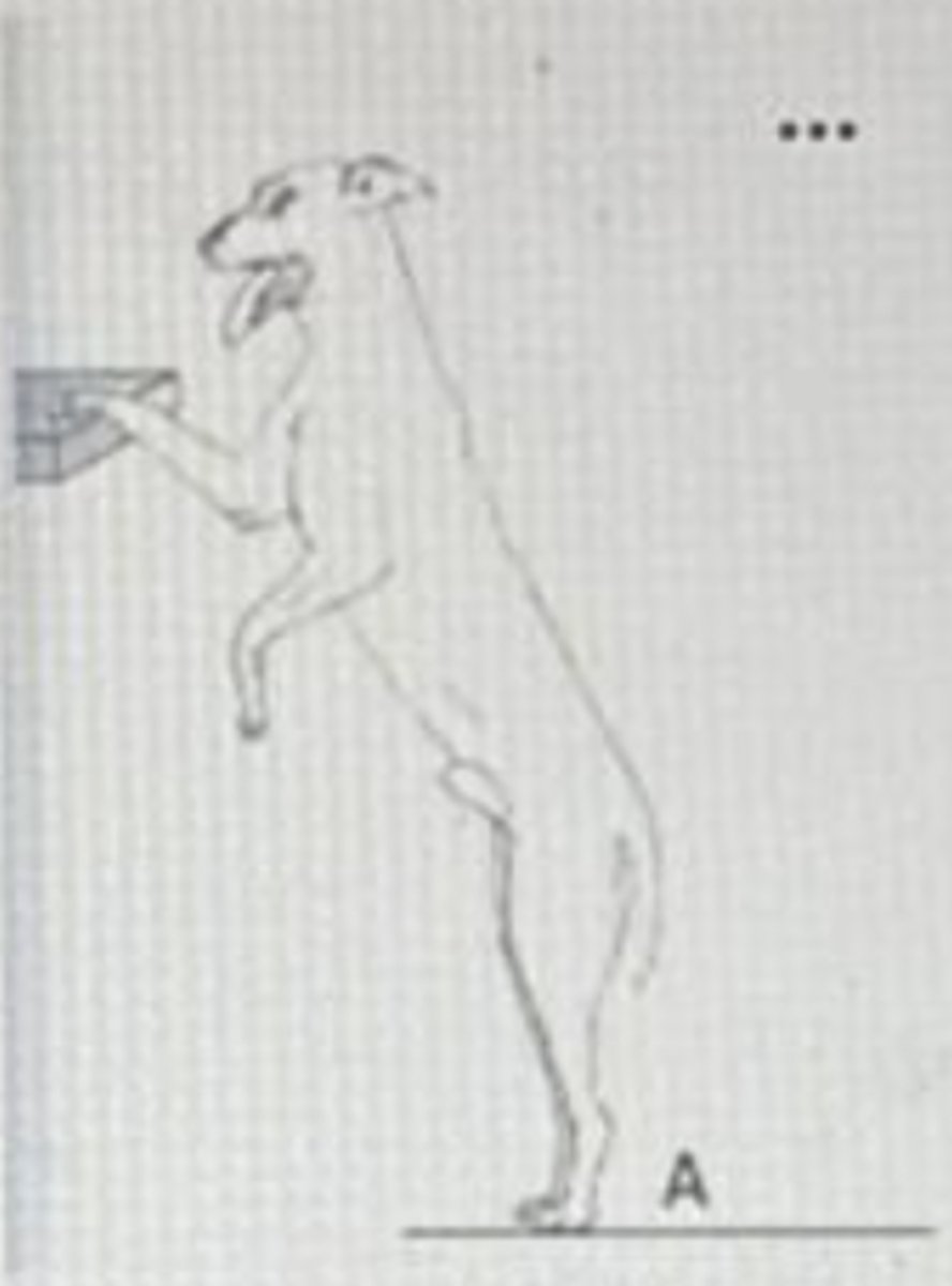
The following picture shows a dog which is not able to flex the elbow joint. What nerve could be damaged?
A) Radial nerve
B) Musculocutaneous nerve
C) Median nerve
D) Ulnar nerve
B
Which of the following muscles is the main anteversor muscle of the pelvic limb?
A) Psoas major muscle
B) Pectineal muscle
C) Sartorius muscle
D) Psoas minor muscle
A
Which of the following arteries provides oxygenated blood to the thoracic limb and what origin does it have in equine?
A) Subclavian artery. Origin: Aortic arch
B) Common carotid artery. Origin: Brachiocephalic trunk
C) Subclavian artery. Origin: Brachiocephalic trunk
D) Common carotid artery. Origin: Aortic arch
C
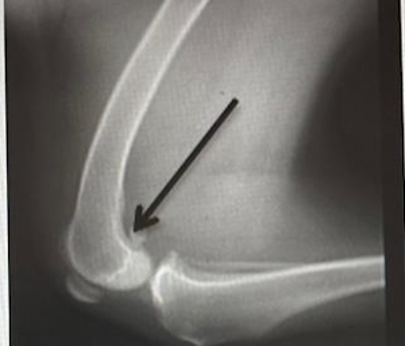
Latral view radiograph of the femur in a dog. What anatomic structure is the arrow pointing at?
A) Patella
B) Gastrochemius sesamoid bones
C) Femoral condyle
D) Patellar ligament
B
What muscle is the following arrow pointing at? And what function does it have?
A) Gracilis muscle. Abduction
B) Pectineus muscle. Adduction
C) Sartorius muscle. Extensor muscle
D) Adductor muscle. Adduction
B
Which of the following gluteal muscles are superficial?
A) Middle gluteal muscle
B) Deep gluteal muscle
C) Superficia gluteal muscle
D) Answers A and C are correct
D
Regarding the patellar ligament. Choose the correct answer.
A) In equine and ruminants the patellar ligament has four different portions
B) The patellar ligament is formed by the tendon of the quadriceps muscle of the thigh
C) The patellar ligament extends from the patella to the lateral tibial condyle
D) Carnivore have the medial patellar ligament
B
Which of the following ligaments in the elbow joint is only present in carnivores?
A) Olecranon ligament
B) Radiouinar ligament
C) Lateral colateral ligament
D) Answers A and B are correct
D