MIC102 Lecture 1
1/6
There's no tags or description
Looks like no tags are added yet.
Name | Mastery | Learn | Test | Matching | Spaced | Call with Kai |
|---|
No analytics yet
Send a link to your students to track their progress
7 Terms
Compare the relative scale of different types of microbes
Viruses (even though not technically living)
20-300 nanometers
Bacteria
0.2-5 micrometers
Archaea
0.1-15 micrometers
SIngle-celled Eukaryotes
10-1000 micrometers (some can be seen by human eye -100 micrometers and up)
What is the defining feature of microogranisms?
small size
Which type of living cell is most abundant in and on the human body?
bacterial cells
they outnumber eukaryotic and archaea by 2-3 orders fo magnitude
Which can be a potential energy source for microbes?
CO2
Cellulose
Methane
Infrared radiation
Electricity
All except CO2
CO2 is fully oxidized and can’t be extracted form for energy
Identify the different types of Light Microscopy
Magnifies up to 1000x, best for viewing living cells but can view dead specimens sometimes, can resolve structures ~200 nm apart
Brightfield
to view pigmented or stained specimens (high contrast)
image formed by light passing and then absorbed or scattered by stain or naturally pigmented structures
Phase contrast
good for live unstained cells, can be used to watch cell movement, cell division, and internal structures
to view non-pigmented, transparent-like (low contrast) specimens
Fluorescence
to view cells/structures labeled with a fluorochrome
can detect specific molecules, organelles, or strcutures
needs fluorscent labelling
Identify the types of Electron Microscopy
Magnifies up to 500,000X, can resolve structures -0.1 nm apart
best for viewing small structures
Usually only works with dead cells due to processing requirements, but has high resolution
no color usually for two types covered
Scanning EM
scans the exterior of a specimen to reveal the topography and fine details in 3d
requires samples that are highly processed (thin, fixed, stained with conductive metal, etc.)
Transmission
used to view internal structures of specimens or small specimens
specimens may be sliced or stained in cross sections to view
dense areas scatter electrons more and show up darker in image
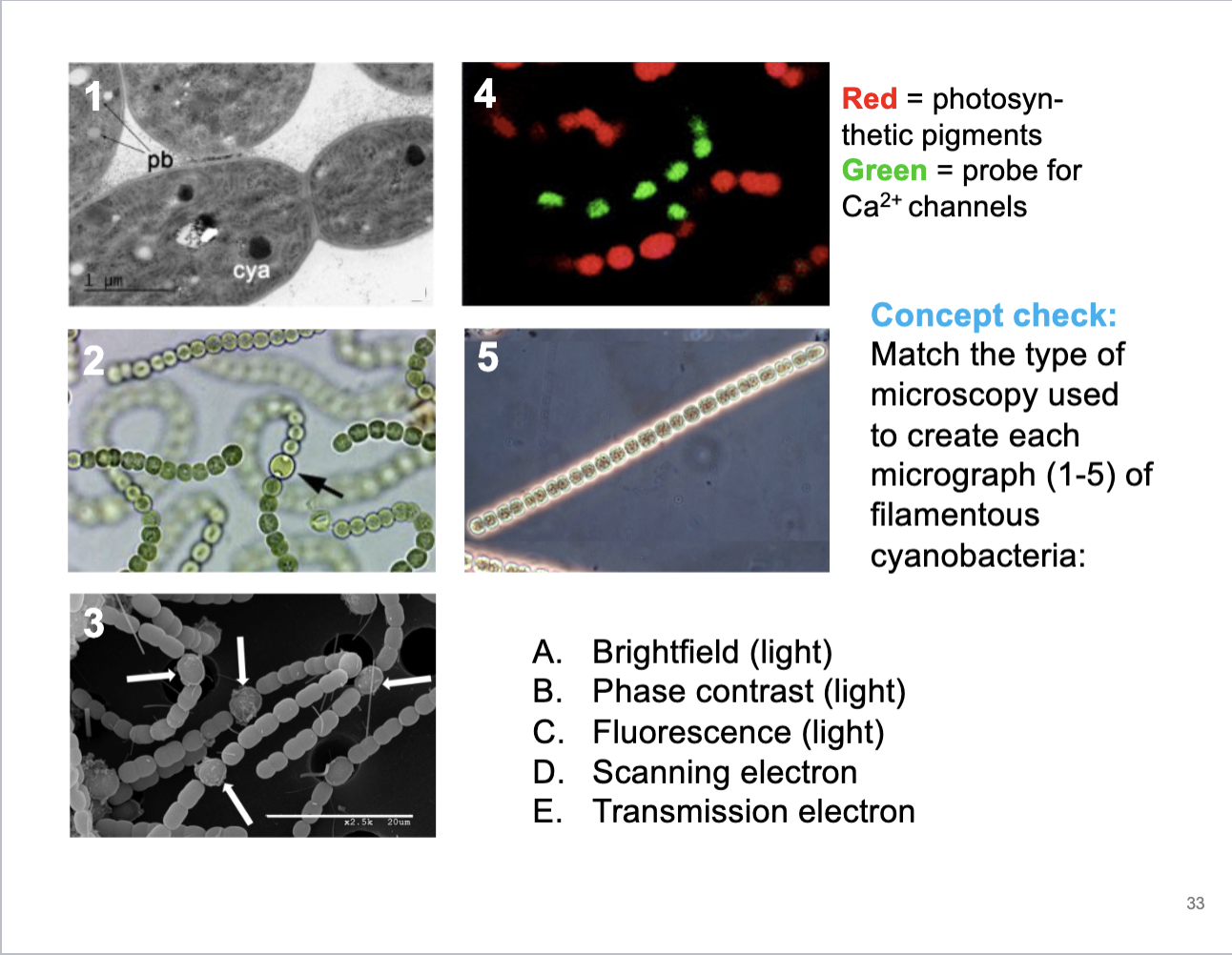
Match the type of microscopy used to create each micrograph of filamentous cyanobacteria
1) E - Transmission, due to extreme resolution and small imagery, high magnification, internal structures of organism, black adn white
2) A - Brightfield - microbe shows clear pigment and high contrant, but resolution is moderate, true color of organisms and very bright background (hence name of microscopy type)
3) D - Scanning electron - Can tell due to the lack of coloring in the images and how it has high resolution showing the topology of the bacteria, surface only view, blacka dn white, 3D cell appearance
4) C - Fluorescence, due to the fluorescent nature of the photo, how they pop out, differnet colors
5) B - Phase contrast - microbe seems almost transparent and see through and background is darker than microbe itself, brihgt pahse halos around cells, dark background and strucutres in cytoplasm