First-aid - GI - Pathology
 Call Kai
 Learn
Learn Practice Test
 Spaced Repetition
 Match
Match Flashcards
 Knowt Play
1/325

There's no tags or description
Looks like no tags are added yet.
Name | Mastery | Learn | Test | Matching | Spaced | Call with Kai | Chat |
|---|
No analytics yet
Send a link to your students to track their progress
326 Terms
Aphthous ulcers are aka what
Canker sores
What are aphthous ulcers
Common oral lesions that appear as painful, shallow, round to oval ulcers covered with a yellow-ish exudate

Diagnosis:
Patient presents with painful oral lesions, upon examination, you note that they are covered by a white-yellow exudate
Aphthous ulcers - Canker sores
Recurrent Aphthous Stomatitis
Recurrent Aphthous Ulcers, associated with:
Behcet Syndrome
IBD
Celiac Disease
HIV
SLE
Mnemonic: BICHS
Squamous Cell Carcinoma of the Oral Cavity Presentation
Usually affects the tongue, and presents as a nonhealing ulcer with irregular margins and raised borders
Leukoplakia may also be noted and
Erythroplakia are precursor lesions
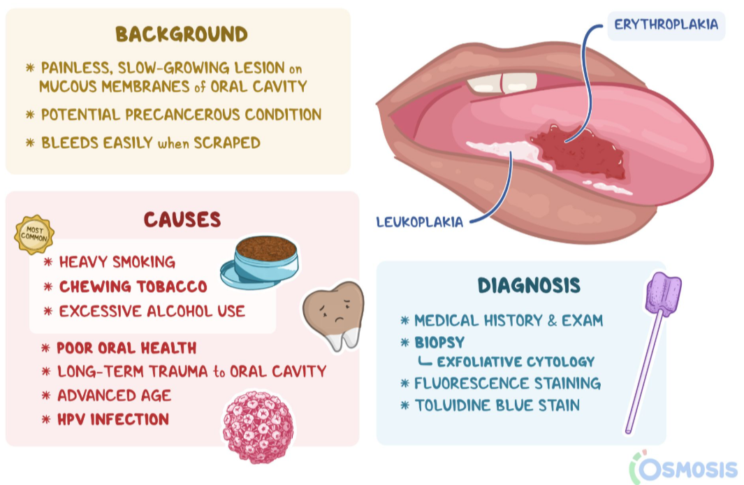
What is the most common malignancy of the oral cavity
Squamous cell carcinoma

Squamous Cell Carcinoma of the Oral Cavity Associations
Tobacco
Alcohol
HPV-16
Sialothiasis - Definition
Stone formation in major salivary gland ducts (= Parotid, submandibular, sublingual)
Sialothiasis - Pathophysiology
Why Do the stones form?
Salivary stasis due to dehydration or trauma (fibrosis → obstruction)
Most common in submandibular gland duct because:
Saliva is more alkaline + more mucus
Saliva has to travel a bit uphill → Stasis
The duct is narrower
Why is the pain pre/periprandial?
Because we start secreting saliva at the moment we start smelling or imagining food, which is before we start eating the food, and all throughout the meal
Sialothiasis - Presentation
Pre/peri-prandial pain and swelling in the affected area
Sialadenitis - Definition
Inflammation of the salivary glands due to:
Obstruction
Infection (S. aureus/ mumps)
Immune-mediated mechanisms (Sjögren syndrome)
Describe how you can get from Sialothiasis to Sialadenitis
Obstruction → Stasis → S. Aureus overgrowth → Sialadenitis
Discuss general Salivary gland tumor Benignity and Malignancy (Likelihood stuff)Wh
Salivary gland tumors are usually benign.
Most commonly found in the parotid gland (Most benign ones)
Tumors found in the submandibular/ sublingual and other minor glands are more likely to be malignant
What is the classic presentation for benign salivary gland tumors
They typically present as a painless mass or painless swelling
What symptoms are more suggestive of malignant involvement of salivary gland tumors
Facial paralysis and painful masses
Pleomorphic Adenoma - General + Histology
Most common salivary gland tumor
It’s a benign mixed tumor, composed of chondromyxoid stroma and epithelium
It may undergo malignant transformation
Recurrence of Pleomorphic Adenoma after treatment
Only recurs if incompletely excised or if ruptured intraoperatively
Other name for Warthin Tumor
Papillary cystadenoma lymphomatosum
Warthin tumor - Description + Associations
Benign cystic tumor with germinal centers, may be bilateral or multifocal. Typically found in smokers
Mnemonic: Warriors from Germany love smoking
Mucoepidermoid carcinoma - Description
Most common malignant tumor. Mucinous and Squamous components
List the main causes of acute gastritis
Chronic NSAIDs use (Especially in alcoholics and those who take NSAIDs daily like in Rheumatoid arthritis)
Burns (Hypovolemia)
Brain injury (Increased vagal tone)

How can chronic NSAID use cause acute gastritis
NSAIDs → COX inhibition → ↓ PGE₂
Normally, PGE₂:
↑ mucus
↑ bicarbonate
↑ mucosal blood flow
Therefore:
↓ PGE₂ → ↓ mucosal protection → gastric erosions
How can Burns cause acute gastritis (Curling ulcers)
Burns cause severe hypovolemia due to massive fluid loss → Decreased gastric perfusion → Severe mucosal ischemia that causes loss of mucosal integrity → Erosions and ulcers - Curling ulcers
A 45-year-old obese woman presents with dysphagia, chronic intermittent cough, and chest burning for several months. Her symptoms are worse after large meals or lying down. Her diffculty swallowing has intensifed recently, which prompted her to schedule an appointment. Tums helped with the burning sensation in the past, but her symptoms are getting worse. She has not lost weight. She recently started verapamil for hypertension. On physical exam, oropharynx is clear without erythema or exudates, lungs are clear to auscultation, and she has mild tenderness to palpation in the epigastric region.
GERD
How can Brain injury cause acute gastritis
Brain injury can cause a Cushing ulcer due to high vagal stimulation which will lead to Increased Acetylcholine production → Increased H+ production
*Note: vagal stimulation can make it so there’s acid secretion even when there’s no food in the GI Tract
Achalasia etiology and pathophysiology
Failure of LES to relax due to degeneration of NO and VIP-secreting neurons in the Auerbach/Myenteric Plexus of the esophageal wall
Achalasia presentation
Progressive dysphagia to solids and liquids
*Note: Achalasia and diffuse esophageal spasm, which present with simultaneous solid and liquid dysphagia.
A 35-year-old woman presents with progressive difficulty swallowing both solids and liquids. Her symptoms have worsened over the past 5 years. She initially only had symptoms with solids, and she progressively has had symptoms with liquids. She also notes regurgitation of undigested food. She denies weight loss. In the past, she has had multiple trials of PPIs without improvement of her symptoms.
Achalasia
Achalasia Risk Factors
Chagas disease (Trypanosoma Cruzi)
Extraesophageal Malignancies
Can be Idiopathic tho
Achalasia potential complications
Achalasia is associated with an increased risk of esophageal cancer.
How do you diagnose achalasia
Manometry: Will reveal uncoordinated peristaltic movements with increased LES resting pressure.
Barium Swallow: Will show dilated esophagus with area of distal stenosis (“bird’s beak”)
Achalasia Treatment
Surgery, endoscopic procedures (e.g. Botulinum toxin injection)
GERD etiology
It’s due to a transient decrease in LES tone
GERD classic presentation
Classically presents as: Heartburn, Regurgitation, Dysphagia
Symptoms of laryngopharyngeal reflux
In laryngopharyngeal GERD, patients may also present with: Chronic cough, lump in throat sensation, constant throat clearing, hoarseness
GERD complications
Erosive esophagitis
Esophageal strictures
Barett Esophagus
Esophagitis presentation
Odynophagia and/or Dysphagia
List the different types of Esophagitis
Reflux/Errosive Esophagitis
Pill Esophagitis
Infectious Esophagitis
Corrosive Esophagitis
Eosinophilic Esophagitis
What condition commonly arises secondary to GERD
Reflux/Erosive esophagitis
List pills that cause Pill esophagitis
Bisphosphonates, tetracyclines, NSAIDs, ferrous
sulfate, potassium chloride
Infectious causes of Esophagitis
Candida (most common) - white pseudomembranes
CMV
HSV-1 - punched out ulcers
All associated with immunosuppression
Plummer-Vinson syndrome presentation
Plummer DIES
Dysphagia
Iron-deficiency anemia
Esophageal webs
Increased risk for esophageal SCC
Name a condition associated with Plummer-Vinson syndrome associated
Glossitis (It’s due to the iron-deficiency causing atrophy of the tongue’s mucosa)
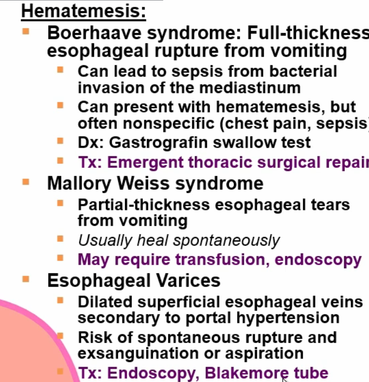
Mallory-Weiss syndrome etiology
Partial thickness, longitudinal lacerations of the gastroesophageal junction
- Confined to mucosa/submucosa, due to severe vomiting
Mallory-Weiss presentation
Hematemesis
Abdominal/back pain (±)
Mallory-Weiss risk factors
Alcohol use
Bulimia nervosa
Esophageal varies etiology
Dilated submucosal veins in lower 1/3 of esophagus 2° to portal hypertension
Esophageal varices presentation and cause
Presents as hematemesis. Secondary to cirrhosis
Distal esophageal spasm etiology
Spontaneous, no peristaltic (uncoordinated) contractions of the esophagus with normal LES pressure
Distal esophageal spasm presentation
Dysphagia & Angina-like chest pain
Distal esophageal spasm Diagnostics
Barium swallow will reveal Corkscrew Esophagus
Manometry is diagnostic

Distal esophageal spasm treatment
Nitrates and CCBs
Esophageal smooth muscle atrophy and fibrosis with weakened LES pressure and distal motility
Scleroderma esophageal involvement
Scleroderma esophageal involvement

Complications of Esophageal involvement in scleroderma
Acid reflux → Dysphagia and Stricture
Barret esophagus and aspiration
Etiology of esophageal perforation
Most commonly, following esophageal instrumentation, but can also be due to spontaneous rupture, foreign body ingestion, trauma, and malignancy
Esophageal perforation findings
Pneumomediastinum & Subcutaneous emphysema
(signs include crepitus in the neck region or chest wall) can indicate dissecting air.
Boerhaave syndrome
transmural, usually distal esophageal rupture due to violent retching (from vomiting)
Hematemesis DDx
What is Barrett esophagus
Intestinal metaplasia characterized by replacement of nonkeratinized stratified squamous epithelium with intestinal epithelium
What is the known cause of Barett esophagus, and what are its complications
Cause: Chronic GERD
Complication: Highly associated with development of esophageal adenocarcinoma
Dumping syndrome
Hyperosmolar food (often sugary) moves too quickly from the stomach to small intestine. Typically occurs after stomach or esophageal surgery
How does esophageal cancer present, and what makes it an agressive cancer
It typically presents with progressive dysphagia to solids and then to liquids and weight loss.
It’s aggressive due to the lack of serosa in the esophageal. Patients also typically present with advanced disease
In which part of the esophagus is squamous cell carcinoma typically observed
Upper 2/3
Esophageal squamous cell carcinoma risk factors
Alcohol, hot liquids, caustic strictures, smoking, achalasia, nitrosamine-rich foods (processed meats, smoked fish, and certain alcoholic beverages, particularly beer)
Esophageal adenocarcinoma risk factors
Chronic GERD, Barrett esophagus, obesity, tobacco smoking
In which part of the esophagus is adenocarcinoma typically observed
Lower 1/3
Which esophageal cancer is more common worldwide, and which is more common in America
SSC → Worldwide
Adeno → America
What is chronic gastritis
Mucosal inflammation, often leading to atrophy (hypochlorhydria → hypergastrinemia) and intestinal metaplasia (→ risk of gastric cancers)
Classic causes of chronic gastritis
H. Pylori
Autoimmune (Anti-IF and Anti-H+/K+ ATPase)
H pylori chronic gastritis basic rundown
Affects antrum first and spreads to body of stomach.
Peptic Ulcer disease
H. pylori is associated with which cancer
MALT lymphoma
Autoimmune chronic gastritis
Anti-IF, and Anti-H+/K+-ATPase on parietal cells → *Increased risk of Pernicious anemia
*Affects body/fundus
Autoimmune gastritis affects which part(s) of the stomach
Affects body/fundus
What is Menetrier disease
Hyperplasia of gastric mucosa p hypertrophied rugae (“wavy” like brain gyri). Causes excess mucus production with resultant protein loss and parietal cell atrophy with r acid production.
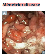
Menetrier disease presentation
Weight loss, Anorexia, Vomiting, Epigastric pain, Edema (due to protein loss; “WAVEE”).
Protein loss because too many amino acids are used in mucin production + Increased permeability of gastric mucosa → loss of protein into stomach)
Parietal cell atrophy is because increased mucin in stomach compresses parietal cells → Atrophy
Common gastric tumors
Gastric adenocarcinoma
Gastric lymphoma
GI stromal tumors (KIT or PDGFRA)
Carcinoid tumors
Gastric cancer classical symptoms
Weight loss
Early satiety
Abdominal Pain
Obstruction
Sudden-onset Acanthosis Nigricans or Leser-Trélat sign (Sometimes)
WEAPON
Intestinal-type Gastric Adenocarcinoma pathophysiology
Chronic H. pylori infection → Chronic mucosal injury (Atrophy/achlorhydria) → Intestinal metaplasia → Dysplasia → Adenocarcinoma
Appearance of Intestinal-type gastric adenocarcinoma
It looks like an ulcer with raised margins
Intestinal-type gastric adenocarcinoma Risk factors
H. pylori
Chronic gastritis
Nitrosamines
Tobacco smoking
Achlorhydria
Chronic gastritis
Diffuse-type gastric adenocarcinoma etiology
It involves E-Cadherin mutations → Cells don’t stick together → They individually infiltrate the gastric wall
These cells also tend to accumulate mucin and become known as Signet-ring cells due to their appearance

Findings in Diffuse-type gastric adenocarcinoma
Signet-ring cells
Stomach wall grossly thickened and leathery = Linitis plastica
Tumor infiltration + fibrosis → Stiff non-compliant stomach = Early Satiety
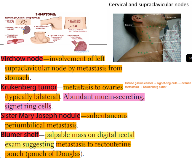
Discuss possible sites of gastric cancer metastasis
Virchow node = Metastasis from stomach to left supraclavicular nodes
Krukenberg tumor = Metastasis to ovaries (In Diffuse-type gastric cancer, since it’s common to find signet ring cells)
Sister Mary Joseph Nodule: Subcutaneous periumbilical metastasis
Blumer Shelf: Palpable mass of DGE, suggesting metastasis to pouch of Douglas
Typical causes for gastric ulcers
H. pylori (70%)
NSAIDs
Due to decreased mucosal protection against gastric acid
Typical causes for duodenal ulcers
H. pylori (90%)
Zollinger-Ellison syndrome
Due to decreased mucosal protection or due to increased gastric acid secretion
Gastric and duodenal ulcers risk of malignancy
Gastric: High risk, should be biopsied to rule out malignancy
Duodenal: Generally benign
Gastric vs duodenal ulcer pain characteristics
Gastric: Pain greater with food → Weight loss
Duodenal: Pain decreases with meals → Weight gain (food buffers acid
Ulcer complications
Hemorrhage
If on lesser curvature = Bleed from left gastric artery
If on posterior wall of duodenum = Bleed from gastroduodenal artery
Obstruction: Pyloric outlet obstruction, from fibrosis, can also be duodenal obstruction
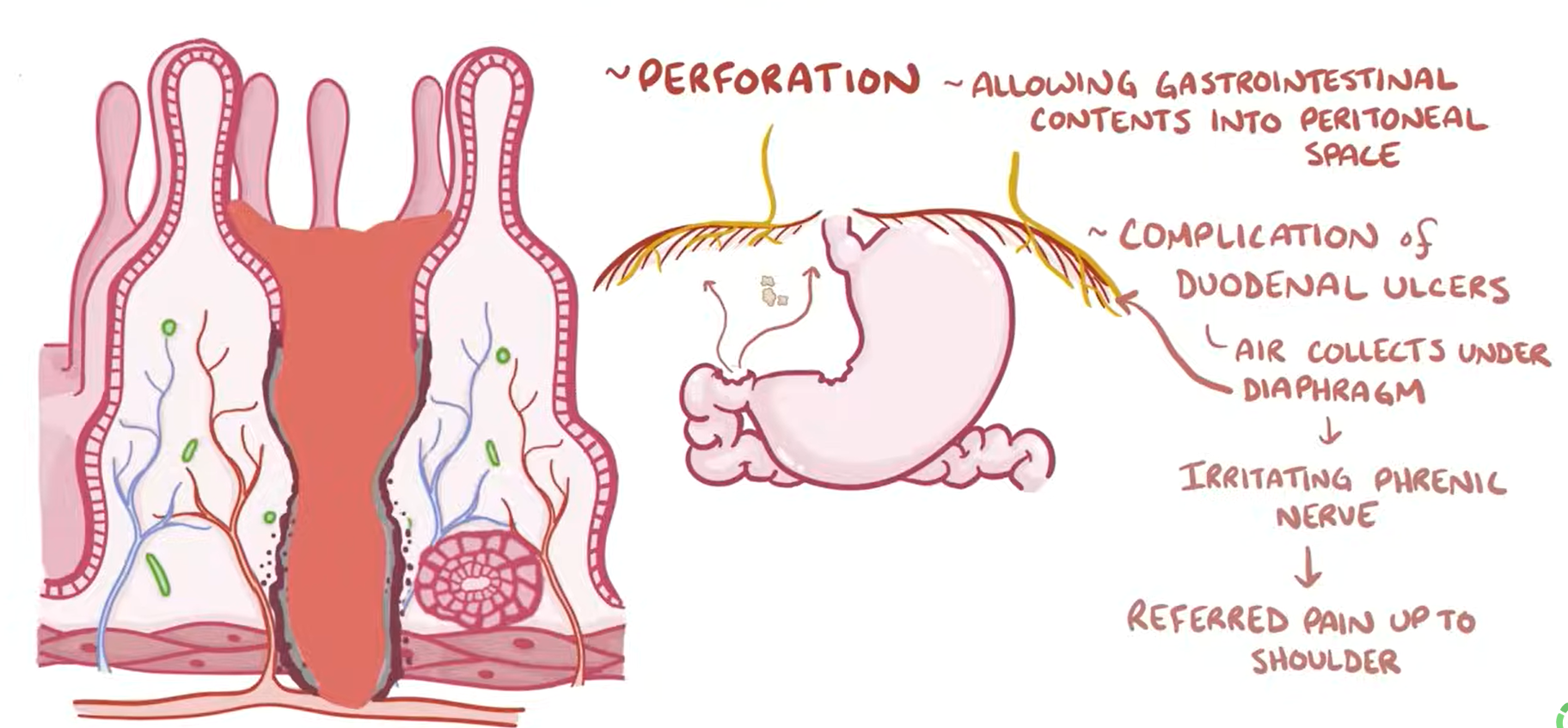
Perforation; Anterior duodenal ulcers more common, will see:
Pneumoperitoneum
Referred shoulder pain due to phrenic nerve irritation
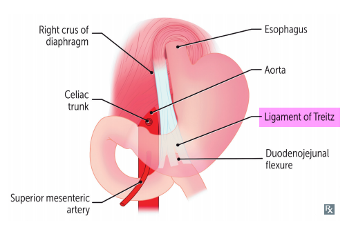
How are GI bleeds classified as Upper or Lower
Depends on if above or below the ligament of Treitz
Upper vs Lower GI bleeds symptoms
Upper: Hematemesis/ melena
Lower: Hematochezia

What symptoms should make you think of malabsorption syndromes
diarrhea
steatorrhea
weight loss
weakness
vitamin and mineral deficiencies
Screening tests if you suspect malabsorption syndromes
Screen for fecal fat with Sudan stain
Use D-Xylose test if you suspect abnormal absorption
Which allelic variants is Celiac disease associated with?
HLA-DQ2 & HLA-DQ8 (Northern-European descent)
I ate [8] too [2] much gluten at [D]airy [Q]ueen
In celiac, the intolerance is against which protein
Gliadin (gluten protein found in wheat, barley, rye)
Which parts of the intestines are affected in Celiac Disease
Distal duodenum and/or proximal jejunum → malabsorption and steatorrhea.
Celiac disease potential Complications/Associations
Dermatitis herpetiformis
Decreased bone density
Iron-deficiency anemia
Increased risk for malignancy (T-cell lymphoma)
Celiac disease Serology
Serology: ⊕ IgA anti-tissue transglutaminase (IgA tTG), anti-endomysial, and anti-deamidated gliadin peptide antibodies.

Celiac disease Histology
Histology: Loss of Villi, mucosal atrophy, Crypt hyperplasia, intraepithelial lymphocytosis
Lactose Intolerance etiologies
Lactase deficiency
2ndary lactose intolerance = If severe gastroenteritis -> Damage to villi, and lactase sits on villi tip -> temporary lactose insufficiency (Secondary Intolerance)