Quiz #1
 Call Kai
 Learn
Learn Practice Test
 Spaced Repetition
 Match
Match Flashcards
 Knowt Play
1/214

There's no tags or description
Looks like no tags are added yet.
Name | Mastery | Learn | Test | Matching | Spaced | Call with Kai | Chat |
|---|
No analytics yet
Send a link to your students to track their progress
215 Terms
What are x-rays?
A type of electromagnetic radiation
What is electromagnetic radiation?
A form of energy created by the movement of electrically charged particles such as electrons
How many types of electromagnetic radiation are there?
7
What is the electromagnetic spectrum?
Radiowaves, microwaves, infrared, visible light, UV, x-rays, and gamma rays
Each type of electromagnetic radiation is characterized by what?
Wavelength and frequency
What is wavelength?
The distance between two consecutive peaks
What is frequency?
The number of waves that form in a given length of time measured in wave cycles per second or hertz (Hz)
A shorter wavelength has a _______ frequency
Higher
X-rays are characterized by a….
Shorter wavelength and a higher frequency
When the wavelength is shorter, what happens to each electrically charged particle?
They have more energy
A longer wavelength has a ______ frequency
Lower
The x-ray tube is made of what two electrodes?
Cathodes (-) and Anodes (+)
Electrons are located where and contain what?
The cathode and a negative charge
Due to the electrons’ negative charge they are pulled to where?
The positive anode (A+)
When the electrons interact with the positive anode, a large amount of energy is produced in the form of….
X-rays and heat
How much heat is produced when the electrons interact with the positive anode?
99%
How many x-rays are produced when the electrons interact with the positive anode?
1% roentgen radiation
X-rays may change ______ but always travel in a ______ path.
Direction and straight
X-rays originate from where and spread apart as they travel in straight lines, gradually moving further and further apart from one another, i.e. they begin to fan out.
A single point of origin
Where is the source of electrons to be electrically charged?
Cathode
The cathode directs electrically charged electrons toward where?
The anode (A+)
The cathode contains what?
A wire filament made of tungsten that is heated by a low-energy circuit
Electrons are released when the wire filament is
Heated
The heat does what within the atoms of the tungsten filament?
Excites electrons
The heated electrons in the wire eventually exceed their binding energy and are available to what?
Accelerate across the tube to the anode
The number of electrons that are produced depends on what?
The amount of heat applied to the filament
Tungsten has a very high melting point which is ideal for x-ray production because….
Tungsten has a high atomic number which means the atoms have a higher number of electrons to give up when charged.
Because of tungsten’s high melting point it also does what to the life of the x-ray tube?
Extends it
The anode is the target for what?
The electrically charged electrons to strike
The area that the electrons strike is known as the what?
Focal spot
The size of the focal spot determines the what of the image?
Sharpness
The smaller the focal spot the….
Sharper the image
The anode draws heat….
Away from the target
The anode can withstand temperatures as high as….
1832°
Stationary anode (dental or portable radiograph units)
Not designed to withstand high heat
Rotating anode
Designed to handle higher heat signatures and are typically found in stationary units
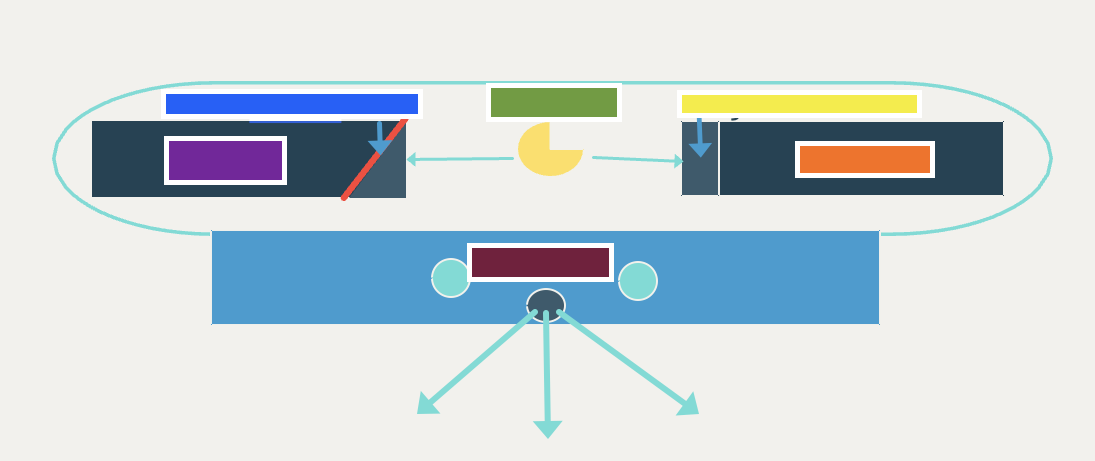
Blue
Focal spot on target

Purple
Anode +

Green
Electrons

Yellow
Tungsten filament

Orange
Cathode -
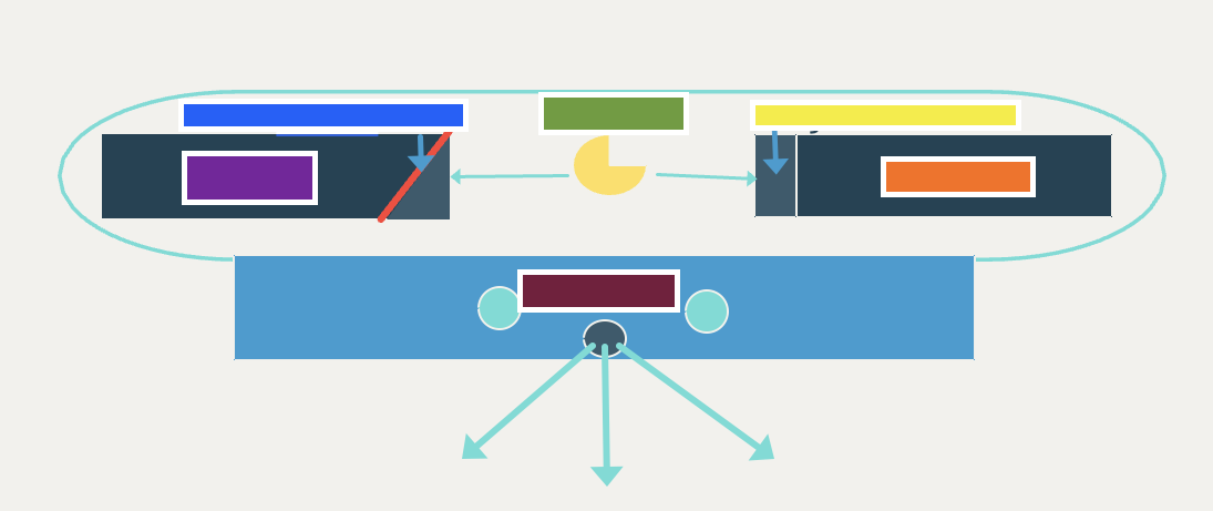
Maroon
Collimator
The tube stand does what?
Supports the x-ray tube
Tube stand models vary
Portable handheld units, to tabletop stands, to large stands mounted from the ceiling
What do the shutters do in the collimator?
Controls the size of the primary x-ray field
Includes a visible light source to ‘view’ the x-ray field size
Collimator
The collimator assists with what
The accurate patient positioning (technique) and safety personnel (ALARA)
Absorbs scatter radiation to improve the quality of the image
The grid
The grid is positioned where?
Between the patient and the image receptor
The grid is built into what?
Potter-Bucky diaphragm
Grid patterns may be linear or crossed but which is most common?
Crossed
The grid ratio refers to what?
The height of the lead strips in the grid compared to the space between them
Higher grid ratios are more efficient because….
They absorb more scatter
Older machines required that grids be used only when….
The tissue thickness of the area to be imaged measured at least 10 cm
Digital radiograph machines are efficient which allows for…
The use of the grid for all images taken
The Potter-Bucky diaphragm has what….
Adjustable positioning to accommodate patients of different sizes
The Potter-Bucky Diaphragm contains what?
A built in grid to absorb scatter
Where is the Potter-Bucky diaphragm located?
Under the x-ray table or vertically in a wall mounted unit
Portable Potter Bucky diaphragms require what?
A person to manually support the cassette
Control panel has what?
On/off switch, Voltage compensator/High voltage circuit (surge protector), Low-Voltage circuit, Kilovoltage peak (kVp), Miliamperage and Time selector (mAs), Exposure pedal or button, and warning light
What is the Voltage compensation/High voltage circuit?
Basically a surge protector
What is the Low-Voltage circuit?
Applies heat to tungsten filament in miliamps
What is the kilovoltage peak selector?
Acceleration
What is the miliamperage and time selector?
Number of x-rays and time of active x-ray production
The x-ray field must be always directed….
Perpendicular or 90° to the patient and the image receptor
The Source of the Image Distance (SID) should always be how far?
40 inches
The anatomy of interest must be ______ with the image receptor/table top.
Parallel
The anatomy of interest must be positioned as ______ to the tabletop as possible.
Close
What are the anatomical landmarks?
Centering, collimation, and measurement
What do you center?
The x-ray field over the middle of the area of interest
What needs to be included when collimating?
The entire area of interest so that the entire area can be properly evaluated radiographically
What do you measure?
The thickest area within the collimated field to set the machine for appropriate density and contrast
What results in artifacts on the radiograph that interfere with interpretation?
Technical errors
What errors may result in artifacts?
positioning, preparation, and measurement
Patient position artifacts
magnification, foreshortening, elongation, and artifactual changes in density and contrast
Patient preparation artifacts
Food and feces, patient motion, leashes or collars, human anatomy
Patient measurement artifacts
Using the wrong landmark, measuring patient while sitting or standing, using the technique chart incorrectly
Always remember to include what in the area of collimation?
Positioning marker
Intentional artifacts include what?
Gunshot/birdshot, splints, casts, pins, screws, plates, and microchips
Radiographic density refers to the degree of what on the x-ray?
Brightness or darkness
What changes the brightness or darkness on the x-ray?
mAs
Radiographic contrast refers the difference in what on the x-ray?
Radiographic density between two areas
What changes the radiographic density between two different areas on the x-ray?
kVp
When disease is present in an organ or tissue what does it impact?
The radiographic density and contrast, as well as the visualization of surrounding anatomical structures.
What are common abnormal findings in an x-ray?
Fluid, air/gas, enlarged organs, displaced organs
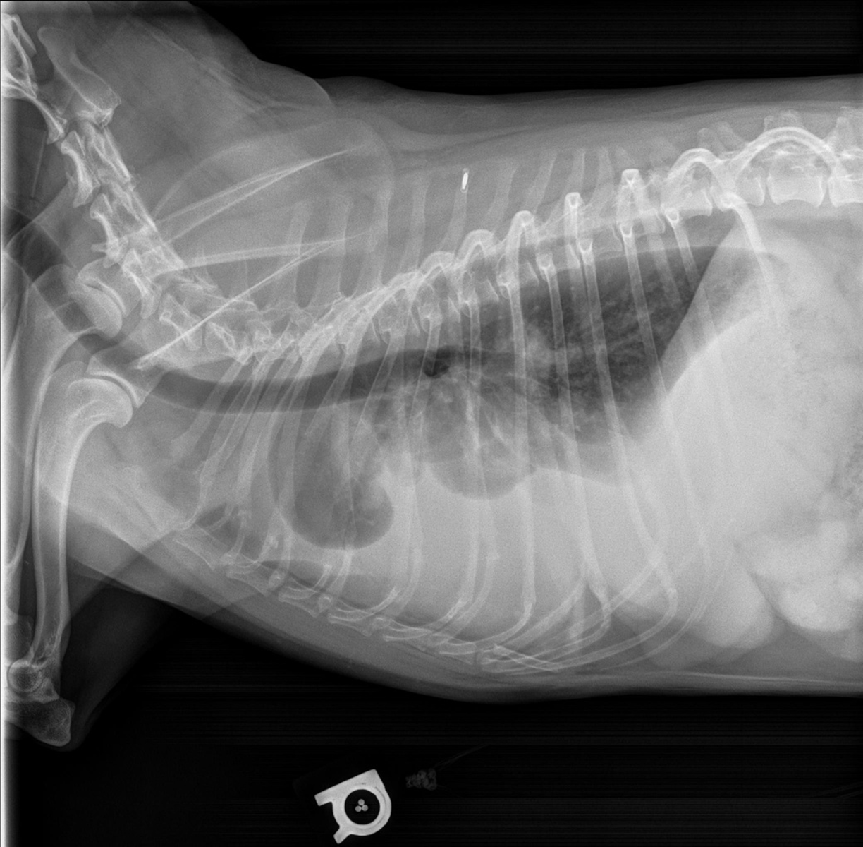
What is wrong?
Pleural effusion
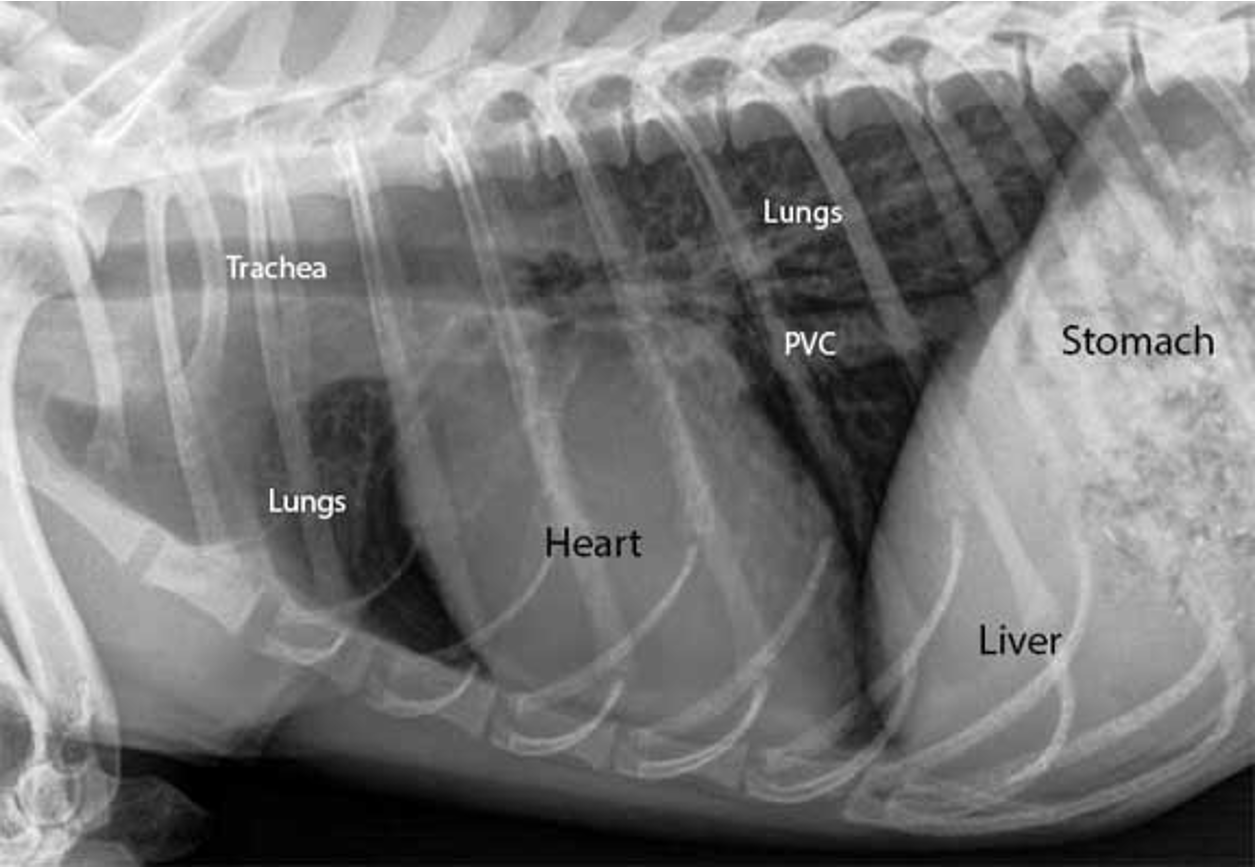
What is wrong?
Enlarged heart

What is wrong?
Pyometra

What is wrong?
GDV
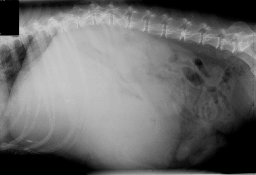
What is wrong?
Hepatomegaly
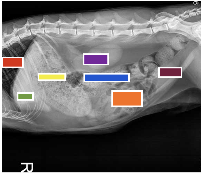
Red
Lungs

Green
Liver

Yellow
Stomach

Purple
Kidneys

Blue
Large Intestine

Orange
Small Intestines

Maroon
Bladder
What is the anode heel effect?
Variation of the number of x-rays i.e. intensity of the x-rays of the primary x-ray field as they diverge from the point of origin.
The number of x-rays i.e. intensity of the x-rays are greatest where?
Closest to the cathode