RHEUMATOLOGY
 Call Kai
 Learn
Learn Practice Test
 Spaced Repetition
 Match
Match Flashcards
 Knowt Play
1/75

There's no tags or description
Looks like no tags are added yet.
Name | Mastery | Learn | Test | Matching | Spaced | Call with Kai | Chat |
|---|
No analytics yet
Send a link to your students to track their progress
76 Terms
Four cardinal signs of inflammation
Erythema
Warmth
Pain
Swelling
Differentiate articular vs nonarticular disorders

When is synovial fluid aspiration indicated in MSK disorder
Acute monoarthritis (monoarthritis in patients with chronic polyarthritis)
Cases of trauma with effusion
Infectious or crystal-induced arthropathy suspected
How to assess viscosity of fluid

Study assessment of MSK complaint

Study interpretation of Synovial fluid aspiration and analysis

PIP joint hypertrophy
Bouchard’s nodes
DIP joint hypertrophy
Heberden’s nodes
Pathologic sine qua non of osteoarthritis
Hyaline articular cartilage loss
Morning stiffness in Osteoarthritis
Brief morning stiffness <30 mins
Radiographic finding in osteoarthritis
Sinovial fluid analysis
Joint space narrowing
Osteophytes (bone spurs)
Synovial fluid analysis non-inflammatory - WBC <2000mm3
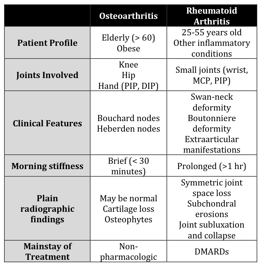
Difference in joint affectation of RA and OA
Morning stiffness
DIP
PIP
MCP
RF and anti CCP
Joint fluid leukocyte count
DIP affected in OA, RA not

Treatment of OA

Medication and dose for OA
Oral NSAIDS 4
Topical NSAIDS 4
Intraarticular injections

Earliest joints involved in RA
Small joints of the hands and feet (EXCEPT DIP)
Distribution of RA
Frequent hallmark
Symmetric
Flexor tenosynovitis (trigger fingers)
Piano key movement
3 Pathologic hallmarks of RA
Synovial inflammation and proliferation
Focal bone erosions
thinning of articular car
Most common hematologic abnormality of RA
Most common pulmonary manifestation
Most common cardiac abnormality
Normochromic normocytic anemia
Pleuritis
if with dry cough, progressive SOB in RA - think of interstitial lung disease
MC valvular abnormality
Joint finding in RA
Pannus formation - abnormal, aggressive growth of thickened synovial tissue filled with inflammatory cells and new blood vessels that invades and destroys adjacent joint cartilage and bone.
Joint deformities seen in RA

Xray findings in RA
Periarticular osteopenia - initial
Others:
soft tissue swelling
Joint space loss
Subchondral erosions
Synovial fluid analysis in RA
Inflammatory: WBC 5000-50,000/uL
Overwhelming cell type: neutrophil
Diagnostic for RA
Rheumatoid factor and Anti-citrullinated peptide (anti-CCP antibodies)
Anti-CCP - single most accurate test - more specific than RF
Treatment of choice for RA 3
DMARD - Disease modifying anti-rheumatic agents
slows or prevent structural progression
Conventional DMARDs
Methotrexate with folic acid - first choice for the treatment of early ra
Leflunomide, hydroxychloroquine, sulfasalazine
Biologic DMARDS
Anti-TNF agents (infliximab, etanercept)
Anakinra (IL-1_
Abatacept Rituximab tocilizumab
Glucocorticoids
low moderate dose to control disease before full effect of DMARDS - given in acute flares
Use:
Initial disease control before onset of fully effective DMARD therapy
Acute disease flares
Inadequate response to DMARD therapy
NSAIDs
Other former core of RA therapy
Adjunctive: symptom control

Most common early clinical manifestation of gout
Acute recurrent gout flare
Medication that can cause/trigger gout flare
Thiazide diuretics - decreases renal excretion of uric acid
What is pseudogout Vs gout
Pseudogout:
Positively birefringent rhomboid shaped calcium pyrophosphate dehydrate (CPPD) crystals
Gout:
negatively birefringent needle-shaped monosodium urate crystals
3 diagnostics for gout

Mainstay for acute gouty attack
NSAID
Colchicine
Glucocorticoid
Most effective NSAID
Indomethacin
Naproxen
Inuprofen
Celecoxib
When to stop colchicine
At first sign of loose stool
4 considerations for urate lowering therapy
2 that SHOULD be initiated
More than 2 acute attacks yearly
Severity and duration of flares
Quality of life
Patient’s willingness to commit to lifelong therapy
Presence of uric acid stones
Tophi or chronic gouty arthritis


How to differentiate psoriatic arthritis vs RA
Psoriatic arthritis is seronegative often with DIP, spine, and sacroiliac joints; distinctive radiographic features; and familial aggregation
Clinical features of PsA
psoriasis precedes joint disease in 70%
Nail changes in fingers/toes in 90%
Characteristic of PsA: shortening of digits because of underlying osteolysis
Back and neck pain
Dactylitis - sausage digit inflammation of an entire digit
Enthesitis - inflammation of etheses (sites where tendons/ligaments insert into bone)
What is the CASPAR criteria

what is ankylosing spondylitis
Chronic inflammatory arthritis primarily affecting the spine and large joints causing pain, swelling, and potential bone fusion
Ankylosing Spondylitis shows striking correlation with what histocompatibility antigen?
HLA-B27
Most common extraarticular manifestation of Ankylosing Spondylitis?
Anterior Uveitis
When to suspect Ankylosing spondylitis
Sacroiliitis on imaging plus presence of spondyloarthritis such as inflammatory back pain and possible anterior uveitis
Young adult, fever, chills, rash (papules and pustules on extensor surface), and articular symptoms
Disseminated gonococcal arthritis
most common route - -hematogenous
Gonococcal vs non-gonococcal arthritis
Joint involvement
Other manifestations
Synovial fluid analysis
Synovial fluid culture
Blood culture

Treatment for non-gonococcal arthritis
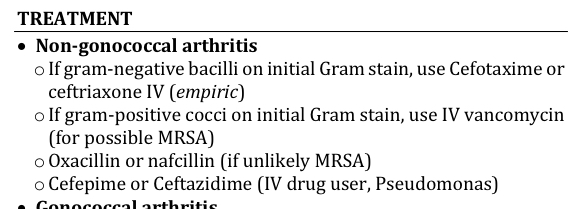
Treatment for gonococcal arthritis

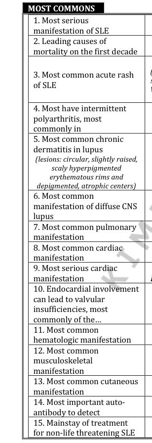
Best initial diagnostic choice for SLE
Antinuclear antibody (ANA)
Most specific test for SLE
Anti-dsDNA (70%) or Anti-Sm (25%)
What test correlates with disease activity of SLE
Anti-dsDNA
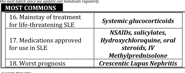
Mainstay treatment for life threatening SLE
Systemic glucocorticoid
Most common chronic dermatitis in lupus
Discoid lupus erythematosus
Systemic Lupus International Collaborating Clinic (SLICC) Criteria for Classification of SLE
5 Clinical
5 Immunologic
Presence of any 4 criteria (must have at least 1 in each category) = SLE

Clinical Manifestations of SLE
BOSS BRAS

Nephritis in lupus
Asymptomatic, so urinalysis should be requested in SLE suspects
Understanding SLE

ANA in SLE
>98% positive during course of disease, repeated negative tests by IF suggest NOT SLE
Treatment of SLE
Non-Life threatening
mainstay
Arthritis/arthralgia
Lupus dermatitis
Life threatening
mainstay
Lupus nephritis
Severe thrombocytopenia
Maintenance

Classification of Lupus Nephritis
I-VI

Meds that can cause Drug-induced lupus
LUPUS Makes My HIPS Extremely Painful:
Methyldopa
Minocycline
Hydralazine
Isoniazid
Phenytoin
Sulfa Drugs
Etanercept
Procainamide




A&B
B
C
D
D

A
E
C
B
Best treatment for Acute Rheumatic Fever
Aspirin
Best antibiotic for secondary prophylaxis of ARF
Benzathine Penicillin G
Definitive test for ARF
NONE
Evanescent migrating rash + first degree AV block
Rheumatic fever
Latent period in ARF between sore throat and ARF
Around 3 weeks (1-5 weeks) between precipitating group A streptococcal infection and the appearance of clinical features of ARF
Percent of ARF that proceed to RHD
60%
joint pain in ARF
ARF is unlikely if joint pain persists for more than 1-2 days after starting salicylates
Arthritis in ARF
Polyarthritis is migratory, asymmetric, and always affect large joints (knees, ankles, hips, elbows)
Hallmark of RHD
Valvular damage
JONES CRITERIA

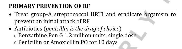
Primary prevention of RF
Secondary prevention of RF
Treatment of RF
