Lab 4: Body Circulations (models)
 Call Kai
 Learn
Learn Practice Test
 Spaced Repetition
 Match
Match Flashcards
 Knowt Play
1/71

There's no tags or description
Looks like no tags are added yet.
Name | Mastery | Learn | Test | Matching | Spaced | Call with Kai | Chat |
|---|
No analytics yet
Send a link to your students to track their progress
72 Terms
Ascending aorta
the ascending part of the aorta as it emerges from the left ventricle
Branches: L. & R. Coronary aa.
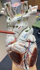
Aortic arch
a curved blood vessel from which arteries branch to the head and neck.
Branches: Brachiocephalic trunk; L. Common carotid a.; L Subclavian a.

Thoracic aorta
part of the aorta the descends from the aortic arch through the thorax to the diaphragm
Branches: Intercostal aa.

Abdominal aorta
continuation of the thoracic aorta that runs through the abdominal cavity
Branches: Inferior Phrenic aa.; Common Iliac aa.
Celiac trunk; Superior & Inferior Mesenteric aa.; Renal aa.; Gonadal aa.

Brachiocephalic trunk
The first large artery arising from the aortic arch. Serves the neck, head, and right forelimb.
Branches: R. Common Carotid a.; R. Subclavian a.

R. Common Carotid a.
Serves head and neck.
Branches: R. Internal Carotid a.; R. External Carotid a.

R. Subclavian a.
Serves right upper limb.
Branches: Thyrocervical trunk; Costocervical trunk; Internal Thoracic a.; Vertebral a.

Thyrocervical trunk
Serves thyroid gland & scapular muscles.

Vertebral a.
Serves posterior brain.
Branches: Basilar a.

R. External carotid a.
Serves outer skull and face.
Branches: Superficial Temporal a.; Facial a.; Occipital a.

R. Internal Carotid a.
Serves anterior brain (~2/3).
Branches: Middle Cerebral a.; Anterior Cerebral a.

L. Common Carotid a.
Serves head and neck.
Branches: L. Internal Carotid a.; L. External Carotid a.

L. Subclavian a.
Serves left upper limb.
Branches: Thyrocervical trunk; Costocervical trunk; Internal Thoracic a.; Vertebral a.

Superior Vena Cava
A vein that is the second largest vein in the human body and drains blood to the right atrium of the heart from the upper half of the body.
Branches: Brachiocephalic vv.; Azygos v.

L. Brachiocephalic v.
Drains left head, neck and upper limb.
Branches: Internal Jugular v.; Subclavian v.

R. Brachiocephalic v.
Drains right head, neck and upper limb.
Branches: Internal Jugular v.; Subclavian v.

L. Internal Jugular v.
Drains the dural venous sinuses of the brain.
Branches: dural venous sinuses

L. External Jugular v.
Drains the superficial aspects of the skull and neck.

L. Subclavian v.
Drains the right upper limb.
Branches: Axillary v.; External jugular v.

Internal thoracic a. & v.
Serves anterior thoracic wall and mammary glands.
Branches: Anterior intercostal aa.

Intercostal aa./vv.
Serves and drains the thoracic cavity wall.

Azygos v.
Drains the thoracic cavity wall and part of upper abdominal wall.
Branches: Inercostal vv.; Hemiazygos v.; Accessory hemiazygos v.

Hemiazygos v.
Drains thoracic and abdominal walls.
Branches lumbar and intercostal vv. on lower left side.

Inferior Phrenic aa./vv.
Serves inferior side of the diaphragm.

Internal Carotid a.
Serves anterior aspects of brain.

Middle Cerebral a.
Serves lateral aspects of frontal, parietal and temporal lobes' cortices.

Anterior Cerebral a.
Serves medial aspects of frontal and parietal lobes' cortices.

Anterior Communicating a.
Connects anterior cerebral aa.

Basilar a.
Formed by vertebral arteries coming together.
Branches: Superior cerebellar aa. (really all cerebellar aa.); Posterior Cerebral aa.

Posterior Cerebral aa.
Serves posterior and inferior aspects of temporal and occipital lobes' cortices.

Posterior Communicating aa.
Joins internal carotid a. to posterior cerebral a. on the same side.

Superior Cerebellar aa.
Serves the upper region of the cerebellum and brain stem.

Circle of Willis
A circulatory anastomosis that supplies blood to the brain and surrounding structures. (highlighted in blue)

Superior sagittal sinus
A venous sinus located in the midline at the superior margin in the falx cerebri, between the two cerebral hemispheres.
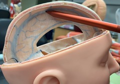
Inferior sagittal sinus
A venous sinus located within the inferior margin of the falx cerebri, drains into straight sinus.

Straight sinus
Connects inferior sagittal sinus to confluence of sinuses

Transverse sinus
Drains confluence of sinuses to sigmoid sinus

Sigmoid sinus
Continuation of the transverse sinus drains into internal jugular v.

Axillary a.
Serves the upper limb.
Branches: Brachial a.

Brachial a.
Serves the arm, foream, wrist and hand.
Branches: Anterior & Posterior circumflex humeral aa.; Radial a.; Ulnar a.
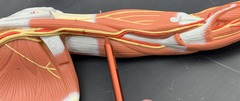
Anterior circumflex humeral a.
smaller of two circumflex aa.
-runs laterally deep to coracobrachialis m. and biceps brachii m.
-encircles surgical neck of the humerus and anastomoses with posterior humeral circumflex aa.

Posterior circumflex humeral a.
-larger of the two circumflex humeral aa.
-encircles surgical neck of the humerus and anastomoses with anterior humeral circumflex a.
-passes medial to humerus and pierces quadrangular space to perfuse deltoid m., teres minor m., teres major m., and long head of triceps brachii m.

Cephalic v.
Superficial vein that drains the lateral aspect of the arm.

Basilic v.
Superficial vein that drains the medial aspect of the arm.

Radial a.
Serves the lateral forearm and deep palmar arch.
Branches: Deep palmar arch

Ulnar a.
Serves the medial forearm and superficial palmar arch.
Branches: Superficial palmar arch

Digital aa.
Serves the fingers (or toes in the foot)
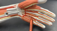
Superficial palmar arch
Serves the hand and fingers.
Branches: Digital aa.

Common iliac a.
Serves the lower limb and pelvis.
Branches: External iliac a.; Internal Iliac a.

Internal iliac a.
Serves internal organs of the pelvis.
Branches: Superior Gluteal a.; Inferior Gluteal a.

Superior gluteal a.
Serves muscles of gluteal region, skin of the buttock.

Inferior gluteal a.
Serves muscles of gluteal region, skin of the buttock.

External iliac a.
Serves the lower limb.
Branches: Femoral a.

Femoral a.
Serves the thigh and leg.
Branches: Deep femoral a.; Popliteal a.

Deep femoral a.
Serves hamstring group. Branches off femoral a.
Branches: Lateral Circumflex Femoral a.

Lateral circumflex femoral a.
Serves anterior thigh muscles.

Great Sapphenous v.
Drains superficial areas of medial side of leg and thigh. Joins femoral v. to form external iliac v.

Popliteal a.
Continuation of femoral a after passing through adductor hiatus; serves knee.
Branches: Anterior & posterior tibial arteries; Genicular aa.

Superior medial genicular a.
Serves inner, upper side of knee.

Superior lateral genicular a.
Serves upper, outer side of knee.

Inferior medial genicular a.
Serves lower, inner side of knee.

Inferior lateral genicular a.
Serves lower, outer side of knee.

Posterior tibial a.
Serves posterior compartment of leg and plantar foot.
Branches: Fibular a.

Anterior tibial a.
Serves anterior compartment of leg and dorsal foot.

Fibular a.
Branches off posterior tibial a. Serves lateral compartment of leg.

Tunica intima
The innermost layer of a blood vessel (A).
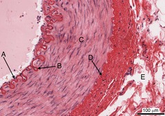
Internal elastic lamina (IEL)
EL between tunica intima and tunica media (B).

Tunica media
The middle (C) and thickest layer of tissue of a blood vessel wall, composed of elastic tissue and smooth muscle cells that allow the vessel to expand or contract in response to changes in blood pressure and tissue demand.

External elastic lamina (EEL)
EL between tunica externa and tunica media (D).

Tunica externa
Outer layer (E) of a blood vessel which connects it to surrounding tissues, composed to DICT.

Artery histology (B)
-NO valves
-Thicker, tunica media, smooth muscle, and elastic fibers in wall
- More resilient
-Lumen appears smaller than veins and retains shape

Vein histology (A)
-Contain valves, prevent back flow of blood.
-Thinner, tunica externa is thickest
-Easily collapses or tears
- Lumen looks larger, flat and distorted
