Computed Tomography (CT)
 Call Kai
 Learn
Learn Practice Test
 Spaced Repetition
 Match
Match Flashcards
 Knowt Play
1/18

There's no tags or description
Looks like no tags are added yet.
Name | Mastery | Learn | Test | Matching | Spaced | Call with Kai | Chat |
|---|
No analytics yet
Send a link to your students to track their progress
19 Terms
What is computed tomography (CT)?
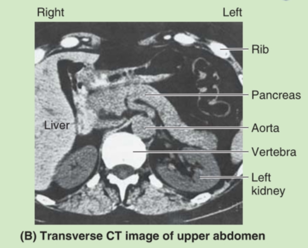
An imaging technique that uses rotating X-rays and computer processing to produce cross-sectional (transverse) images of the body.
What do CT images resemble?
Transverse anatomical sections (cross-sectional slices) of the body.
How are CT images produced?
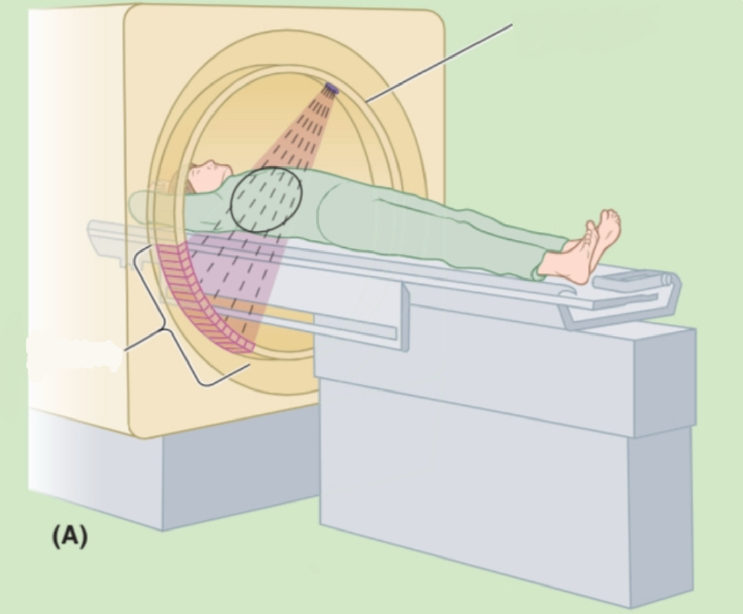
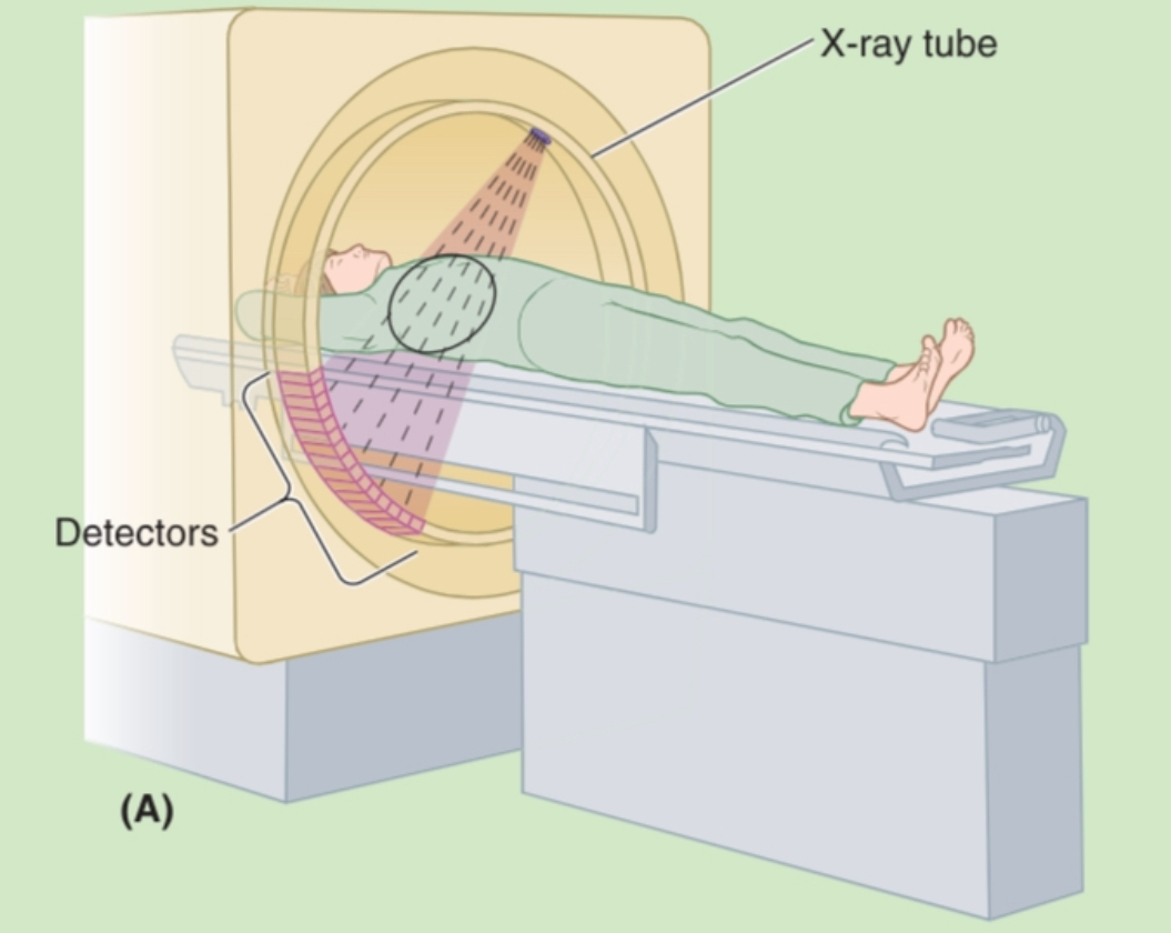
An X-ray tube rotates around the body while detectors measure the amount of radiation passing through different tissues. A computer reconstructs these measurements into images.
What rotates around the patient during a CT scan?
X-ray tube
X-ray detectors
What determines how much X-ray radiation is absorbed during CT?
The tissue composition, particularly the amount of:
Bone
Fat
Water
Why do different tissues appear differently on CT?
Different tissues absorb different amounts of X-rays based on their density and composition.
What does a computer do in CT imaging?
It reconstructs the measured X-ray data into:
2D cross-sectional images
3D reconstructions
What are the two main image formats produced by CT?
Two-dimensional slices
Three-dimensional reconstructions
What orientation are CT images usually displayed in?
As if you are standing at the patient’s feet looking toward their head.
Patient’s right = your left.
What is one major advantage of CT over conventional radiography?
CT eliminates much of the superimposition of structures by producing cross-sectional images.
Why is CT considered superior to plain radiography for many conditions?
Because it provides detailed cross-sectional anatomy and allows 3D reconstruction.
What type of beam is used in CT?
A fan-shaped beam of X-rays.
What is reconstructed from CT slices?
Three-dimensional images of anatomical structures.
Complete the CT process:
X-ray tube → ______ → Computer → ______
X-ray tube
↓
Detectors measure transmitted X-rays
↓
Computer reconstruction
↓
2D slices and/or 3D images
Give 3-4 differences between conventional radiography and CT
Conventional radiography
Single projection
2D image
Superimposition of structures
CT
Rotating X-ray beam
Cross-sectional slices
Minimal superimposition
Can produce 3D reconstructions
Label
Remember:
CT = transverse (cross-sectional) image
Right side of image = patient’s right
Left side of image = patient’s left
Oki
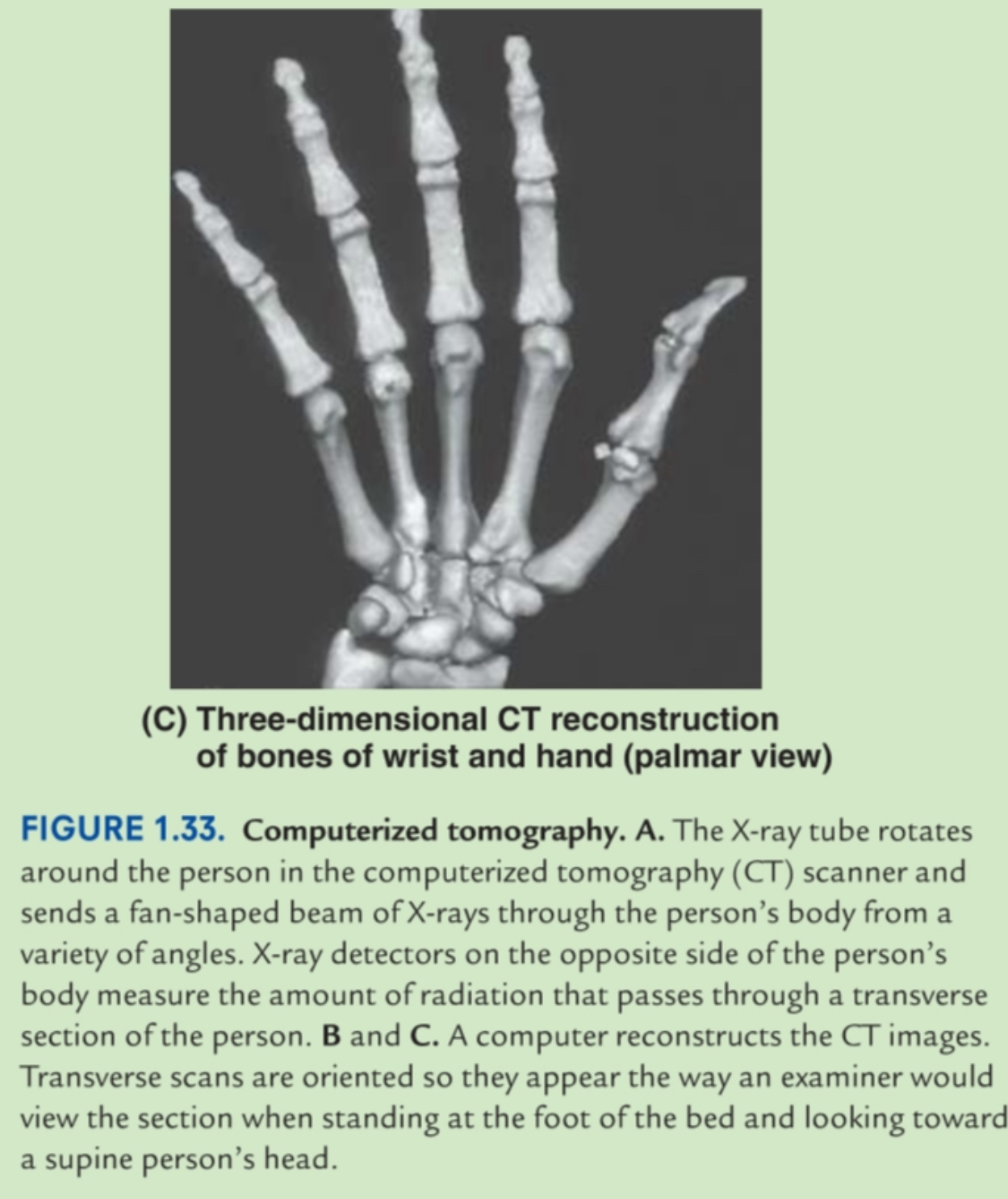
CT reconstructed into 3D model
Perf
CT Cheat Sheet
Uses X-rays ✔
Rotating X-ray tube
Produces transverse slices
Excellent anatomical detail
Computer reconstructs:
• 2D slices
• 3D images
Little superimposition
Patient viewed from feet toward head