Cardio Physiology 3 - Action Potential, ECG, Excitation-Contraction Coupling
 Call Kai
 Learn
Learn Practice Test
 Spaced Repetition
 Match
Match Flashcards
 Knowt Play
1/60

There's no tags or description
Looks like no tags are added yet.
Name | Mastery | Learn | Test | Matching | Spaced | Call with Kai | Chat |
|---|
No analytics yet
Send a link to your students to track their progress
61 Terms
Two types of action potentials in the heart
Fast and slow action potential
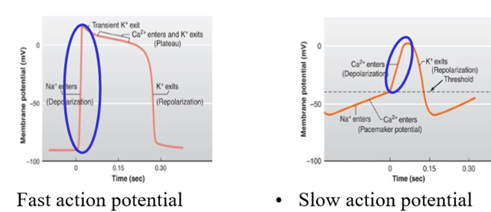
Where are fast action potentials found
- Contractile myocytes in the atrial myocardium
- Ventricular myocardium
- Parts of the heart conducting system (bundle of His, bundle branches, purkinje fibers)
Where are slow action potentials found
Sinoatrial node, Atrioventricular node
What does it mean when the action potential is fast or slow?
Describes how quick the membrane potential changes in the depolarization (fast = rapid depolarization. Slow = slower depolarization)
What causes the different rates of depolarization
The types of ions/ion channels involved in depolarization
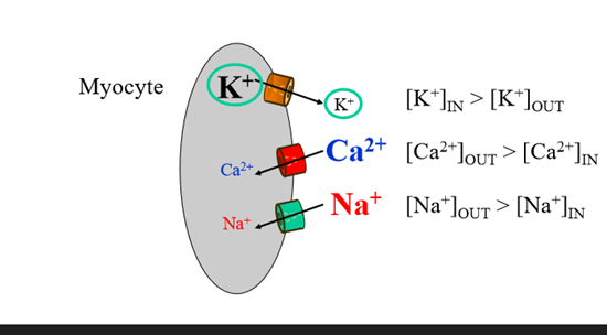
What causes the phases of cardiac action potentials
Changes in permeability to Na+, K+, Ca2+
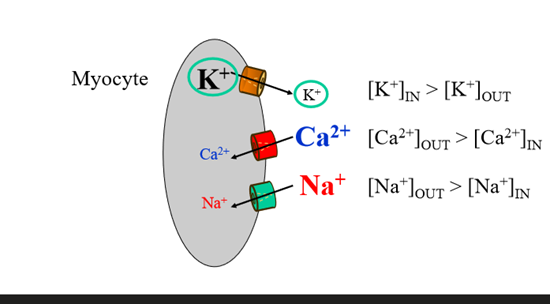
Key feature of the slow action potential
Pacemaker potential - slow depolarization to threshold membrane potential.
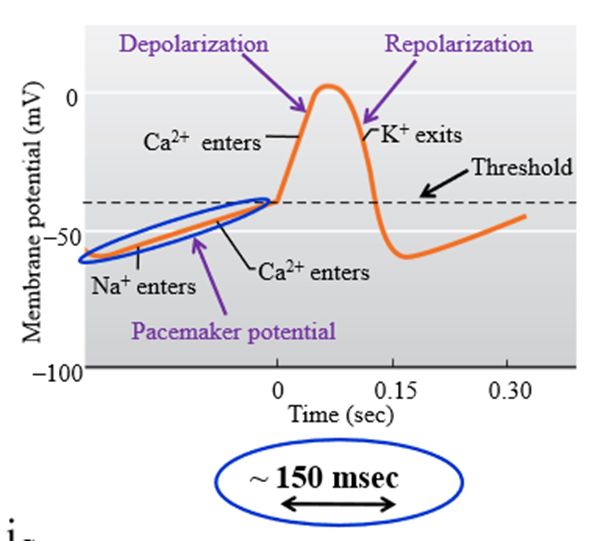
What is the use of the pacemaker potential
Allows sinoatrial node cells to generate regular spontaneous action potentials without influence from nerves or hormones. Allows for generation of the heartbeat
3 ionic conductions involved in pacemaker potential
1. Progressive reduction in K+ permeability (channels close to prevent K+ from leaving)
2. F-type channels - sodium channels open in negative membrane potential (F = funny channel)
3. T-type channel - transient (briefly opening) channels allow Ca2+ to enter the cell
How does the slow action potential depolarization stage occur
L type (long lasting) calcium channels open slowly and remain open, allowing calcium into the cell.
- Calcium allows for slower depolarization than sodium
How does the slow action potential repolarization occur
Opening of voltage gated K channels to allow potassium to leave. L type channels close, preventing calcium channels from opening
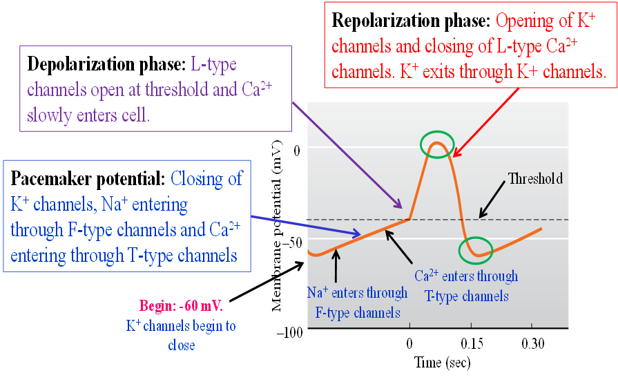
Steps of slow action potential (of SA and AV nodes)
1. Pacemaker potential (K+ channels close, Na+ enters F-type channels, Ca2+ enters T channels)
2. Depolarization phase (L-type channels open at threshold and Ca2+ enters cells)
3. Repolarization phase (Opening of K+ channels to let out K+ and closing of L-type Ca2+ channels)'
What happens to the AVN if SAN is damaged
May generate action potential to drive the ventricles at a lower rate (40-60 BPM)
Key features of the fast action potential
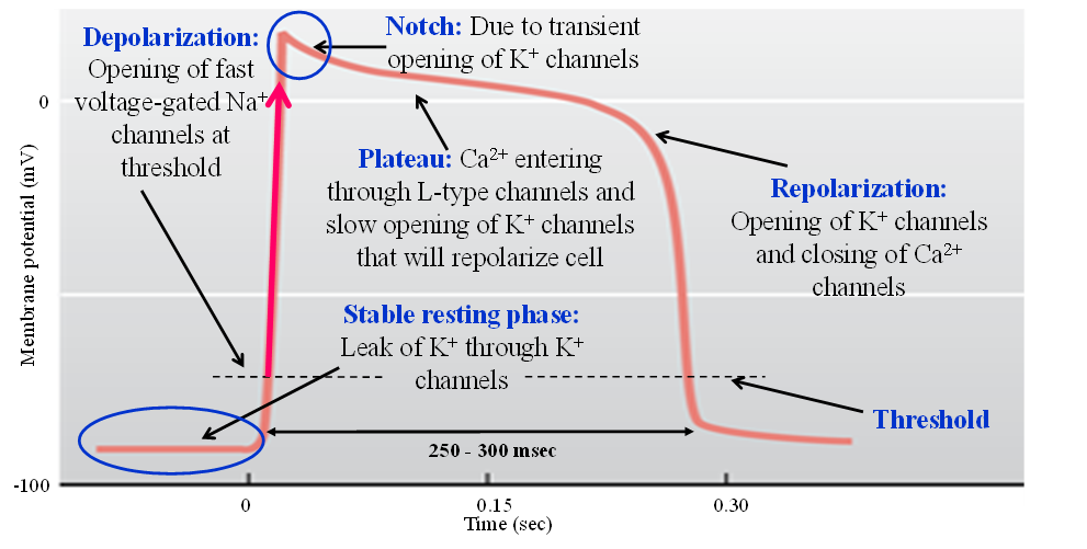
Fast depolarization, plateau at repolarization phase
Key difference in activation for fast and slow activation
Slow activation - no reliance on stimulus
Fast activation - requires action potential from conducting myocytes
Steps of fast action potential
1. Stable resting phase - leak of calcium through K+ channels
2. Depolarization - opening of fast gated Na+ channels at threshold
3. Notch - transient opening of K+ channels that slightly repolarize
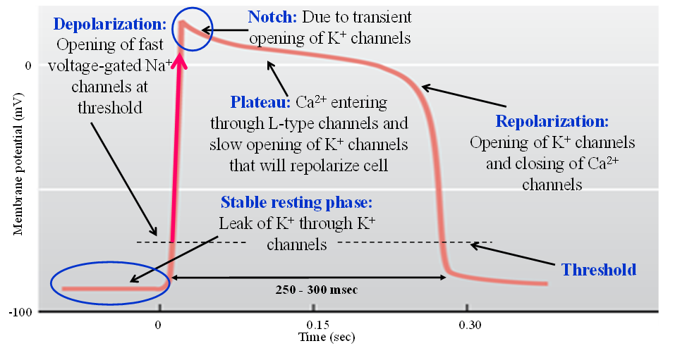
4. Plateau - Ca2+ enters L type channels balances with K+ leaving channels
5. Repolarization - Opening of K+ channels and closing of L type Ca2+ channels
Is the fast action potential fast or slow in length?
Slow - due to plateau phase, affecting refractory period
Comparison between SA and AV node action potentials
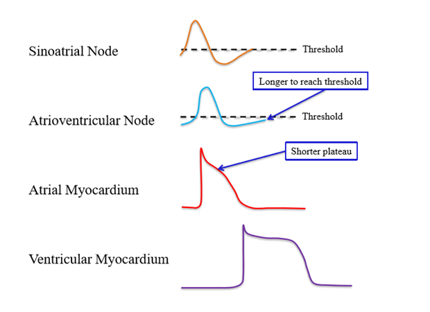
SA and AV are both slow action potentials
- AV takes longer to reach threshold due to longer pacemaker phase
Comparison between Atrial and Ventricular Myocardium nodes
Both are fast action potentials
- Atrial myocardial muscle is shorter length, as it has a shorter plateau
Electrocardiogram (ECG, EKG)
Measure of the currents generated in the extracellular fluid by the changes occurring in many cardiac cells
Strength of electrical signal in ECG
Weaker than the heart voltage, as the electric signal has to travel to the skin surface (100 mV --> 1mV)
Use of ECG
Can diagnose problems with heart's conducting system - cannot diagnose mechanical issues
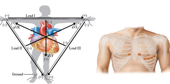
12 Lead ECG
12 leads attach all over the body, allows for different angles of heart electrical activity
- Established electrode pattern results in specific tracing pattern
ECG graph
Voltage as a function of time. Consists of P wave, QRS complex, T wave
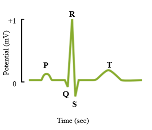
P wave
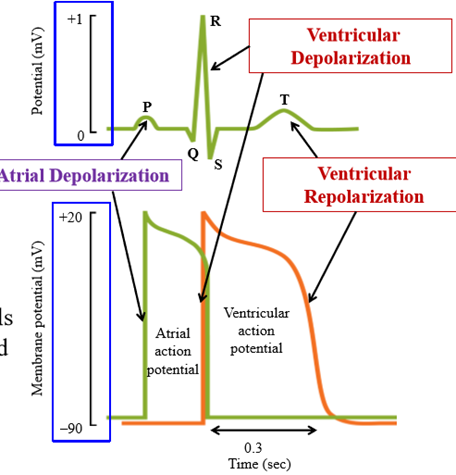
First wave on ECG, represents atrial depolarization
QRS complex
Wave consists of 3 peaks, representing rapid ventricular depolarization
What occurs during QRS complex
ventricular depolarization + atrial repolarization (too small for an electrical event to be recorded)
T wave
After QRS complex, represents ventricular repolarization
Comparison of Action potentials and ECG
Action potential timing lines up with ECG waves (except no atrial repolarization)
AV node block
Conduction between atria and ventricles is partially impaired
Normal ECG
P wave, QRS compelx, T wave
- Shows synchrony between atria and venctricles: atria contracts and then ventricles contract
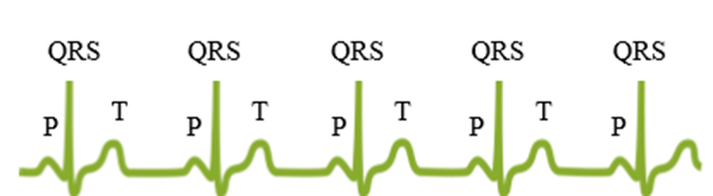
Partial AV node block ECG
Damaged AV node permits only every other atrial impulse to be transmitted to ventricles
- Every second P wave is not followed by QRS complex and T wave
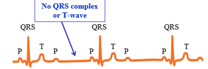
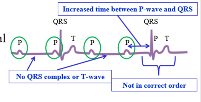
Complete AV node block ECG
Depolarization of atria not transmitted to ventricles - no synchrony between atrial and ventricular electrical activities
- Missing QRS/T, or order is messed up
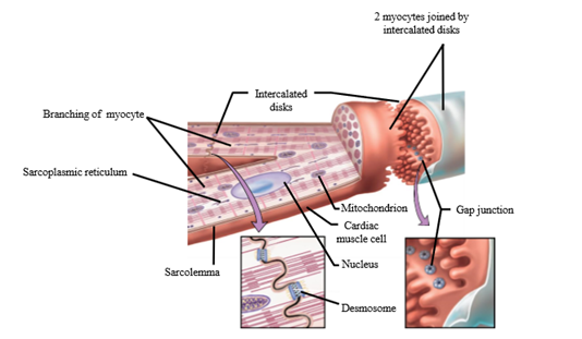
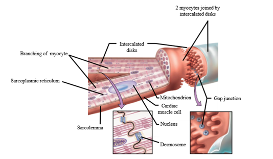
Intercalated disk
Where the membranes of two adjacent myocyte are intertwined (has desmosomes and gap junctions)
Sarcolemma
Plasma/cell membrane of cardiac muscle
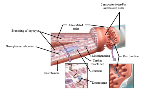
Sarcoplasmic reticulum
Special type of SER that stores/pumps Ca2+
Importance of calcium int he ehart
Used in excitation-contraction coupling
What are myofibrils
Bundles of sarcomeres
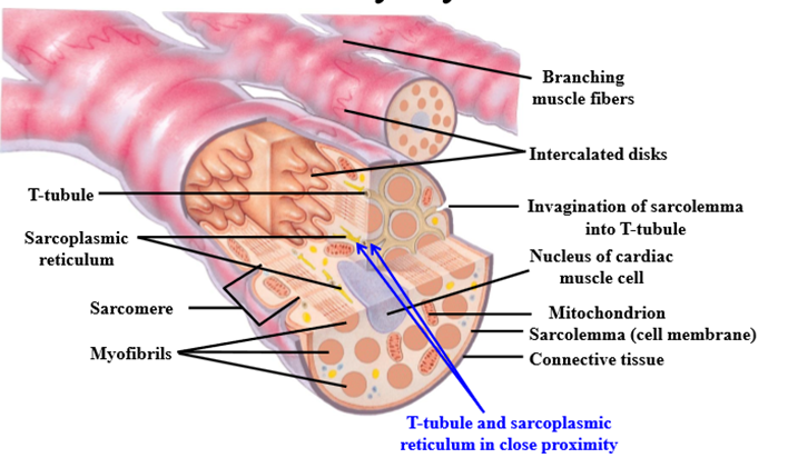
Sarcomere
Contractile unit of muscle, contains thick (actin) and thick (myosin) filaments
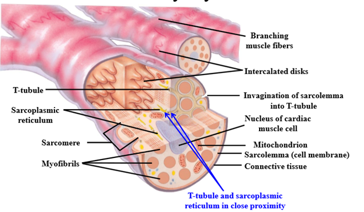
How does cardiac muscle get striated appearance
Orderly arrangement of actin and myosin
T-tubules
Invaginations of the sarcolemma - transmit action potentials to the interior of the muscle fiber
Excitation contraction coupling
Process by which the arrival of an action potential at the cell membrane leads to contraction of muscle cell
What ion controls contraction of heart muscles
Calcium
Which AP phase starts Excitation contraction coupling
Plateau phase - extracellular calcium enters the cytoplasm of cardiac muscle cells
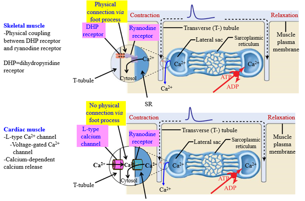
L type calcium channel in ECC
Modified DHP receptors. Channels on the T tubules that allow calcium to entry into the muscle cell
Effect of calcium entering the L type calcium channel in ECC
Calcium binds to ryanodine receptors, which releases calcium from the SR into the cytoplasm
Calcium induced calcium release
Ca2+ causes its calcium release from the SR following binding to the ryanodine receptor
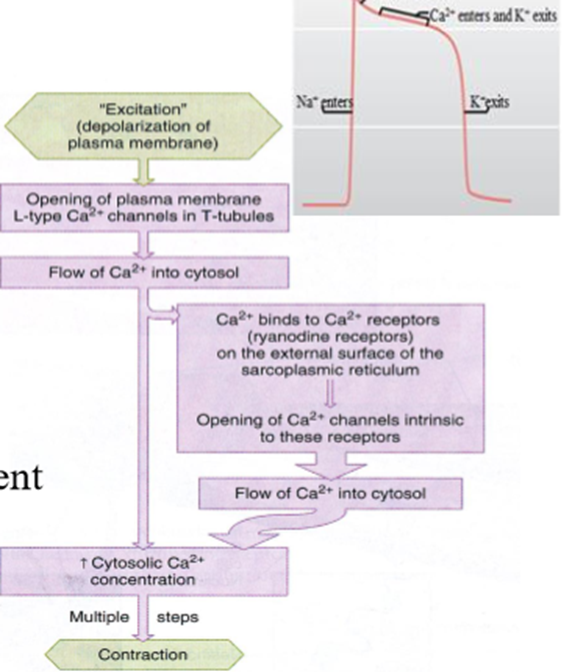
Steps of excitation contraction coupling
1. Excitation - depolarization of plasma membrane spreads down T-tubules
2. Opening of plasma membrane L-type Ca2+ channels in T-tubules
3. Calcium flows into cytosol
4. Calcium binds to ryanodine receptors on external surface of sarcoplasmic reticulum
5. Flow of Calcium into cytosol
6. Increase in cytosolic calcium concentration
7. Cross bridge cycling = contraction (shortening of sarcomere)
Cross bridge cycling steps
1. High amount of calcium due to excitation contraction coupling --> binds to troponin binding sites
2. Troponin shape changes, causing tropomyosin to move away from myosin binding sites on actin
3. Energized head on myosin binds to actin binding sites of myocytes = leads to heart muscle contraction
Difference between DHP receptor on skeletal muscle and cardiac muscle
Cardiac muscle - No physical coupling of L type channel to ryanodine receptor. L type channel is voltage gated, and causes calcium induced calcium release
Skeletal muscle - Physical coupling of DHP receptor to ryanodine receptor via the foot process. DHP receptor changes confirmation via action potential, allowing calcium to move out of SR. Calcium independent calcium release
Why is relaxation of the ventricular myocardium important?
Ventricles only fill with blood when relaxed (diastole)
How is a muscle contraction ended
Removal of calcium from troponin binding site on thin filament
How are calcium levels reduced to pre-release levels
- L type Calcium channels close to reduce influx of calcium into the cell, SR is no longer stimulated to release Ca2+ into the cytoplasm
- SR contains Ca2+ ATPase - pump calcium back into SR from cytosol
- Ca2+ removed from myocyte by Na+/Ca2+ exchanger in sarcolemma
How does reduced calcium how in relaxation
Less calcium = less binding to myocyte troponin. Cross bridge cycling occurs
Which method removes the most calcium from cytosol?
Ca2+-ATPase
Ca2+ ATPase
Pumps calcium back into SR by using ATP
Na+/Ca2+ exchanger
Found in the sarcolemma - calcium leaves the cell in exchange for sodium to enter the cell
Refractory period
Period of time during/after action potential in which an excitable membranes cannot be re-excited no matter how strong the stimulus
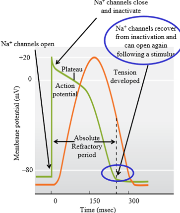
Why is the refractory period so long for cardiac muscles?
Fast action potential has the long plateau phase
What causes the refractory period in myocytes
Inactivation of fast voltage-gated sodium channels that open during the depolarization phase of the action potential. Do not open again until the muscle cell returns to negative potentials
What is the use of the myocytes long refractory period?
Prevents tetanus