B3.3 Muscle and Motility HL
1/12
There's no tags or description
Looks like no tags are added yet.
Name | Mastery | Learn | Test | Matching | Spaced |
|---|
No study sessions yet.
13 Terms
skeleton (2)
exoskeleton ( chitin )
endoskeleton ( bones)
joint (2)
hinge joint ( elbow and knee)
one plant of movement
bend & straight
ball and socket joint ( hips, shoulder)
large range of movement
protraction, retraction , abduction, adduction , rotation
measure joint
goniometer
most allowing movement joint
synovial joint - human hip joint
Bone (Femur & Pelvis) | Cartilage | Synovial fluid | Ligaments | Muscles | Tendons |
function
bone- anchorage for muscle & ligants, guide movement
muscle - provide force for movemet
cartilage - smooth connective tissuet that covers the end of bone to reduce friction
synovial fluid - lubricate joint reduce friction
ligaments - slight elastic tissue - attaches bones to bones
tendons - non elastic tissue - attaches muscle to bone
skeletal muscle
attacth bones - cause movement of animal body
It consists of large multinucleated cells called muscle fibers.
There are also mitochondria between the myofibrils.
level of organisation
muscle fibres → myofibris → microfillaments → sacromere
wra around myofibrils
sacroplamatic reticulum
skeletal muscle & electrical impluse
Skeletal muscles are voluntary muscles that requires electrical impulse from the brain.
Electrical impulse sent from brain through motor neuron → neuromuscular junction.
Each motor neuron has a set number of muscle fibers that it control called a motor unit.
motor unit + function
contraction of skeletal muscle
include single motor neuron & muscle fibres
muscle fibre contract when stimulated by motor neuron
stimulus pass through neuron , through synapse- neuromuscular junction to muscle fibre
require neurotransmitter : acetylcholine ( basically just the normal neurotransmitter process)
sacromere
two protein filaments
subunit of myofibrils
between two Z-lines
myosin
thick , dark bands
head that forms cross bridge by binding to actin
actin
thin, light bands
lengthening and shortening of sarcomere
attach at the end of Z lines
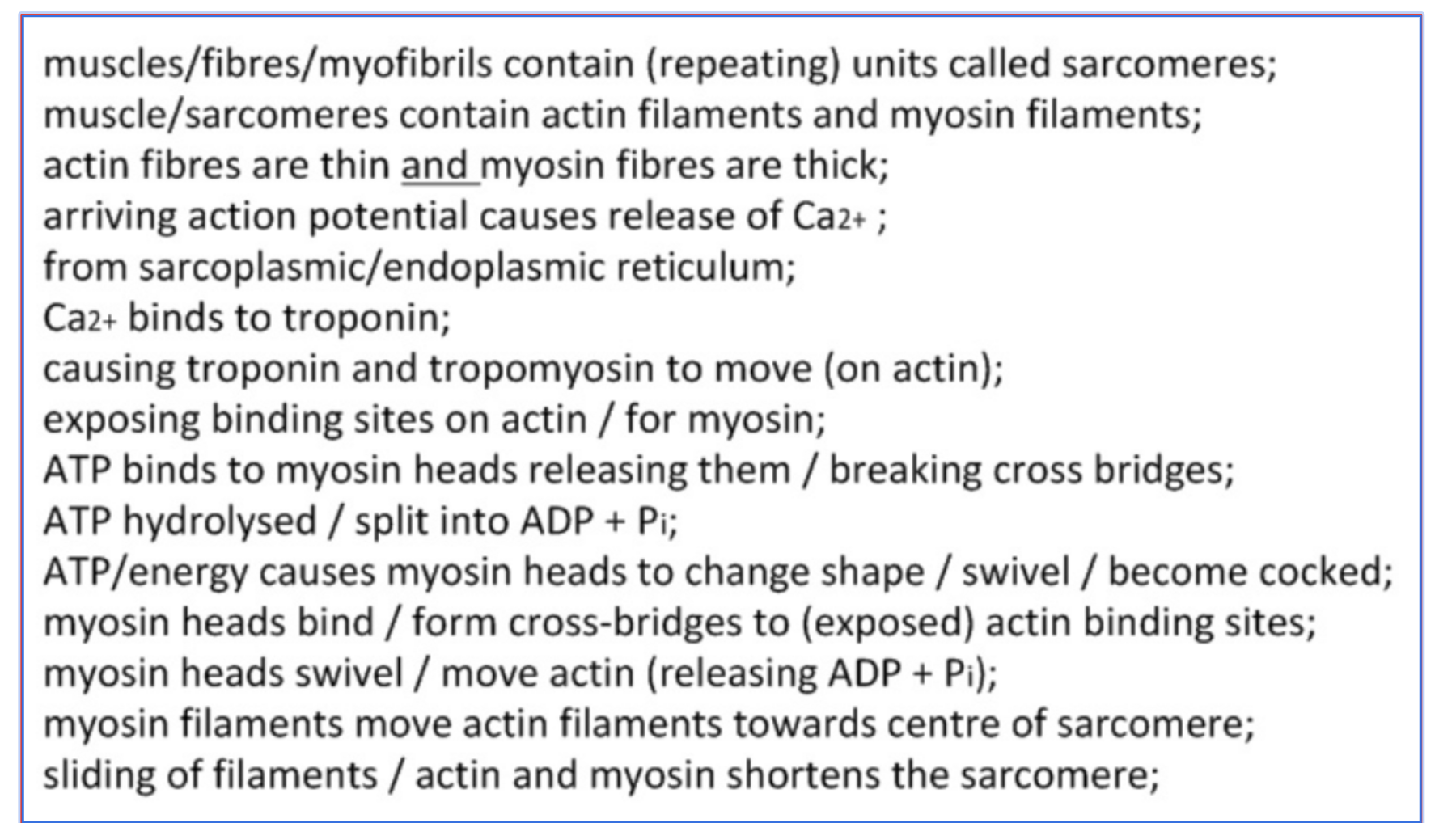
crose bridge cycle
When a nerve impulse arrives at the neuromuscular junction,calcium ions are released from the sarcoplasmic reticulum
calcium bind with troponin, change shape, move tropomyosin to expose the myosin-binding site on actin
Myosin heads bind to actin, forming crossbridges
Myosin releases ADP and Pi, causing the power stroke - pulls the actin filament towards the centre of the sarcomere
ATP binds to myosin, breaking the crossbridges
ATP is hydrolysed to ADP and Pi, provide energy that “cock” the myosin head away from the center
Myosin heads bind to actin at a new binding site further along the sarcomere
The cycle continues until Calcium is pumped back into the sarcoplasmic reticulum, or there is no ATP available
titin
contraction of antagonistic muscle → creates energy → needed to lengthen a muscle, which stretches titin. titin recoils → release energy → adds to the force of contraction in that muscle (provide supplemental force)
prevent overstretching of sacromere
holds myosin filaments in place
Explain how a skeletal muscle contracts