NUR 305 Neurological System I
1/116
There's no tags or description
Looks like no tags are added yet.
Name | Mastery | Learn | Test | Matching | Spaced |
|---|
No study sessions yet.
117 Terms
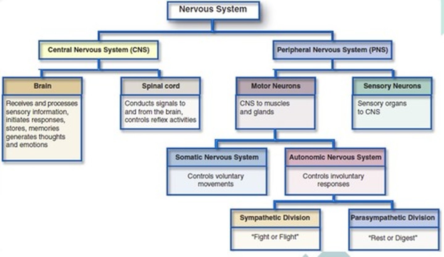
The Nervous system consists of
Central Nervous System (CNS)
Brain
Receives and processes sensory information, initiates responses, stores memories, generates thoughts and emotions.
Spinal Cord
Conducts signals to and from the brain, controls reflex activities.
Peripheral Nervous System (PNS)
Motor Neurons
CNS to muscles and glands
→
1. Somatic Nervous System
Controls Voluntary Movements
2. Autonomic Nervous System
Controls Involuntary Movements
→
Sympathetic Division
Fight/Flight
Parasympathetic Division
Rest/Digest
Sensory Neurons
Sensory organs to CNS
What 3 things is the Nervous System responsible for?
Movement
Sensation
Cognition
What does the Central Nervous System consist of?
Brain and Spinal Cord
What does the Peripheral Nervous System consist of?
Motor Neurons and Sensory Neurons
Where does the Somatic Nervous System stem from?
Motor Neurons
Where does the Autonomic Nervous System stem from? What divisions exist under the Autonomic Nervous System?
Motor Neurons
Sympathetic and Parasympathetic
What are the functions of Neurons?
Generate action potentials.
Through Na+/K+ pumps
Action potentials are specific/exclusive to neurons.
Transmitter cells.
Carry messages to and from the brain and spinal cord.
What are the functions of Glial Cells?
Description
Support and protect neurons.
Do not generate action potentials (exclusive to neurons) but have a resting membrane potential.
Produce CSF.
Contains proteins, glucose, and electrolytes
What are the functions of Glial Cells in the CNS?
Astrocytes → Recycle glutamate into vesicles.
Microglia → Immune system of brain (most active at night)
Oligodendroglia → Produce myelin in the CNS
Ependymal Cells → Located on roof of lateral ventricles (spaces) in brain (There are 4 ventricles in the brain) → Generate CSF
What are the functions of Glial Cells in the PNS?
Schwann Cells → Produce myelin in the PNS
Satellite Cells → Supportive function
Electrical Impulses
Info passed between neurons by chemicals.
Can be excitatory (most) or inhibitory.
Action potentials are sent or inhibited.
Along the axons, the information passes electrically.
Neurons can synapse with:
Neurons
Muscles
Glands
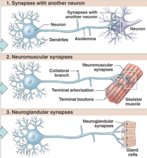
Synaptic Transmission
A small burst of neurotransmitters is released.
The neurotransmitters stimulate or inhibit action potentials.
Neurotransmitters are chemical messengers that are either ____ by enzymes or _____. They are also usually _____ for the next transmission.
destroyed; reabsorbed; recycled
In the neuromuscular junction (the area where nerves synapse with muscles) the neurotransmitter is _____?
Acetylcholine
Dopamine
Description
Promotes smooth, coordinate muscle movement.
Deficiency
Involved in Parkinson’s Disease.
Excess
Involved in Schizophrenia.
Norepinephrine & Epinephrine
Description
Stimulates adrenergic receptors in sympathetic nervous system (causes vasoconstriction)
Keeps vasomotor tone → keeps BP stable
Adrenergic receptors located in medial muscle layer of arteries.
Receptors
Alpha 1 & 2
Beta 1 (HR, rhythm)
Beta 2 (Inc pumping/contraction of heart) / (Bronchodilation)
Beta 3
Serotonin
Mood regulation, inhibits pain.
Acetylcholine
Description
Stimulates cholinergic receptors in parasympathetic nervous system & neuromuscular junction.
Deficiency
Due to damaged receptors
Involved in Myasthenia Gravis.
GABA/Glutamate
Excitatory
Involved in addiction
Involved in award system
Recycled by astrocytes
Endorphins/Enkephalins
Endogenous opioid system.
“Runner's high” feeling
Substance P
Pain transmission
Melatonin
Sleep regulation
Myelin
Lipoprotein that increases speed of conduction.
Especially on nerves w/ large axons
Some small nerves are myelinated too.
Present in CNS
"Unmyelinated" Axons
Smaller nerves, slower transmission.
Visceral pain
Ex) GI pain.
What are characteristics of Neurons?
Do not have the ability to divide.
Losses due to aging or injury cannot be replaced.
Once the NS matures, by age 2, that’s it.
Not all cell death results in loss of functioning.
Other areas will pick up the functions is one area is damaged w/ time.
Undamaged neurons in the brain will assume functions of damaged neurons (“plasticity”).
Neuroplasticity easier in childhood.
Require a constant oxygen and glucose supply.
>5-6 mins w/o O2 leads to death.
Neurons are vulnerable to hypoxia and hypoglycemia.
See Point 5 above
What are injuries to the NS that can be fixed?
Severed Peripheral Nerves
Can regenerate to a point to reestablish connections.
What are injuries to the NS that cannot be fixed?
Severed Brain and Spinal Cord Axons
Result in paralysis
Loss of sensation below the area of damage.
How long the brain can go without oxygen?
5-6 minutes MAX
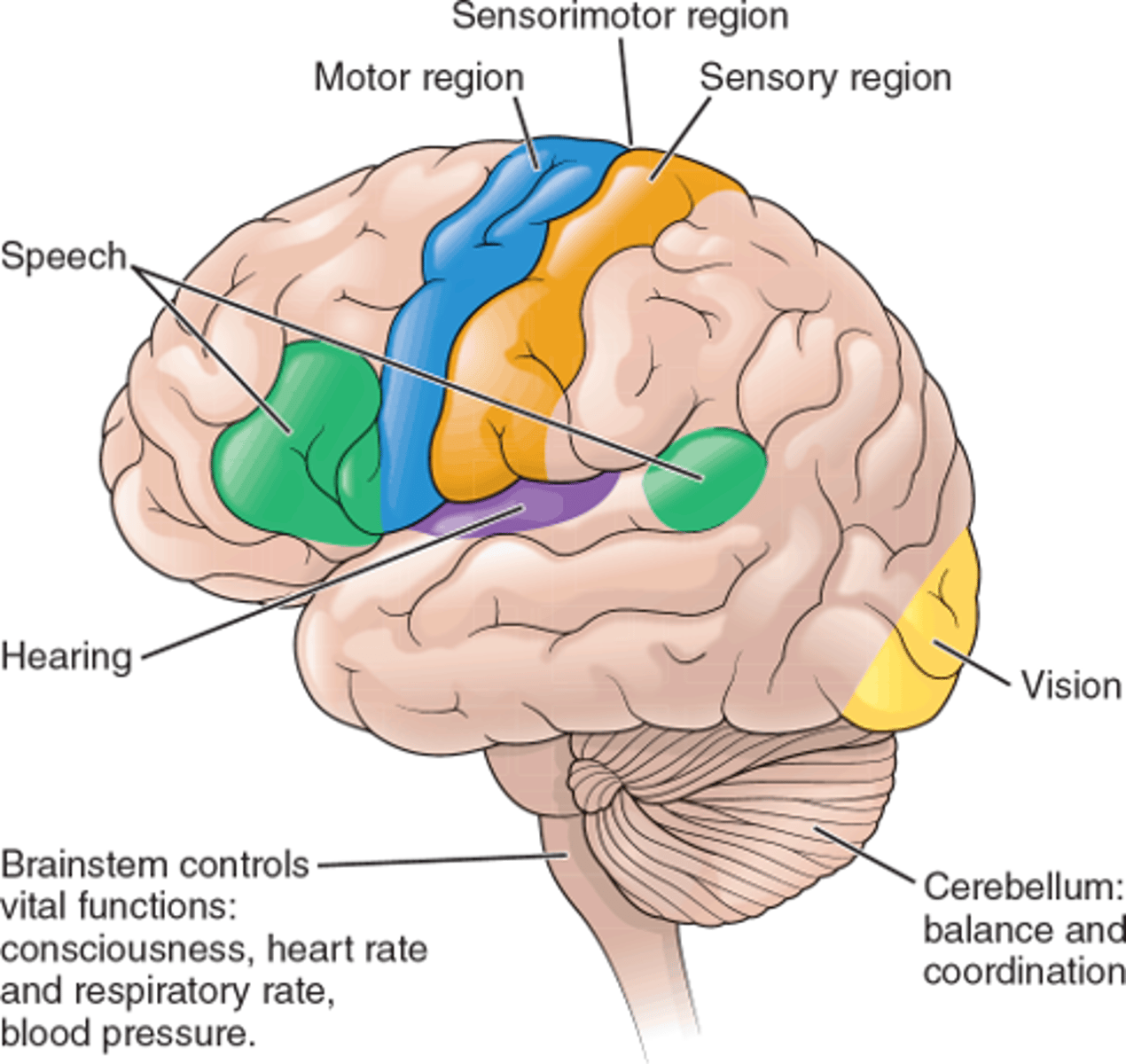
What does the cerebrum contain?
Frontal lobe
Parietal lobe
Occipital lobe
Temporal lobe
Cerebrum Function
Cognition & Abstract thinking.
What does the Diencephalon contain?
Thalamus
Hypothalamus
Cerebellum Function
Balance and coordination.
Thalamus Function
Relay station for motor and sensory signals to the cerebral cortex.
Carries/coordinates different areas of the brain.
Plays a role in motor activity, emotion, memory, arousal, and other sensorimotor association functions.
Regulates consciousness, sleep, and alertness.
Brain Stem Function
Autonomic function
What does the Brain Stem contain?
Midbrain
Pons
Medulla Oblongata
Hypothalamus Function
Releases precursor/tropic hormones
Thermoregulation
Most individuals with damage to the left hemisphere develop Aphasia. What is Aphasia?
A disorder that makes it difficult to speak or understand language.
What is the difference between Expressive and Receptive Aphasia?
Expressive Aphasia
Person knows what they want to say, but what they say doesn’t make sense.
Receptive Aphasia
Person cannot understand what is being said to them, but can speak normally.
If you hit the back of your head on the occipital region, which sense would you expect to lose?
Vision
Ipsilateral vs. Contralateral Nerves
Ipsilateral
Nerve fibers that run on the same side of the body they originate on.
Ex. Nerve fiber starts on right hemisphere, is expressed on right side of body.
Ex. Optic nerves
(20%) of corticospinal and spinothalamic nerve fibers.
Contralateral
Nerve fibers that run on the opposite side of the body they originate on.
Ex. Nerve fiber starts on left hemisphere, is expressed on right side of body.
The majority of corticospinal and spinothalamic nerve fibers (80%)
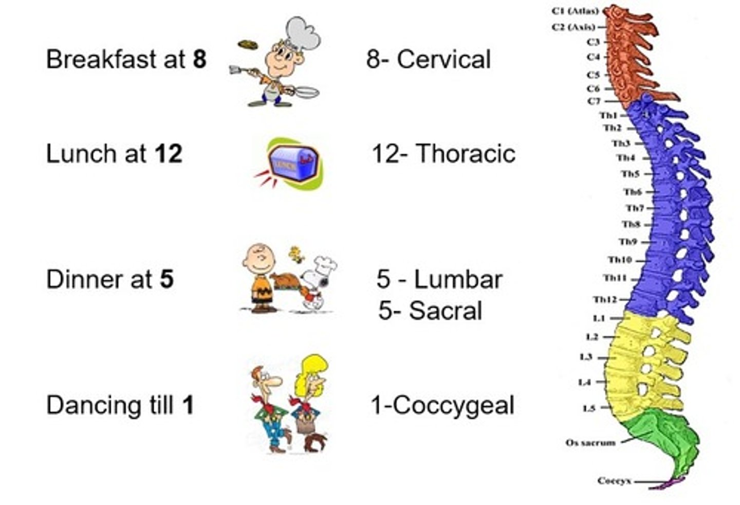
Cervical Nerves
8 pairs
Thoracic Nerves
12 pairs
Lumbar Nerves
5 pairs
The Brain Regions
If you have a stroke on the right hemisphere, in most people the deficits will be on the left. Why is this true?
Most nerves are contralateral.
The Meninges
Layers of membranes that envelop and protect the CNS.
There are 3 meningeal membrane layers.
Dura mater arachnoid mater, pia mater.
Between each meningeal membrane layer there is a potential space.
Skull (Epidural Space) Dura Mater (Subdural Space) Arachnoid Mater (Subarachnoid Space) Pia Mater
The middle meningeal artery runs very close to the skull bone above the dura mater.
Middle meningeal artery commonly ruptures and causes an epidural hematoma.
First sign is loss of consciousness.
What leads to a Subdural Hematoma?
There is a network of veins within the subdural space.
Head trauma often causes rupture of subdural veins, which causes a subdural hematoma.
T/F: CSF is contained beneath the arachnoid membrane within the subarachnoid space.
True
3 Meninges
Dura Mater: outer layer, lines skull
Arachnoid Mater: middle layer, contains blood vessels
Pia Mater: inner layer, covers brain
3 Potential Meningeal Spaces
Epidural: outside dura.
Subdural: between dura and arachnoid.
Subarachnoid: deep to arachnoid, filled with CSF.
Loss of CSF from nose/ears may indicate subarachnoid bleed.
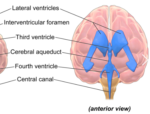
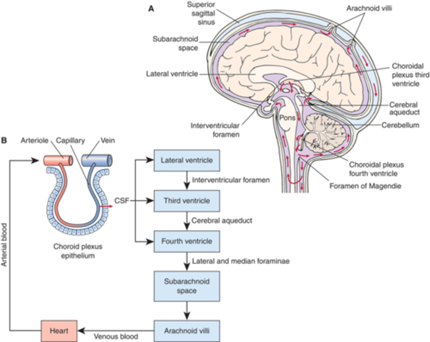
Cerebrospinal Fluid (CSF)
Made in choroid plexuses (roofs of ventricles).
Filtration of plasma from capillaries through ependymal cells
Takes electrolytes, glucose from plasma.
Cushions and nourishes brain.
Useful for diagnosing meningitis (cloudy CSF), bleeds, MS (multiple sclerosis).
Excessive accumulation of CSF → Hydrocephalus
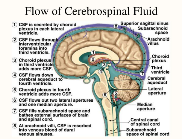
The Flow of Cerebrospinal Fluid
Created/Filtered in: 4 Ventricles
Flow
Lateral Ventricle → Third Ventricle → Fourth Ventricle → Subarachnoid Space
Mean Arterial Pressure
MAP = diastolic pressure + 1/3 (systolic BP-diastolic BP)
Intracranial Pressure (ICP)
Normal Range
0-15 cmH20 or mmHg
Cerebral Perfusion Pressure (CPP)
Description
Pressure it takes to completely perfuse the brain.
Bleeds can happen if CCP is high.
Calculation
CCP = MAP - ICP
Optimal Range
70-90 mmHg
A high ICP = A ___ CPP
low
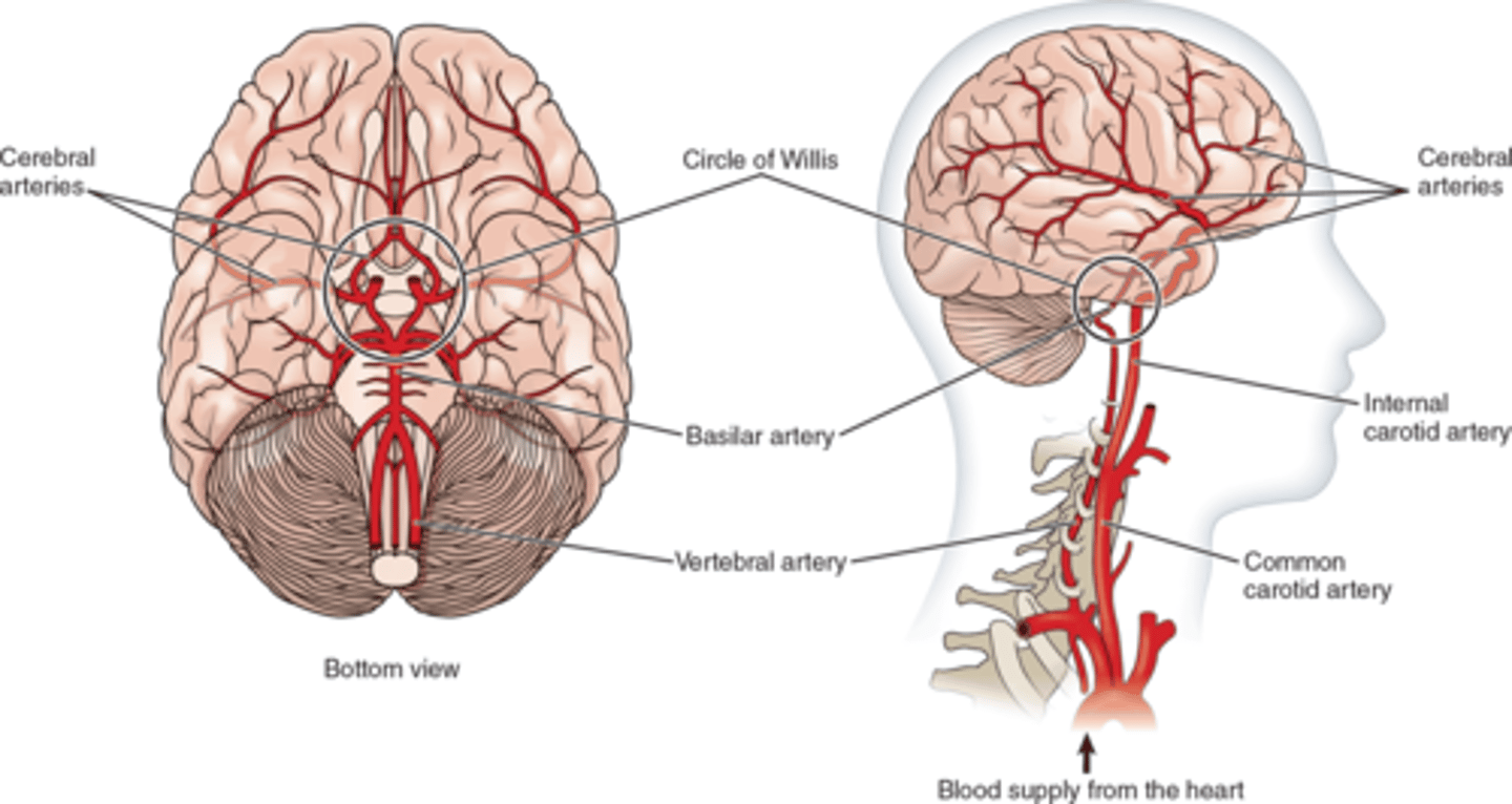
Blood Supply to the Brain
Supplied by the internal carotid (2, anterior) and vertebral (2, posterior) arteries.
Arterial circle (Circle of Willis) helps ensure flow.
New veins can form w/ time from here if damage were to occur.
Blood flows through the brain at a rate of: 800 to 1000 mL per minute.
CO2 level affects CNS blood flow.
Increases in CO2 will increase cerebral blood flow & arterial blood pressure.
Causes vasodilation (leaky blood vessels)
Can worsen cerebral edema.

Anterior Cerebral Artery
Supplies frontal lobes.

Middle Cerebral Artery*
Supplies the Frontal lobe.
Supplies the lateral surface of the temporal and parietal lobes.
Includes motor, sensory, speech centers.
Most frequently occluded artery In a stroke.
Lenticulostriate Arteries
Small, deep penetrating arteries that branch from the middle cerebral artery.

Posterior Cerebral Artery
Supplies the temporal & occipital lobes of cerebral hemispheres.
If lack of blood flow, can affect vision.
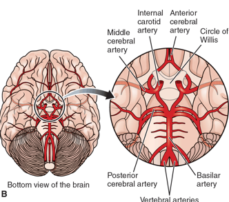
Circle of Willis
The middle cerebral artery supplies a large area of brain tissue.
When occluded, it causes deficit of a major region of the brain.
Most strokes involve a branch of the middle cerebral artery.
Autoregulation of Cerebral Blood Flow
The arteries that comprise the circle of Willis in the brain normally maintain a constant flow of blood within the brain.
Baroreceptors help regulate pressure in the brain.
Optimizes blood flow when BP is low — peripheral vessels constrict, cerebral vessels dilate.
When does Autoregulation of Cerebral Blood Flow not work?
When BP is either too high or too low.
Autoregulation is overridden
Ex) 50/40 mmHg
Ex) 240/120 mmHg
Cranial Nerves
12 of cranial nerves branch directly from the base of the brain.
Some only carry sensory fibers.
Some only carry motor fibers.
Some carry both.
Each nerve travels from the brain through the foramen ovale to its destination.
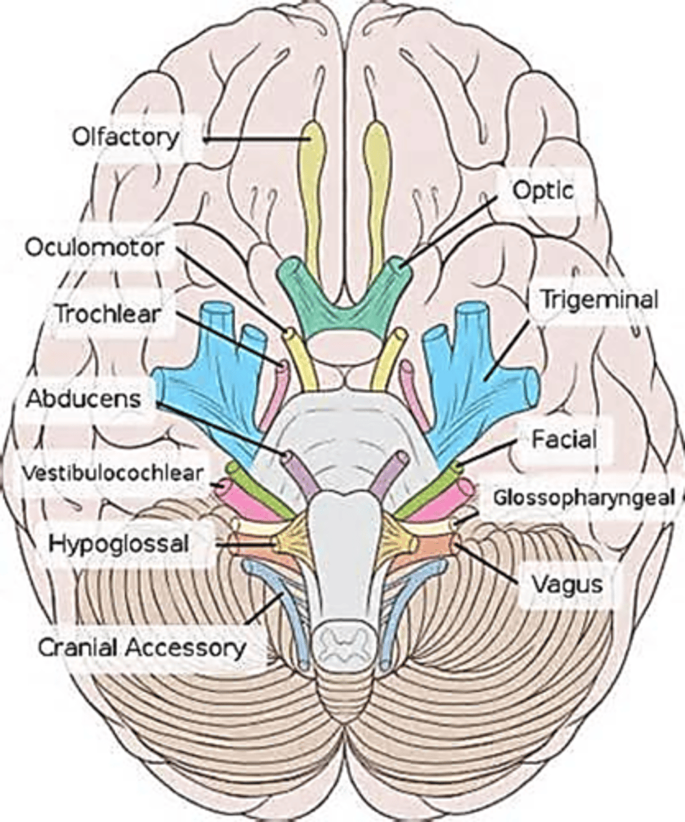
T/F: All of the cranial nerves mostly supply the head and neck.
False; Most of the cranial nerves mostly supply the head and neck. The Vagus nerve (CN X) is the exception. The vagus nerve has nerve fibers in the heart, respiratory (accessory) muscles, stomach, and gallbladder.
T/F: Cranial nerves can act as antennae that can sense changes in intracranial pressure.
True
CN I
Olfactory Nerve | Sensory
CN II
Optic Nerve | Sensory
CN III
Oculomotor Nerve | Motor
CN IV
Trochlear Nerve | Motor
CN V
Trigeminal Nerve | Both
CN VI
Abducens Nerve | Motor
CN VII
Facial Nerve | Both
CN VIII
Acoustic Nerve | Sensory
CN IX
Glossopharyngeal Nerve | Both
CN X
Vagus Nerve | Both
CN XI
Spinal Accessory Nerve | Motor
CN XII
Hypoglossal Nerve | Motor
Application of the Glasgow Coma Scale (GCS)
Used in patients that are unconscious or have altered levels of consciousness / mental status.
Assesses:
Motor response
Eye opening
Verbal response.
Assess this, vital signs, and pupil response.
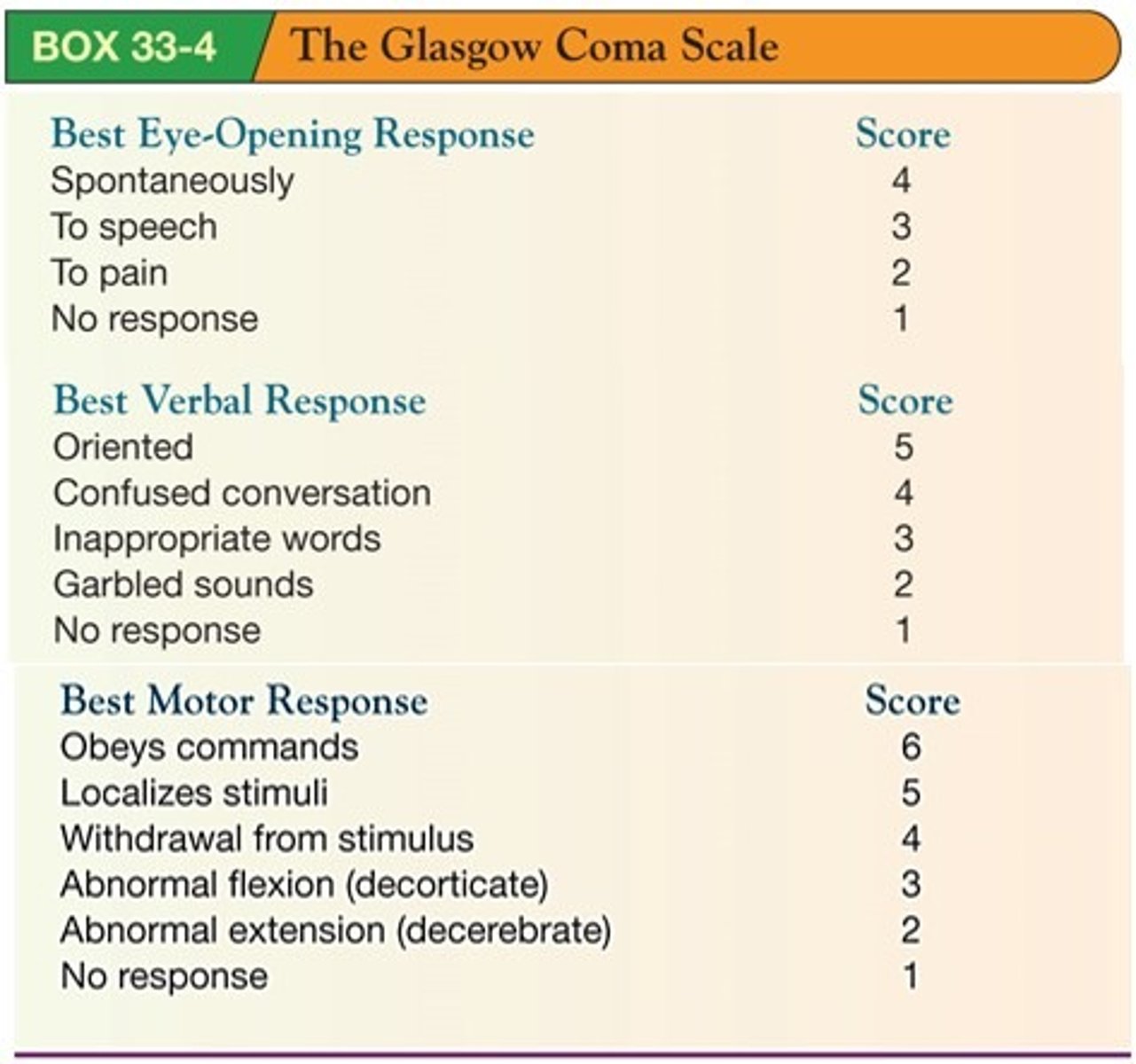
Level of Impairment of GCS*
Mild: 13-15
Moderate: 9-12
Severe: 3-8
What does the Spinal Cord consist of?
White matter
Gray matter
Ascending Fibers/Afferent Tracts
Descending Fibers/Efferent Tracts
White Matter (Spinal Cord)
Comprised of nerve fiber tracts/pathways.
Gray Matter (Spinal Cord)
Neurons
Interneurons connect the CNS to the PNS
Ascending Fibers/Afferent Tracts (Spinal Cord)
Carry sensory information from body back to the brain.
Afferent/Ascending → Body UP to Brain
Descending Fibers/Efferent Tracts (Spinal Cord)
Carry motor impulses from the brain to the PNS.
Descending/Efferent → Brain DOWN to body.
What are the detailed components of the Peripheral Nervous System?
Nerves
Bundles of nerve fibers.
Each fiber is part of the neuron.
Work like an “information” highway
31 Spinal Nerve Pairs
8 cervical
12 thoracic
5 lumbar
5 sacral
1 coccygeal
Ganglia
Collections of nerve cell bodies outside the CNS.
Spinal Nerves
Arise from dorsal and ventral surfaces of the spinal cord.
Each spinal nerve is comprised of a dorsal root + a ventral root.
Each “root” formed from 6–8 rootlets.
Somatic (Voluntary) PNS
31 pairs of spinal nerves
12 pairs of cranial nerves
Automatic (Involuntary) PNS
Controls the Following
Smooth muscles
Arteries, GI tract, lungs
Adrenergic receptors
Cardiac muscle
Glands
Spinal Cord Nerves Mneumonic
Breakfast at 8
Cervical (8)
Lunch at 12
Thoracic (12)
Dinner at 5
Lumbar (5) + Sacral (5)
Dancing till 1
Coccygeal (1)
Levels of Injury and Extent of Paralysis
Injury at C 3-5 or Higher
Requires assistance mechanical ventilation to support breathing.
Loss of GI, walking, arm movement, breathing.
Quadriplegia
Injury at C6
Quadriplegia
Loss of GI function and walking
Injury at T6
Paraplegia
Loss of GI function and walking
May have shoulder movement
Injury at L1
Affects walking and bowel/bladder if complete injury
Paraplegia
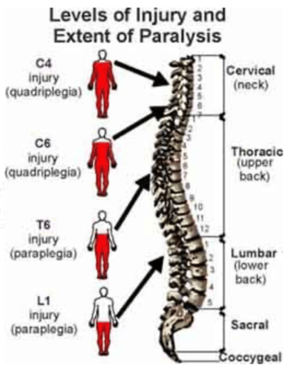
Injury at T6 or Higher - What should we be concerned about?
Breathing
Afferent Nerves are _____ nerves.
sensory
Input = _____. To the brain = ____.
sensory; afferent
Efferent Nerves are _____ nerves.
motor
Output = _____. From the brain = ____.
motor; efferent
Interneurons connect the ____ and ____ neurons in the spinal cord.
sensory; motor
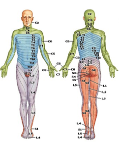
Dermatome
Area of the skin innervated by a given pair of spinal sensory nerves.
The Autonomic Nervous System (ANS)
Responsible for maintaining the internal environment of the body (homeostasis).
Regulates Cardio, Gastro, and Glands
Controls smooth muscles.
Controls unconscious responses such as:
Heart rate
Blood pressure
Intestinal motility