Microbiology - Practical 2
1/116
There's no tags or description
Looks like no tags are added yet.
Name | Mastery | Learn | Test | Matching | Spaced | Call with Kai | Chat |
|---|
No analytics yet
Send a link to your students to track their progress
117 Terms
anaerobic jar
a sealed, air-tight container used in microbiology laboratories to cultivate bacteria that cannot survive in the presence of oxygen (obligate anaerobes). It removes oxygen from the internal environment, allowing these oxygen-sensitive microorganisms to grow on inoculated Petri dishes. [1, 2, 3]
anerobic jar errors
1. Catalyst Inefficiency
The Error: The palladium catalyst pellets—which help Hydrogen and Oxygen bind to form water—can become "poisoned" by moisture or Hydrogen Sulfide (\(H_{2}S\)) over time. [1, 2]
The Impact: Without an active catalyst, the chemical reaction inside the jar stalls, and oxygen remains trapped, resulting in zero growth for strict anaerobes. [1, 2]
How to fix: Always reactivate the catalyst by heating it in an oven as recommended by the manufacturer, or replace it if it is visibly degraded. [1]
2. Leaking Seals and Gaskets
The Error: Cracks, deformations, or lack of grease on the jar's O-ring allow atmospheric oxygen to leak into the container.
The Impact: Fresh oxygen enters during incubation, overwhelming the catalyst and spoiling the anaerobic environment.
How to fix: Inspect the O-ring gasket for tears under good lighting and make sure the lid is securely tightened before starting. [1, 2]
3. Mismatched Sachet Volume
The Error: Using an incorrect gas-generating sachet for your jar's volume (e.g., trying to use a sachet meant for a \(2.5 \text{ L}\) jar in a \(7 \text{ L}\) jar). [1]
The Impact: The chemical sachet will not release enough Hydrogen and \(CO_{2}\) to eliminate all the oxygen in a larger vessel, leaving residual oxygen. [1]
How to fix: Ensure the sachet matches the exact volume of your specific jar. For more detailed troubleshooting, refer to this practical guide to cultivating oxygen-sensitive organisms. [1, 2]
4. Overcrowding and Lack of Headspace
The Error: Stacking too many petri dishes into the jar, or pushing the plates all the way up to the lid.
The Impact: This restricts the necessary gas circulation required to mix the atmosphere evenly and compresses the catalyst basket.
How to fix: Always leave at least \(15 - 20\text{ mm}\) of headspace above the top plate and never overfill the container. [1]
5. Ignoring the Indicator Strip
The Error: Proceeding with your experiment without verifying the anaerobic indicator strip (e.g., methylene blue or resazurin). [1, 2]
The Impact: If the jar fails to convert the atmosphere, the indicator strip will remain in its oxidized state (blue or pink). Assuming the jar worked without checking means you will get false negatives for your strict anaerobes. [1, 2, 3]
How to fix: Check the strip within the first \(2\) to \(3\) hours of incubation. It should turn completely colorless (for resazurin) or white/pink (for methylene blue). Read the catalyst and indicator strip guide for a deeper look at verifying \(0.1\%\) oxygen levels. [1, 2, 3, 4]
6. Prolonged Ambient Air Exposure
The Error: Leaving inoculated plates sitting on the bench for a long period of time before sealing the jar.
The Impact: Obligate anaerobes are extremely sensitive to oxygen. Prolonged exposure to ambient air before the jar is sealed and activated can kill the bacteria before the experiment even begins.
How to fix: Load the plates into the jar and seal the lid as quickly as safely possible after inoculation. [1, 2, 3]
anaerobic jar purpose
primary purpose is to cultivate and isolate obligate anaerobes—microorganisms that are killed or unable to grow in the presence of atmospheric oxygen. It provides a sealed, oxygen-free environment for oxygen-sensitive bacteria. [1, 2, 3, 4]
how anerobic jar works
The jar achieves and maintains an anoxic (oxygen-depleted) environment through specific components: [1, 2]
Gas-Generating Sachets: Chemical packets (like GasPaks) are placed inside the sealed jar with the inoculated agar plates. When activated, they consume the residual \(O_{2}\) and typically release \(CO_{2}\) to support bacterial growth. [1, 2, 3]
Palladium Catalyst: Acts to speed up the chemical reaction between hydrogen and the residual oxygen, safely converting it to water vapor inside the jar. [1, 2, 3, 4, 5]
Anaerobic Indicator: Strips or tablets containing dyes like methylene blue or resazurin are placed in the jar. They turn colorless when the oxygen is successfully removed, confirming the environment is ready for incubation. [1, 2]
anaerobic jar procedure
1. Label plate with whether or not it is aerobic or anaerobic. Label plate with bacteria, name, and date. (idk if yall are also making a quadrant)
2. Using inoculating loop, streak the plate with your bacteria.
3. Place inoculated agar plate in the incubator if it is to be grown in an aerobic environment. Place the plate in the anaerobic jar if it is to be incubated in an anerobic environment.
anaerobic jar expected results
successful removal of atmospheric oxygen (creating an anoxic environment) combined with specific patterns of bacterial growth depending on the microbes' oxygen requirements. [1, 2]
Growth inside the jar depends on the classification of the organism you are culturing: [1, 2, 3]
Obligate Anaerobes: (e.g., Clostridium, Bacteroides). Expected Result: Growth inside the anaerobic jar; NO growth when incubated in normal atmospheric oxygen. [1, 2, 3, 4, 5]
Facultative Anaerobes: (e.g., E. coli, Staphylococcus). Expected Result: Strong growth in both the anaerobic jar and in regular aerobic conditions. [1, 2, 3, 4, 5]
Aerotolerant Anaerobes: (e.g., Streptococcus pneumoniae). Expected Result: Growth occurs in both environments, though they do not use oxygen for their metabolism. [1, 2, 3, 4]
Obligate Aerobes: (e.g., Pseudomonas aeruginosa). Expected Result: NO growth inside the anaerobic jar; robust growth in standard aerobic conditions. [1, 2, 3, 4, 5]
Microaerophiles: (e.g., Campylobacter). Expected Result: Little to no growth in the anaerobic jar, as they typically require a reduced-oxygen but non-anoxic environment (e.g., via a Candle Jar or specialized gas pack). [1, 2, 3]
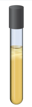
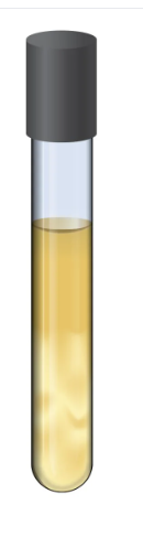
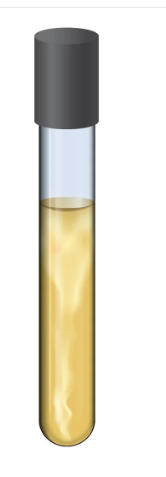
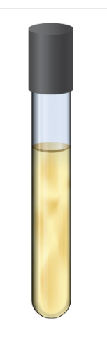
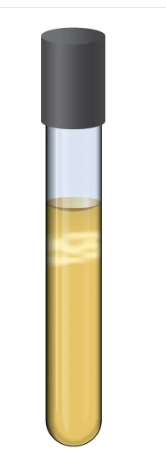
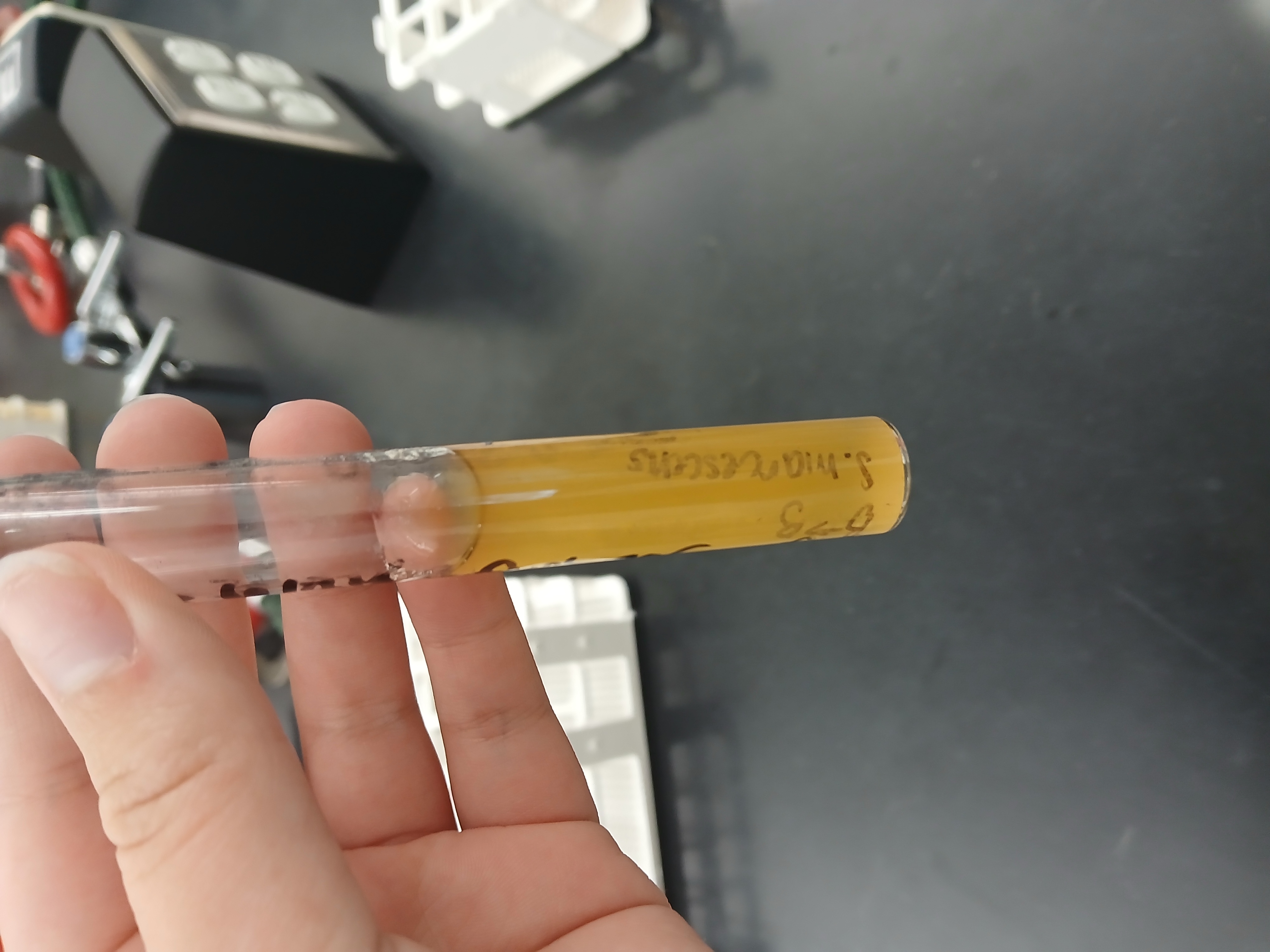
Fluid Thioglycollate Medium (FTM)
a versatile, semi-liquid microbiological culture medium used primarily to determine the oxygen requirements of bacteria and to test for sterility in pharmaceutical and biological products. It establishes a natural oxygen gradient in the tube—from fully aerobic at the top to anaerobic at the bottom. [1, 2, 3, 4, 5]
how FTM works
elies on specific chemical components to create its unique environment and allow microbiologists to interpret bacterial growth: [1, 2]
Oxygen Gradient: A small amount of agar in the broth prevents rapid oxygen diffusion, maintaining a high-oxygen zone at the top and an anaerobic zone at the bottom. [1, 2, 3]
Reducing Agents: Ingredients like sodium thioglycollate and L-cystine actively bind free oxygen to keep the redox potential low (creating a perfect environment for anaerobes). [1, 2, 3]
Redox Indicator: Resazurin is included as a visual indicator. It turns pink in the presence of oxygen (typically the top portion) and remains colorless in anaerobic areas (the bottom portion). [1, 2]
FTM errors
1. Oxygen Gradient Destruction (The "Broad Pink Zone" Error)
The Mistake: FTM uses resazurin as an oxygen indicator, which turns pink-purple in the presence of oxygen and is colorless in its absence. If more than one-third of the tube is pink, too much oxygen has diffused into the medium, or the tube was shaken excessively. [1, 2, 3]
The Error: This creates a toxic environment for strict anaerobes. Obligate anaerobes that normally grow at the bottom will either be killed or will not grow at all, yielding a false-negative result. [1, 2, 3, 4]
The Fix: Boil the tubes in a water bath to drive out oxygen, and allow them to cool slowly before inoculating. [1]
2. Poor Inoculation Technique
The Mistake: When using an inoculating needle, moving the needle side-to-side or altering the path during insertion and removal disturbs the physical medium. [1, 2]
The Error: This introduces excess oxygen deep into the tube and disturbs the agar matrix. It leaves a visible "tornado" or streak of growth, making it look falsely like a facultative anaerobe or aerotolerant organism instead of a strict anaerobe or obligate aerobe. [1, 2, 3]
The Fix: Insert and withdraw the needle along the exact same central axis. Never stir the medium. [1, 2]
3. Capping Tubes Too Tightly
The Mistake: Closing the culture tube lids completely tight before incubation.
The Error: FTM requires a balance of atmospheric oxygen at the top. Tightening caps restricts oxygen, preventing strictly aerobic bacteria from growing properly at the top of the tube, which can result in weaker or misinterpreted growth patterns. [1, 2, 3]
The Fix: Ensure caps are slightly loose (finger-tight but "backed off" a quarter turn) to allow necessary gas exchange, unless your specific laboratory protocol mandates otherwise. [1, 2]
4. Contamination of Control Tubes
The Mistake: Finding bacterial growth in an uninoculated control tube.
The Error: This indicates that the FTM tube was contaminated before inoculation, or that poor aseptic technique was used (e.g., leaving caps off, non-sterile tools). It completely invalidates the results.
The Fix: Discard the contaminated batch, check the incubator and your environment for contamination, and strictly adhere to sterile practices. [1, 2, 3, 4, 5]
5. Early Observation / Not Allowing Recovery
The Mistake: Reading the tubes too early or failing to wait for the required 7-14 day incubation period, particularly for slow-growing or stressed microorganisms.
The Error: The bacterial lag phase can cause the growth patterns (turbidity) to take several days to become visibly apparent. Early readings result in false negatives.
The Fix: Follow standard compendial incubation periods (such as those outlined in USP standards) and document visual turbidity at multiple intervals. [1, 2, 3, 4, 5, 6, 7]
FTM purpose
determine an organism's aerotolerance—its ability to survive and grow at varying levels of oxygen. [1]
FTM procedure
1. Take thioglycolate tube and label it with your name, date, and bacteria that will be in use.
2. Inoculate thioglycolate tube with bacteria using asceptic technique. Mix in sterile agar broth.
3. Tightly close inoculated tube and inoculate overnight at 37 degrees Fahrenheit.
FTM expected results
Microorganism Classification [1, 2, 3, 4, 5] | Expected Growth Location | Result Description |
|---|---|---|
Obligate Aerobe | Top only | Growth is strictly at the oxygen-rich surface (pellicle). Bacteria cannot survive without oxygen. |
Obligate Anaerobe | Bottom only | Growth is restricted to the lower, oxygen-depleted zones. Oxygen is toxic to these organisms. |
Facultative Anaerobe | Throughout the tube | Turbidity (cloudiness) is present from top to bottom, but growth is usually densest near the top because aerobic respiration yields more energy. |
Microaerophile | Just below the surface | Growth appears as a thin, distinct band a little below the pink surface layer. They require oxygen but cannot tolerate atmospheric levels. |
Aerotolerant Anaerobe | Throughout the tube | Growth is evenly spread throughout the medium. These bacteria do not use oxygen but are not |
strict aerobe
strict anaerobe
facultative anaerobe
aerotolerant anaerobe
microaerophile
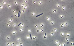
endospore stain
a differential microbiological staining technique used to detect the presence, shape, and location of endospores in bacterial cells. It helps distinguish these highly resistant, dormant spores from actively growing "vegetative" cells. [1, 2, 3]
how endospore stain works
Primary Staining with Steam: The heat-fixed bacterial smear is flooded with malachite green and steamed for about 5 minutes. The steam acts as a mordant, swelling the tough outer keratin coat of the endospores so the dye can penetrate. [1, 2, 3, 4]
Decolorization: The slide is rinsed with water. Since malachite green is water-soluble and does not bind strongly, it easily washes out of the thin walls of the vegetative cells, rendering them colorless. The endospores, however, trap the dye and remain green. [1, 2, 3, 4]
Counterstaining: Safranin is applied for about a minute to dye the colorless vegetative cells. [1, 2]
endospore stain errors
An endospore stain error usually means either the spores aren't staining, or the vegetative cells are the wrong color. The most common mistake is failing to apply continuous heat, which is necessary to drive the stain through the spore's tough outer protein layer. [1, 2, 3, 4]
endospore stain purpose
to visually distinguish dormant bacterial endospores from actively growing vegetative cells. It is a crucial differential staining technique used in microbiology to identify spore-forming bacteria (such as Bacillus and Clostridium species) from non-spore formers. [1, 2, 3, 4]
endospore stain procedure
1. Place two loopfuls of bacteria onto your slide. Spread it around gently. Let it air dry.
2. Fix the bacteria to the slide by passing it two times through the bunsen burner.
3. Place a strip of bibulous paper on top of the smear.
4. Place the slide with bibulous paper on top of steaming beaker, heating on a hot plate.
5. Drop malachite green on top of bibulous slide. Let sit for 5 minutes.
6. Take slide off of steaming beaker.
7. Let slide cool off until it is cool enough to touch.
8. Remove bibulous paper.
9. Hold slide at a 45 degree angle and apply decolorizer. Let decolorizor run down slide until green is no longer seen.
10. Apply safranin. Let sit for 1 minute.
11. Shake off any excess and use bibulous paper to dry.
12. View slide under microscope under the oil immersion lense.
endospore stain expected results
endospores will be clear!
acid fast stain
a differential microbiological test used to identify bacteria that have a waxy, lipid-rich cell wall, specifically mycolic acid. This waxy layer makes the cells highly resistant to routine staining methods like the Gram stain. [1, 2, 3, 4]
This test is primarily used in clinical microbiology to detect the presence of pathogenic bacteria such as: [1, 2]
Mycobacterium tuberculosis (the bacteria responsible for tuberculosis)
Mycobacterium leprae (the bacteria that causes leprosy)
Nocardia species [1, 2, 3, 4]
how an acid fast stain works
The staining process relies on the physical property of acid-fastness—the ability to resist decolorization by acids during staining procedures. It involves three core steps: [1]
Primary Stain (Carbol Fuchsin): A dark red/magenta dye is applied to the slide and heated. The heat melts the waxy mycolic acid layer, allowing the dye to penetrate the bacterial cell. [1, 2, 3, 4]
Decolorizer (Acid-Alcohol): A harsh mixture of acid and alcohol is washed over the slide. Because acid-fast bacteria have a thick, waxy barrier, the dye stays trapped inside. Non-acid-fast bacteria, which lack this waxy layer, have the primary stain washed out and become colorless. [1, 2, 3, 4]
Counterstain (Methylene Blue): A contrasting blue dye is applied. The decolorized, non-acid-fast cells absorb the blue dye. [1, 2]
acid fast stain errors
Common Procedural Errors
Over-decolorizing: Leaving the acid-alcohol decolorizer on for too long can strip the hot pink/red carbolfuchsin stain even from acid-fast bacteria. [1, 2, 3, 4]
Under-decolorizing: Not using enough acid-alcohol will leave the stain on non-acid-fast bacteria, causing them to falsely appear acid-fast (red/pink). [1, 2, 3, 4, 5]
Overheating the smear: Boiling the carbolfuchsin during the steaming step or overheating the slide during heat-fixation can physically rupture the bacterial cell walls, preventing them from holding the primary stain. [1, 2, 3, 4]
Heat-fixing a wet slide: Passing a wet smear through the flame essentially boils the cells, causing the bacteria to burst and lose their ability to retain the dye. [1, 2, 3, 4]
Thick smears: Making the bacterial smear too thick makes it difficult for the decolorizer to wash the primary stain out of non-acid-fast cells. [1, 2, 3]
Using old or dead cultures: Bacteria from old cultures may have weakened cell walls and can lose their acid-fast properties, even if they are acid-fast species. [1, 2]
Preparation & Cross-Contamination Errors
Stain precipitation: If carbolfuchsin isn't filtered properly, the stain can form crystals that look like clumps of acid-fast bacilli under the microscope. [1, 2]
Cross-contamination: Reusing slides or accidentally transferring bacteria from one positive slide to a negative one via the oil immersion lens can cause false-positives. [1]
Debris and artifacts: Stained food particles, fibers, or scratch marks on the slide can be mistaken for acid-fast organisms by inexperienced eyes. [1, 2]
acid fast stain purpose
The primary purpose of the acid-fast stain in microbiology is to identify bacteria that have a waxy, lipid-rich cell wall (specifically containing mycolic acid). These unique walls make the bacteria highly resistant to standard stains (like the Gram stain) and decolorization by harsh acid-alcohol solutions. [1, 2, 3]
acid fast stain expected results
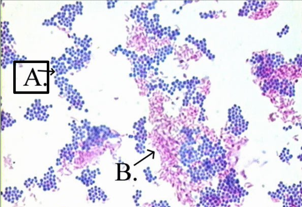
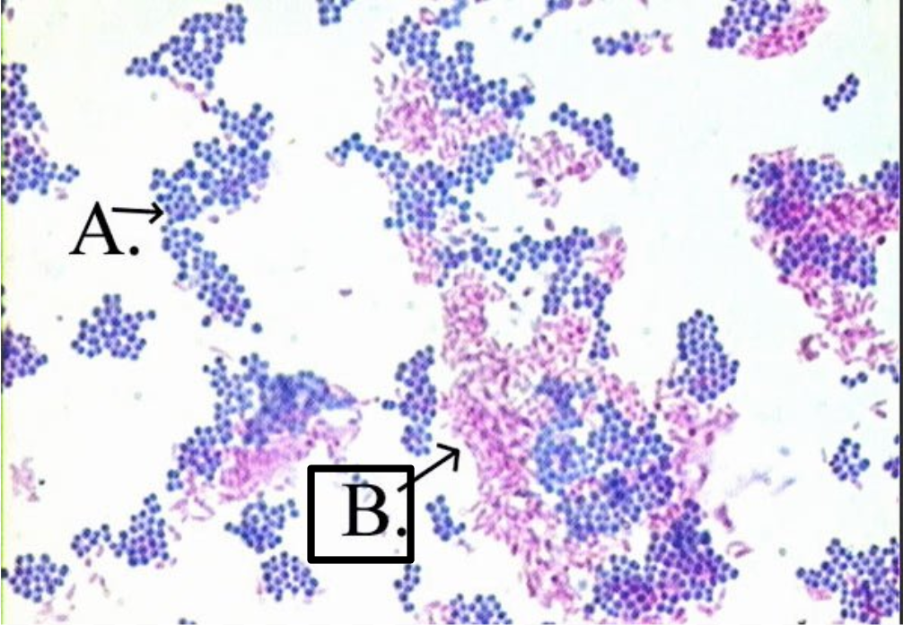
The acid-fast stain (AFB) is a differential stain used to identify bacteria with waxy, lipid-rich cell walls (like mycolic acid) that resist standard staining. Expected results are: [1, 2]
Acid-Fast Bacteria: Appear bright fuchsia, hot pink, or red. They are often seen as slender, slightly curved rods (bacilli), which may occur singly, in small groups, or with a beaded appearance.
Non-Acid-Fast Bacteria: Appear blue.
Background Material: Appears blue. [1, 2, 3, 4, 5]
non-acid fast organism
appear blue
They lose the primary stain during decolorization and take up the counterstain (e.g., methylene blue).
acid fast organisms
Appear bright fuchsia, pink, or red. They resist decolorization and retain the primary stain (carbol fuchsin).
manitol salt agar
a selective and differential growth medium used in microbiology. It selects for salt-tolerant bacteria (like Staphylococcus) while inhibiting others, and it differentiates between species based on their ability to ferment the sugar mannitol, which causes a visual color change in the agar. [1, 2, 3]
how manitol salt agar works
High Salt (7.5% NaCl): Acts as the selective agent. It prevents the growth of most Gram-negative and non-salt-tolerant Gram-positive bacteria, allowing only halotolerant species (like Staphylococcus) to thrive. [1, 2, 3, 4]
Mannitol: Acts as the differential agent. It is a fermentable sugar alcohol. [1, 2]
Phenol Red: Functions as a pH indicator. It is red at neutral pH, turns pink in alkaline conditions, and changes to bright yellow at acidic pH levels (below 6.8). [1, 2]
![<ul><li><p><span><strong>High Salt (7.5% NaCl):</strong> Acts as the <strong>selective agent</strong>. It prevents the growth of most Gram-negative and non-salt-tolerant Gram-positive bacteria, allowing only halotolerant species (like <em>Staphylococcus</em>) to thrive.</span> [1, 2, 3, 4]</p></li><li><p><span><strong>Mannitol:</strong> Acts as the <strong>differential agent</strong>. It is a fermentable sugar alcohol.</span> [1, 2]</p></li><li><p><span><strong>Phenol Red:</strong> Functions as a <strong>pH indicator</strong>. It is red at neutral pH, turns pink in alkaline conditions, and changes to bright yellow at acidic pH levels (below 6.8).</span> [1, 2]</p></li></ul><p></p>](https://assets.knowt.com/user-attachments/101d8677-42be-42f1-8410-866122467631.jpg)
manitol salt agar interpreting results
Growth vs. No Growth:
Growth: The bacteria can tolerate 7.5% salt (typically Staphylococcus or Micrococcus species).
No Growth: The bacteria cannot tolerate high salt levels (most other bacteria are inhibited). [1, 2, 3, 4, 5]
Color Change (If growth occurs):
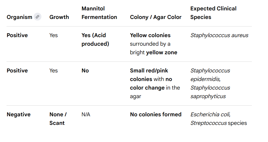
Yellow (Positive for Mannitol Fermentation): The bacteria ferments mannitol to produce acidic byproducts. These acids lower the pH, turning the phenol red indicator bright yellow. This is highly characteristic of pathogenic Staphylococcus aureus.
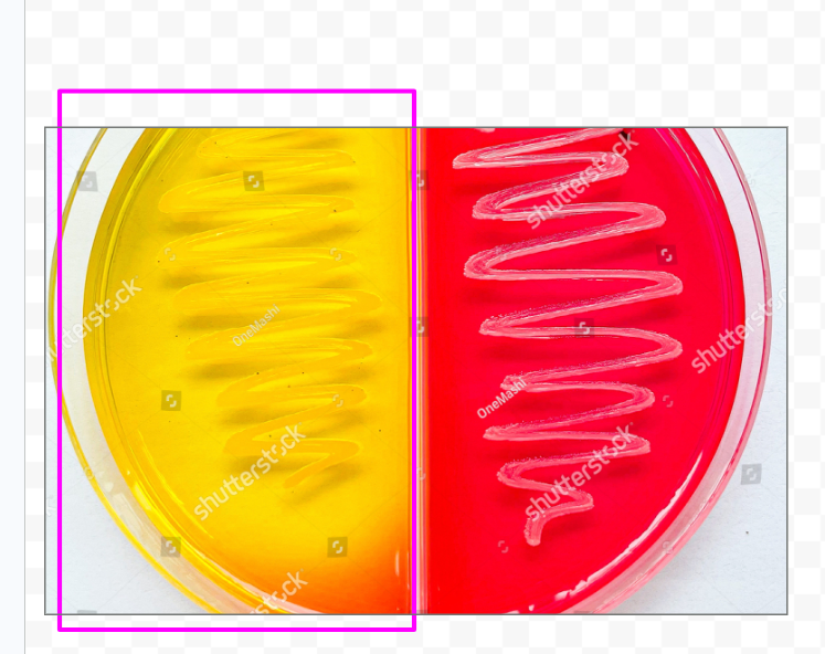
Red/Pink (Negative for Mannitol Fermentation): The bacteria grow but do not ferment mannitol. The color remains red or pink, which is typical for non-pathogenic species like Staphylococcus epidermidis. [1, 2, 3, 4, 5, 6]
manitol salt agar common applications
Isolating and identifying Staphylococcus aureus from clinical and non-clinical specimens.
Screening for Methicillin-resistant Staphylococcus aureus (MRSA), as many resistant strains are mannitol-fermenters.
Analyzing food and dairy products for the presence of staphylococci. [1, 2, 3, 4]
manitol salt agar errors
common experimental errors often relate to timing, improper incubation, or misinterpreting the phenol red pH indicator. [1, 2, 3, 4]
False-Negative (No Yellow Color):
Over-incubation: If left in the incubator for too long (e.g., beyond 48 hours), the bacteria may exhaust all available mannitol. They will then begin breaking down peptones for energy, producing alkaline byproducts that neutralize the acid, turning the yellow color back to red.
Delayed fermentation: Some strains may be slow to ferment. Plates should be re-incubated overnight rather than discarded immediately. [1, 2, 3]
False-Positive (Unexpected Yellow Color):
Non-staphylococci growth: Some salt-tolerant organisms (like Enterococcus or certain Micrococcus species) can survive the salt and ferment the mannitol, which may mimic Staphylococcus aureus. [1, 2]
No Growth / Inhibited Growth:
Improper Inoculation: The high salt concentration (7.5% \(NaCl\)) is highly restrictive. If no growth occurs, the organism may simply not be salt-tolerant, which is considered a valid negative result (not an error).
Inoculum Carryover: If the inoculum was transferred using another medium, carryover nutrients or pH-altering compounds might interfere with the initial reactions. [1, 2, 3, 4]
manitol salt agar purpose
The purpose of Mannitol Salt Agar (MSA) in microbiology is to selectively isolate salt-tolerant bacteria (like Staphylococcus species) and differentially identify those that can ferment the sugar mannitol (such as pathogenic Staphylococcus aureus) from mixed clinical or environmental samples. [1, 2]
manitol salt agar selectivity
MSA contains a high concentration of sodium chloride (usually 7.5%). [1]
What it does: This high-salt environment inhibits the growth of most Gram-negative and non-salt-tolerant bacteria.
What grows: It allows halophilic (salt-loving) or halotolerant organisms—primarily Staphylococcus, Micrococcus, and Enterococcus species—to thrive. [1, 2, 3]
manitol salt agar differentiation
Once the salt-tolerant bacteria grow, the agar helps microbiologists tell them apart using its two specific ingredients: [1, 2, 3]
Mannitol: A fermentable carbohydrate sugar.
Phenol Red: A pH indicator that is red at a neutral pH (around 7.4) but turns yellow at acidic pH levels (below 6.8). [1, 2, 3]
Positive for Mannitol Fermentation
The bacteria ferment the mannitol, producing acidic byproducts. This drop in pH causes the phenol red to turn the colony and surrounding agar bright yellow. [1, 2]
Staphylococcus aureus
![<p><span>The bacteria ferment the mannitol, producing acidic byproducts. This drop in pH causes the phenol red to turn the colony and surrounding agar bright <strong>yellow</strong>.</span> [1, 2]</p><p></p><p>Staphylococcus aureus </p>](https://assets.knowt.com/user-attachments/0f14c32c-6e07-4aa8-889d-e4eb49c443c6.png)
negative for manitol fermentation
The bacteria grow but do not ferment the mannitol. The agar surrounding the colonies will remain red or pink. [1, 2, 3]
![<p><span>The bacteria grow but do not ferment the mannitol. The agar surrounding the colonies will remain <strong>red or pink</strong>.</span> [1, 2, 3]</p>](https://assets.knowt.com/user-attachments/24f977cc-b56c-4e34-8ab9-270542e1d0ff.png)
manitol salt agar procedure
Label back of agar plate with your name and date. On the lid, make three sections and label with which bacteria will be streaked in that area.
2. Asceptically transfer each bacteria to the appropriate area. Use a single streak.
3. Place lid upside down and incubate at 37 degrees celsius.
4. Observe growth and color.
Color Change:
Yellow Zones/Colonies (Mannitol-Positive): Bacteria ferment mannitol, producing organic acids that drop the pH and cause the phenol red to turn yellow (e.g., Staphylococcus aureus).
Pink/Red Agar (Mannitol-Negative): No fermentation occurs. The agar remains pink/red (e.g., Staphylococcus epidermidis). [1, 2, 3]
mannitol salt agar expected results
Staphylococcus aureus
On mannitol salt agar, have Yellow colonies surrounded by a bright yellow zone
this means it ferments mannitol
Staphylococcus epidermidis, Staphylococcus saprophyticus
Small red/pink colonies with no color change in the agar
It does not ferment mannitol
Bile Esculin Medium
a selective and differential microbiological medium used primarily to isolate and presumptively identify Group D streptococci and Enterococcus species. It differentiates bacteria based on their ability to tolerate bile salts and hydrolyze the glycoside esculin. [1, 2, 3, 4]
![<p><mark>a selective and differential microbiological medium used primarily to isolate and presumptively identify Group D streptococci and </mark><em><mark>Enterococcus</mark></em><mark> species</mark>. It differentiates bacteria based on their ability to tolerate bile salts and hydrolyze the glycoside esculin. [1, 2, 3, 4]</p>](https://assets.knowt.com/user-attachments/76e3e8e3-90c8-42be-81bc-cb03eafc913f.jpg)
Bile Esculin Medium characteristics
Selectivity: Contains bile salts (oxgall) which inhibit the growth of most Gram-positive bacteria, except for Enterococci and Group D Streptococci.
Differential Capability: Contains esculin and an iron indicator (ferric citrate). [1, 2, 3]
Bile Esculin Medium interpretation of results
Positive Result: Growth with intense blackening of the medium (e.g., Enterococcus faecalis).
Negative Result: No blackening (or growth without blackening), indicating the bacteria either cannot tolerate bile or cannot hydrolyze esculin. [1, 2, 3, 4]
how Bile Esculin Medium works
is a selective and differential agar used in microbiology to identify Group D Streptococci and Enterococci. It works by using bile salts to inhibit most Gram-positive bacteria, while allowing target organisms to survive and hydrolyze the sugar esculin, which results in a dark brown or black color change. [1, 2]
Bile Salts (Oxbile): Acts as the selective agent. These detergent-like salts destroy the cell membranes of most bacteria, successfully inhibiting the growth of unwanted, non-Group D Gram-positive organisms. [1, 2]
Esculin: Acts as the differential component. This glycoside is broken down by the bacteria into two products: glucose and esculetin. [1, 2, 3, 4]
Ferric Ammonium Citrate: Serves as the chemical indicator. It contains iron (Fe³⁺) ions that react with esculetin to produce the visible color change. [1, 2, 3]
Bile Esculin Medium purpose
The primary purpose of the Bile Esculin Medium (often in the form of Bile Esculin Agar or BEA) is to selectively isolate and presumptively identify members of the genus Enterococcus and Group D Streptococcus. It achieves this through two main mechanisms: [1, 2, 3, 4]
Bile Esculin Medium procedure
1. Label each BE slant tube with your name, date, and organism.
2. Asceptically inoculate one BE slant with each organism.
3. Inoculate one nutrient agar control with each organism.
4. Incubate tubes 24-48 hrs at 37C.
5. A positive test is darkening of at least ½ of the agar.
Bile Esculin Medium expected results
A positive Bile Esculin test results in growth and a dark brown or black discoloration of the medium. A negative test results in either no growth or growth with no color change (the medium remains amber/yellow). [1, 2]
positive bile esculin
growth and a dark brown or black discoloration of the medium
Bacteria that can survive the 4% bile salts and hydrolyze esculin produce esculetin, which reacts with ferric ions in the medium to create a dark pigment. [1, 2, 3]
Positive Result:
Appearance: Diffuse blackening of more than half of the agar slant, or dark halos around colonies on plates.
Indications: Presumptive identification of Group D streptococci (e.g., Streptococcus bovis) and Enterococcus species (e.g., Enterococcus faecalis). [1]
![<p><strong><mark>growth</mark></strong><mark> and a </mark><strong><mark>dark brown or black discoloration</mark></strong><mark> of the medium</mark></p><p></p><p>Bacteria that can survive the 4% bile salts and hydrolyze esculin produce esculetin, which reacts with ferric ions in the medium to create a dark pigment. [1, 2, 3]</p><ul><li><p><span><strong>Positive Result:</strong></span></p><ul><li><p><span><strong>Appearance:</strong> Diffuse blackening of more than half of the agar slant, or dark halos around colonies on plates.</span></p></li><li><p><span><strong>Indications:</strong> Presumptive identification of <strong>Group D streptococci</strong> (e.g., <em>Streptococcus bovis</em>) and <strong>Enterococcus</strong> species (e.g., <em>Enterococcus faecalis</em>).</span> [1]</p></li></ul></li></ul><p></p>](https://assets.knowt.com/user-attachments/6df152f8-2888-49a6-9851-b4d457ca6cbc.png)
negative bile esculin
no growth or growth with no color change (the medium remains amber/yellow). [1, 2]
Appearance: Normal amber/yellow color of the medium with no blackening, regardless of whether bacterial growth occurred.
Indications: Most other streptococci species, which are either inhibited by the bile salts or cannot hydrolyze esculin. [1, 2, 3, 4, 5]
![<p><strong>no growth</strong> or <strong>growth with no color change</strong> (the medium remains amber/yellow). [1, 2]</p><p></p><ul><li><p><span><strong>Appearance:</strong> Normal amber/yellow color of the medium with no blackening, regardless of whether bacterial growth occurred.</span></p></li><li><p><span><strong>Indications:</strong> Most other streptococci species, which are either inhibited by the bile salts or cannot hydrolyze esculin.</span> [1, 2, 3, 4, 5]</p></li></ul><p></p>](https://assets.knowt.com/user-attachments/faa405cc-ac9f-4dec-8d11-5936ce1199ac.png)
catalase test
a foundational microbiology technique used to identify whether a bacteria produces the catalase enzyme. It is primarily used to differentiate between Staphylococcus (catalase-positive) and Streptococcus (catalase-negative). [1, 2, 3, 4]
![<p><mark>a foundational microbiology technique used to identify whether a bacteria produces the </mark><strong><mark>catalase enzyme</mark></strong>. It is primarily used to differentiate between <em>Staphylococcus</em> (catalase-positive) and <em>Streptococcus</em> (catalase-negative). [1, 2, 3, 4]</p>](https://assets.knowt.com/user-attachments/73cc5d71-1f7a-48b8-b412-f15a7fddef78.jpg)
catalase test principle
Bacteria that use oxygen produce toxic byproducts, such as hydrogen peroxide (H₂O₂). To survive, these bacteria produce the enzyme catalase, which neutralizes hydrogen peroxide by breaking it down into harmless water and oxygen gas: [1, 2, 3, 4, 5]
![<p><span>Bacteria that use oxygen produce toxic byproducts, such as hydrogen peroxide (H₂O₂). To survive, these bacteria produce the enzyme <strong>catalase</strong>, which neutralizes hydrogen peroxide by breaking it down into harmless water and oxygen gas:</span> [1, 2, 3, 4, 5]</p>](https://assets.knowt.com/user-attachments/160127fc-0557-4372-9945-237bfaf74f5d.png)
catalase test interpreting results
Positive Result: Immediate and rapid production of oxygen gas bubbles. This indicates the bacteria possess the catalase enzyme.
Negative Result: No bubbles (or very few scattered bubbles) form. This indicates the bacteria lack the catalase enzyme. [1, 2, 3, 4, 5]
catalase test clinical importance
Staph vs. Strep: The test is most famously used to distinguish Gram-positive cocci. Staphylococcus species are catalase-positive, whereas Streptococcus and Enterococcus species are catalase-negative. [1, 2]
Differentiation: It also aids in identifying other genera, such as distinguishing strict aerobes (usually positive) from strict anaerobes (usually negative). [1]
how the catalase test works
detects the presence of the enzyme catalase by testing a bacterial sample's ability to break down hydrogen peroxide (H₂O₂) into water (H₂O) and oxygen (O₂). A positive result is instantly recognizable by the rapid production of visible oxygen gas bubbles. [1]
The Catalase Enzyme: Aerobic bacteria produce catalase to neutralize this toxicity by converting hydrogen peroxide into harmless water and oxygen. [1, 2]
The Chemical Equation: \(2H_2O_2 \xrightarrow{\text{Catalase}} 2H_2O + O_2 (\text{gas bubbles})\) [1]
catalase test errors
improper culture age, contaminated medium carryover, or reagent instability. These mistakes lead to false-positive or false-negative results, which can misidentify organisms like Staphylococcus (catalase-positive) versus Streptococcus (catalase-negative). [1, 2, 3]
catalase test positive
The primary purpose of the catalase test in microbiology is to determine if a bacterium produces the enzyme catalase, which breaks down toxic hydrogen peroxide (H₂O₂) into water and oxygen gas. It is a vital, quick biochemical assay used to differentiate between morphologically similar groups of bacteria and identify specific pathogens. [1, 2, 3]
catalase test procedure
1. Asceptically transfer bacteria to a glass slide.
2. Add a few drops of hydrogen peroxide on top of the transfered bacteria
3. If there is bubbles, the bacteria is catalase positive. No bubbling means catalase-negative.
catalase test expected results
Positive Result: Immediate, vigorous formation of oxygen bubbles (effervescence).
Negative Result: No bubbles, or very minimal/insignificant bubbling. [1, 2]
catalase negative
Examples: Streptococcus species, Enterococcus species, and Clostridium species. [1, 2, 3]
![<p><span><em>Examples:</em> <em>Streptococcus</em> species, <em>Enterococcus</em> species, and <em>Clostridium</em> species.</span> [1, 2, 3]</p>](https://assets.knowt.com/user-attachments/f2b150ec-54f0-412b-9929-d9eea9b6d1c7.png)
catalase positive
examples: Staphylococcus species, Micrococcus species, Bacillus species, and the Enterobacteriaceae family (e.g., E. coli). [1, 2, 3, 4, 5]
![<p><span><em>examples: Staphylococcus</em> species, <em>Micrococcus</em> species, <em>Bacillus</em> species, and the <em>Enterobacteriaceae</em> family (e.g., <em>E. coli</em>).</span> [1, 2, 3, 4, 5]</p>](https://assets.knowt.com/user-attachments/6cf49e33-4968-4a22-9af9-a595b9bfe284.png)
coagulase test
identifies whether a bacterium produces the coagulase enzyme, which clots blood plasma. It is primarily used to differentiate highly pathogenic Staphylococcus aureus (which is coagulase-positive) from less virulent, coagulase-negative staphylococci (CONS), like S. epidermidis. [1, 2, 3, 4]
![<p><mark>identifies whether a bacterium produces the </mark><strong><mark>coagulase enzyme</mark></strong><mark>, which clots blood plasma</mark>. It is primarily used to differentiate highly pathogenic <em>Staphylococcus aureus</em> (which is coagulase-positive) from less virulent, coagulase-negative staphylococci (CONS), like <em>S. epidermidis</em>. [1, 2, 3, 4]</p>](https://assets.knowt.com/user-attachments/66e7e59a-9d49-4f86-a8e9-9de74c2bcf4a.jpg)
how the coagulase test works
The coagulase test identifies pathogenic bacteria by detecting the enzyme coagulase. When mixed with blood plasma, the bacterial coagulase converts soluble fibrinogen into insoluble fibrin. This creates a visible clot, distinguishing highly virulent species like Staphylococcus aureus from non-pathogenic, coagulase-negative staphylococci. [1, 2, 3, 4]
coagulase test purpose
differentiate Staphylococcus aureus (which is coagulase-positive) from other typically harmless or less pathogenic Staphylococcus species (which are coagulase-negative, such as Staphylococcus epidermidis). [1, 2]
Why This Test Matters
Pathogen Identification: S. aureus produces the enzyme coagulase, acting as a major virulence factor that protects the bacteria from host immune defenses. [1, 2]
Clinical Significance: Identifying whether an infection is caused by S. aureus determines the severity of the clinical threat and guides treatment decisions. [1]
coagulase test procedure
1. Draw two circles on a glass slide. Add label (A) for Staphylococcus aureus, (B) for Staphylococcus epidermidis
2. Transfer each organism to its corresponding circle on the slide using a transfer pipet. Place only one drop.
3. Using a transfer pipette add a drop of coagulase plasma reagent to each circle.
4. Mix each with a sterile toothpick. Use a different toothpick for each circle.
5. Rock the slide by hand for about a minute.
6. Read the slide. Look for the formation of small to large clumps as an indication of agglutination. Record your results.
coagulase test expected results
Positive Result: Visible, macroscopic clumping or agglutination of bacteria within 5–10 seconds.
Negative Result: No clumping; the bacterial suspension remains smooth and uniform after 10 seconds.
Note: Slide-negative tests must always be confirmed by a tube test because some S. aureus strains do not produce bound coagulase. [1, 2, 3, 4, 5]
coagulase positive
Visible, macroscopic clumping or agglutination of bacteria within 5–10 seconds.
coagulase negative
The plasma remains milky, smooth, and fluid, with no clots or clumping. This indicates other, non-pathogenic Staphylococci. [1, 2, 3, 4, 5]
![<p><span>The plasma remains <strong>milky, smooth, and fluid</strong>, with no clots or clumping. This indicates other, non-pathogenic Staphylococci.</span> [1, 2, 3, 4, 5]</p>](https://assets.knowt.com/user-attachments/2c409e87-a4e3-4c78-b9c0-d78d286f4521.png)
Eosin Methylene Blue (EMB) agar
a selective and differential microbiological culture medium used to isolate and differentiate Gram-negative enteric bacilli (coliforms). The dyes eosin Y and methylene blue inhibit Gram-positive bacteria and act as pH indicators to distinguish lactose-fermenting microbes. [1, 2, 3]
![<p><span><mark>a selective and differential microbiological culture medium used to isolate and differentiate Gram-negative enteric bacilli (coliforms)</mark>. The dyes eosin Y and methylene blue inhibit Gram-positive bacteria and act as pH indicators to distinguish lactose-fermenting microbes.</span> [1, 2, 3]</p>](https://assets.knowt.com/user-attachments/ee756412-c247-4fdb-b41a-cd536af8d866.jpg)
emb agar characteristics
Selective: The dyes Methylene Blue and Eosin inhibit the growth of most Gram-positive bacteria.
Differential: The medium contains lactose and sucrose to allow differentiation based on fermentation.
Target Microbes: Commonly used in clinical and water quality labs to identify E. coli and other Enterobacteriaceae. [1, 2, 3, 4]
emb agar interpreting results
Colony appearance varies drastically depending on the organism's ability to ferment the sugars within the agar: [1, 2, 3]
Green Metallic Sheen: Vigorous lactose/sucrose fermentation (e.g., Escherichia coli). Acid byproduct lowers the pH, precipitating the dye.
Dark Purple/Black Centers: Moderate lactose fermentation (e.g., Klebsiella pneumoniae).
Colorless / Transparent: Non-lactose fermenters (e.g., Salmonella, Shigella). [1, 2, 3, 4, 5]
EMB agar purpose
Its primary purpose is to inhibit the growth of Gram-positive bacteria and isolate and distinguish Gram-negative enteric bacilli (especially coliforms) based on their ability to ferment lactose. [1, 2, 3, 4]
EMB agar procedure
Label agar plate lid with name and date. Split into quarters and label accordingly with the bacteria that will correspond to that quadrant.
Asceptically transfer each bacteria to its corresponding quadrant.
Incubate lid upside down overnight at 37 degrees Celsius.
Observe the color of growth and record your results.
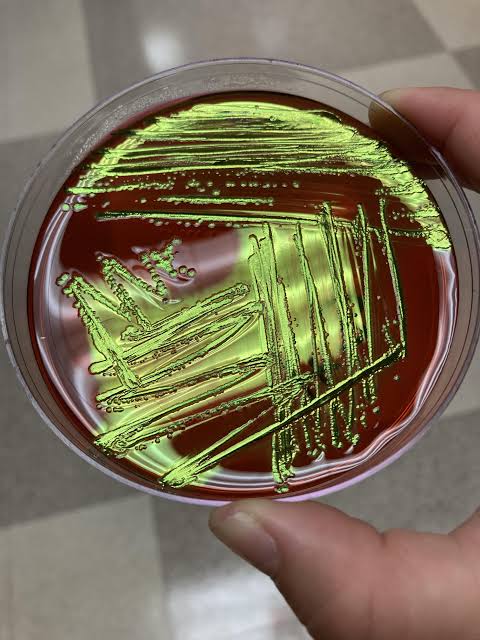
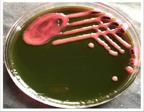
Green Metallic Sheen: Indicates strong lactose fermentation. This is the classic, hallmark appearance of Escherichia coli (E. coli). [1, 2]
Dark Purple / Black Centers: Indicates moderate lactose fermentation, often characteristic of coliforms like Enterobacter aerogenes or Klebsiella pneumoniae. [1, 2]
Colorless / Pink: Indicates non-lactose fermenters, meaning the organism does not ferment lactose. This appearance is typical for pathogens like Salmonella and Shigella. [1, 2, 3]
emb agar expected results
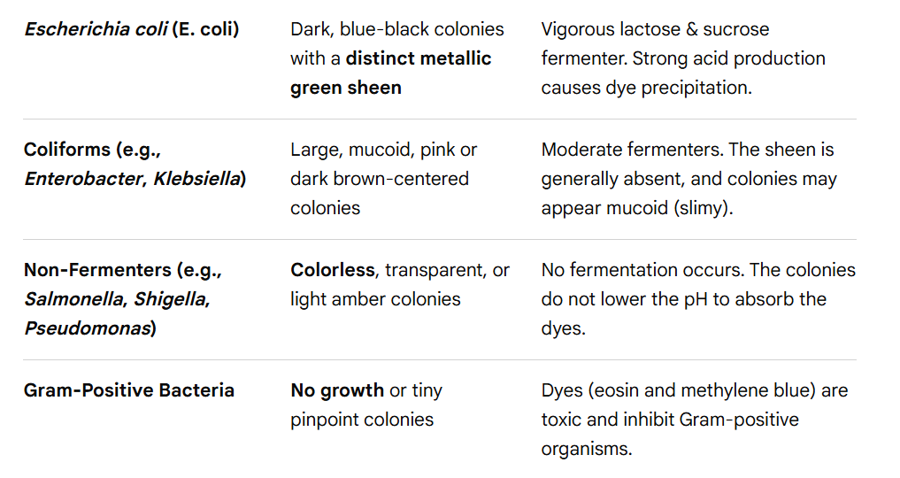
E. coli on EMB agar
Dark, blue-black colonies with a distinct metallic green sheen
Vigorous lactose & sucrose fermenter. Strong acid production causes dye precipitation.
Coliforms (e.g., Enterobacter, Klebsiella) on EMB agar
Large, mucoid, pink or dark brown-centered colonies
Moderate fermenters. The sheen is generally absent, and colonies may appear mucoid (slimy).
Non-Fermenters (e.g., Salmonella, Shigella, Pseudomonas) on EMB agar
Colorless, transparent, or light amber colonies
No fermentation occurs. The colonies do not lower the pH to absorb the dyes.

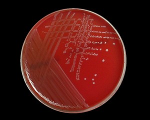
gram-positives on EMB agar
No growth or tiny pinpoint colonies
Dyes (eosin and methylene blue) are toxic and inhibit Gram-positive organisms.
Hektoen Enteric (HE) agar
a selective and differential culture medium used in microbiology to isolate and distinguish between enteric pathogens, particularly Salmonella and Shigella. It is heavily used to process fecal samples, food, and water to diagnose gastrointestinal infections. [1, 2, 3]
![<p><mark>a selective and differential culture medium used in microbiology to isolate and distinguish between enteric pathogens, particularly </mark><em><mark>Salmonella</mark></em><mark> and </mark><em><mark>Shigella</mark></em>. It is heavily used to process fecal samples, food, and water to diagnose gastrointestinal infections. [1, 2, 3]</p>](https://assets.knowt.com/user-attachments/67bd7031-fb82-43d2-947e-6b76137adf3a.jpg)
HE agar selectivity
Bile Salts: HE agar has a high concentration of bile salts. This inhibits the growth of most Gram-positive bacteria.
It allows Gram-negative bacilli (including the normal flora of the intestine and pathogenic targets) to grow. [1, 2, 3, 4]
HE agar differentiation carb fermentation
The Sugars: The agar contains three fermentable carbohydrates: lactose, sucrose, and salicin. [1]
The Indicators: It uses bromothymol blue and acid fuchsin. [1]
The Results:
Non-fermenters (like Salmonella and Shigella) do not ferment these sugars. They use peptone instead, which produces an alkaline environment, turning the colonies blue-green to green.
Fermenters (E. coli, Klebsiella, etc.) quickly ferment one or more of the sugars, producing acid. This lowers the pH and turns the colonies salmon-pink to orange-yellow. [1, 2, 3, 4, 5]
HE agar Differentiation (Hydrogen Sulfide H₂S Production)
The Indicators: HE agar includes sodium thiosulfate (a sulfur source) and ferric ammonium citrate. [1]
The Results:
Some bacteria (like Salmonella) reduce sulfur to hydrogen sulfide gas (H₂S).
The H₂S reacts with the iron, creating a distinct black precipitate in the center of the colony. [1, 2]
HE agar interpreting results
You can identify bacteria based on their distinct colony appearances: [1, 2]
Shigella: Transparent or green colonies; raised with smooth edges. They do not ferment sugars or produce H₂S.
Salmonella: Blue-green to transparent colonies with distinct black centers. They do not ferment sugars but do produce H₂S.
Coliforms (e.g., E. coli): Bright salmon-pink to orange-yellow colonies. They are aggressive fermenters of the sugars. [1, 2, 3, 4, 5, 6]
how HE agar works
a selective and differential culture medium used to isolate and distinguish gastrointestinal pathogens like Salmonella and Shigella from mixed fecal, food, or water samples. It ensures pathogen recovery by inhibiting normal flora and using color indicators to separate fermenters from non-fermenters. [1, 2, 3]
HE agar purpose
Hektoen Enteric (HE) agar is a selective and differential medium primarily used to isolate and differentiate gastrointestinal pathogens like \(Salmonella\) and \(Shigella\) from clinical, food, and water samples. It inhibits normal intestinal flora while allowing pathogens to grow and display distinct visual markers. [1, 2, 3, 4]
HE agar expected results
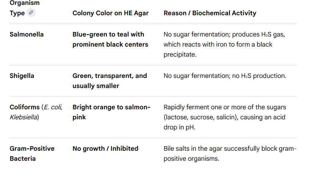
Salmonella on HE agar
Blue-green to teal with prominent black centers
No sugar fermentation; produces H₂S gas, which reacts with iron to form a black precipitate.
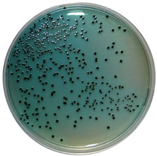
Shigella on HE agar
Green, transparent, and usually smaller
No sugar fermentation; no H₂S production.
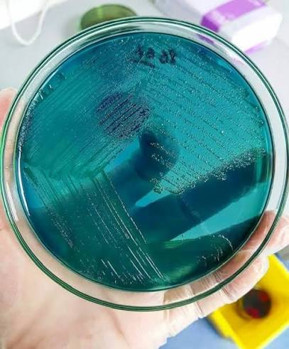
Coliforms (E. coli, Klebsiella) on HE agar
Bright orange to salmon-pink
Rapidly ferment one or more of the sugars (lactose, sucrose, salicin), causing an acid drop in pH.
Gram-Positive Bacteria on HE agar
No growth / Inhibited
Bile salts in the agar successfully block gram-positive organisms.
motility test
determines if a microorganism is capable of self-propelled movement, usually via flagella. The most common method involves stabbing a semi-solid agar medium. Motile bacteria spread outward from the stab line, while non-motile bacteria are confined strictly to it. [1, 2, 3, 4, 5]
how motility test works
The Medium: Unlike standard solid agar plates (which use 1.5% agar), motility test media use a much lower concentration (around 0.4%). This creates a semi-solid, gelatinous environment that restricts non-motile bacteria but is soft enough for motile bacteria to swim through. [1, 2]
Inoculation: A straight, sterile inoculating needle is used to pick up a bacterial colony and stab it directly down the center of the tube, stopping halfway before hitting the bottom. [1, 2]
Incubation: The tubes are incubated at the optimum temperature (typically 35°C to 37°C) for 24 to 48 hours to allow the bacteria to multiply and migrate. [1, 2, 3, 4]
interpreting results for motility test
Positive (Motile): The bacterial growth spreads outward from the central stab line, creating a cloudy, hazy, or "diffuse" zone of growth that may fan out through the entire tube. [1, 2]
Negative (Non-motile): The bacteria only multiply exactly along the line where the needle stabbed them. The surrounding agar remains perfectly clear, and the line of growth has sharply defined, clean edges.
motile positive
The bacterial growth spreads outward from the central stab line, creating a cloudy, hazy, or "diffuse" zone of growth that may fan out through the entire tube. [1, 2]
![<p>The bacterial growth spreads outward from the central stab line, creating a cloudy, hazy, or "diffuse" zone of growth that may fan out through the entire tube. [1, 2]</p>](https://assets.knowt.com/user-attachments/a58fa78d-3691-4a38-936a-a351858ca02b.jpg)
motile negative
The bacteria only multiply exactly along the line where the needle stabbed them. The surrounding agar remains perfectly clear, and the line of growth has sharply defined, clean edges.
motility test possible errors
«Fanning» the needle: If the inoculating needle is not withdrawn along the exact same vertical line used to stab the semi-solid agar, or if it is wiggled around, it creates a wide, jagged track. Non-motile bacteria can grow along this disrupted area, mimicking a motile "cloudy" result and causing a false-positive. [1, 2]
Incomplete stabbing: Failing to stab the medium to about half or two-thirds of its depth may not provide the right microaerophilic environment or depth for the bacteria to demonstrate true movement, often leading to poor growth or a false-negative. [1, 2]
motility test procedure
1. Label the tubes with your initials and the initials of the bacteria.
2. Aseptically inoculate a tube with S. flexneri using a sterile inoculating needle. With the needle, inoculate 3/4 of the way to the bottom of the tube and pull the needle straight back out. Do no inoculate a second time.
3. Repeat the previous step with S. enterica.
4. Place the tubes at 37°C for 24 to 48 hours.
5. Observe the tubes for color and how much color has spread.