AP03: The sphenoid bone and orbit
 Call Kai
 Learn
Learn Practice Test
 Spaced Repetition
 Match
Match Flashcards
 Knowt Play
1/24

There's no tags or description
Looks like no tags are added yet.
Name | Mastery | Learn | Test | Matching | Spaced | Call with Kai | Chat |
|---|
No analytics yet
Send a link to your students to track their progress
25 Terms
CNV
trigeminal nerve
What are the 3 divisions of the trigeminal nerve
CNV1-ophthalmic
CNV2- maxillary
CNV3- mandibular
What are the contents of the optic canal
optic nerve CNII, ophthalmic artery
Function of the optic canal
vision- arterial supply to the orbit
Contents of the foramen rotundum
maxillary division of CNV
Function of the foramen rotundum
sensory innervation to skin over cheeks, part of oral cavity, hard palate, nasal cavity
Contents of the foramen ovale
mandibular division of CNV (trigeminal nerve)
also accessory meningeal artery, emissary vein, lesser petrosal nerve
Function of the foramen ovale
motor innervation to muscles of mastication and others;
sensory innervation to mandibular teeth, skin over mandible and lower lips
Contents of the foramen spinosum
middle meningeal artery and vein
meningeal branch of CNV3 (nervus spinosus)
Functino of the foramen spinosum
neurovascular supply to the meninges
Contents of the superior orbital fissude
CNIII- oculomotor nerve
CNIV- trochlear nerve
CNVI- abducens nerve
ophthalmic division of CNV (frontal, nasociliary and lacrimal branches)
superior ophthalmic veins
Function of the superior orbital fissure
-transmits CN III, IV, VI and V1, allowing motor control of extraocular muscles, parasympathetic control of pupil and accommodation, sensory innervation of the eye and orbit, and venous drainage via the superior ophthalmic vein
Contents of the inferior orbital fissure
infraorbital nerve (CNV2)
zygomatic nerve (CNV2)
infraorbital artery (from maxillary artery)
inferior ophthalmic vein
Function of the inferior orbital fissure
infraorbital nerve provides sensory innervation to the skin inferior to the eye
zygomatic nerve innervates skin over cheeks and temple via branches
Bones found on the roof of the orbit
-orbital plate of the frontal bone
Floor of the orbit
-orbital surface of the maxilla
-zygomatic bone
-palatine bone
Lateral wall of the orbit
-frontal process of the zygomatic bone
-greater wings of the sphenoid
Medial wall of the orbit
-frontal process of maxilla
-lesser wing of sphenoid
-orbitsl plate of ethmoid
- lacrimal bone
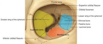
Which soft tissues of the orbit may be damaged if there is a blow-out fracture
eyeball
extraocular muscles
infraorbital nerve
What is a blowout fracture
break in the thin bones of the orbit (usually the floor or inner wall)
Which bony walls of the orbit are likely to be fractured in the event of a blowout fracture?
-floor of the orbit (very thin, likely to fracture)
-medial wall of the orbit (ethmoid bone) is also very thin and may fracture
Clinical symptoms of a blowout fracture
-pain, swelling, bruising (black eye)
-diplopia (double vision) due to cranial nerve palsies (paralysis of cranial nerves) specifically CN III/ oculomotor, IV/trochlear and VI/abducens
What is the optic chiasm
X-shaped structure at the base of the brain where optic nerve fibers from each eye cross over
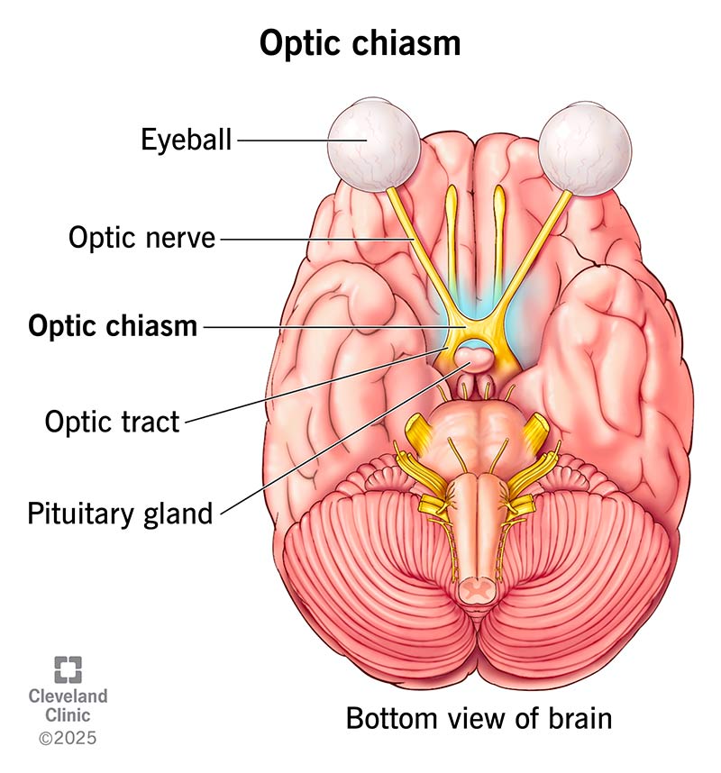
Where is the optic chiasm located
superior to the pituitary gland and the diaphragma sellae
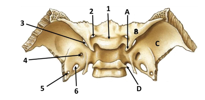
Label this diagram of the sphenoid bone
1- optic groove
2- optic canal
3- superior orbital fissure
4- foramen rotundum
5- foramen spinosum
6- foramen ovale
A- anterior clinoid process
B- lesser wing of sphenoid
C- greater wing of the sphenoid
D- posterior clinoid process