Histology- Exam #1
 Call Kai
 Learn
Learn Practice Test
 Spaced Repetition
 Match
Match Flashcards
 Knowt Play
1/118

There's no tags or description
Looks like no tags are added yet.
Name | Mastery | Learn | Test | Matching | Spaced | Call with Kai | Chat |
|---|
No analytics yet
Send a link to your students to track their progress
119 Terms
What is histology?
The study of tissue
What is a tissue?
a group of cells that share the same function

What is histopathology?
the study of tissue failure
(histo= tissue and pathos= suffering)
Who is Marcello Malpighi?
lived 1628-1694
italian doctor
father of histology
first to discover capillaries
Who was Marie Francois Xavier Bichat?
lived 1771-1802
french anatomist
recognized that organs were composed of multiple tissues
first to name tissues
What are skills that make a good histologist?
observant/attention to detail
organized (multitasking)
strong background in (bio)chemistry
teamwork
patience
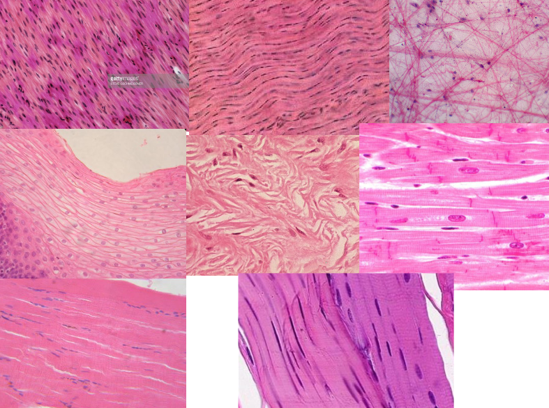
What stain was used for these slides?
H&E (hematoxylin & eosin)
What are the different histology jobs?
clinical/research settings
hospitals
universities
veterinary hospitals
state and federal agencies
e.g. CDC, department of health
professor
private industry- R&D, contract work
pathologists (MD)
What is parental investment?
provisioning of resources to offspring at expense of future offspring
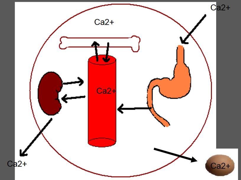
What is Calcium important for?
intracellular communication
nerve transmission
muscle contraction
hemostasis
pH buffer
structural component of bone
What are the types of microscopy?
light (bright-field microscopy)
polarizing
phase-contrast
dark-field
fluorescence
confocal
electron
atomic force
What kinds of microscopy can be used with unstained cells?
phase-contrast
dark-field
Describe light (bright-field) microscopy.
reflective or transmitting
limited to dark or strongly contrasting objects
max resolution is 0.2 μm

Describe polarizing microscopy.
extension of bright-field microscopy but with filters
looking for high ordered objects
can be used to detect silicosis

What kind of microscopy should be used to detect silicosis?
polarizing microscopy
What is amyloidosis and what mode of microscopy is used to detect it?
amyloidosis: abnormal deposition of amyloid proteins in tissue and organs
amyloids are highly-ordered with lots of beta sheets
examples of diseases associated with amyloidosis: alzheimers, type 2 diabetes, parkinsons, atherosclerosis, TSE (prion based disease)
amyloids stained with congo red
detected with polarizing microscopy

Describe phase-contrast microscopy.
makes dense areas more bright
reveals many structures without need for staining
thus can be performed on unstained (i.e. living) cells
can be used for cell culture research
appear as bright and dark areas
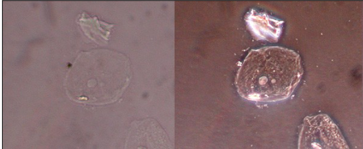
Describe darkfield microscopy.
also used with unstained cells
light directed at specimen from indirect angles. only scattered light reaches objective lens
field of view appears black

Describe fluorescence microscopy.
not usually used for naturally fluorescent molecules
used in cell biology/ histology to:
establish the intracellular distribution of a protein
characterize the dynamics of the protein distribution
suggest potential functions of the protein
immunofluorescence techniques

What is fluorochrome?
dye that fluoresces when excited by UV
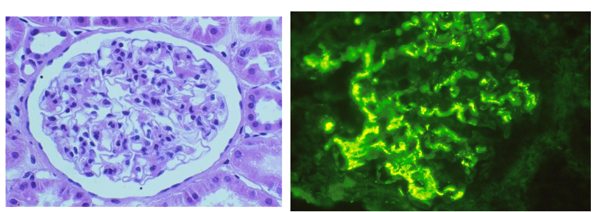
What is the disease we discussed in class that can be detected using immunofluorescence techniques?
IgA nephropathy (Berger’s Disease)
IgA is most commonly found in mucous membranes
nephropathy = pathology of the kidney
most common form of adult glomerulonephritis (inflammation of the glomerulus)
IgA clumps together and causes backup in glomerulus
What are the types of immunistaining?

Describe confocal microscopy.
microscope that increases contrast and resolution by eliminating out of focus light
eliminates out of focus light by only allowing one plane to be visible
give a better “3-D” perspective
in focus = in focal plane

Describe electron microscopy and the two types.
use electrons instead of light
highest resolving power
limitations: skill set
types:
transmission electron microscopy: 2D, allows you to see organelles
scanning electron microscopy: 3D images

What are the steps of tissue slide production?
tissue fixation
tissue processing
dehydration
clearing
embedding
sectioning
mounting
staining
coverslipping
What is tissue fixation and it’s functions?
fixation = tissue preservation, ideally keeps cell in position it was in when you obtained the sample
function:
1. kills tissue to resist:
putrefaction: bacterial attack
autolysis
2. maintain proper relationship between cells and extracellular components (keeps cell from getting smushed)
3. aids in contrast among cell components
4. aids in staining process
How does tissue fixation work? How are fixatives classified?
actions- protein stabilization
renders enzymes inactive
primary method is use of chemicals
classified as:
1. additive or non-additive fixative
and as:
2. coagulant or non-coagulant fixative
What is the difference between an additive and a non-additive fixative?
additive fixative: a chemical that chemically binds to protein structure—link
non-additive fixative: doesn’t bind or link; all non-additives must be coagulants
What are some common fixatives?
non-additives:
alcohols
acetone
acetic acid
these are very fast, but harsh on tissues (causes shrinkage and makes them brittle)
non-coagulants:
formaldehyde
glutaraldehyde (good for electron microscopy)
coagulants:
picric acid (trichrome stains)
alcohols
acetone
What is one of the best fixatives?
10% NBF (neutral buffer formalin)
What are some factors affecting fixation quality?
temperature
size of tissue (easier to fix through small diameter)
volume ratio (should be 10-20x more fixative than tissue)
time
since death
duration of fixation
underfix= staining and decay problems
overfix= hardening
choice of fixative
penetration- goes along with size of tissue
storage
pH
neutral, most fixatives are buffered
osmolality= staining of fixative
What are the steps of tissue processing?
dehydration
clearing
infiltration
embedding
Describe the dehydration step of tissue processing.
process of removing water from tissue to allow for infiltration of embedding media
embedding media, is water insoluble and thus won’t infiltrate hydrated tissues
What are some common dehydrating reagents?
alcohols
ethanol (the best one)
methanol
isopropanol
butanol
acetone
universal solvents- dehydrate and clear (the next step)- faster
dioxan
tertiary butanol
tetrahydrofurane
What is the best dehydrating agent? How is it used?
ethanol
fast procedure
ethanol (95%)
ethanol (absolute)
problems- alcohol > 70% precipitates phosphate salts making sectioning difficult
better procedure
ethanol (60-65%)
ethanol (95%) 2X
salt precipitation
ethanol (absolute) 2X
must be replaced at certain time intervals
Describe the clearing step of tissue processing.
clearing: renders tissues transparent
removal of dehydrating alcohols to allow for proper infiltration of embedding media
commonly used:
xylene- best, aromatic hydrocarbon, quickly dissolves fats
toluene
benzene
chloroform
acetone
What is the best clearing agent?
xylene, but its unsafe
Describe the infiltration step of tissue processing.
process by which clearing agent is replaced by embedding medium
most common embedding media:
paraffin- needs to be melted; wanna go 2-3 degrees C above melting point → any hotter will harden tissue
water-soluble waxes
celloidin
plastics- epoxy resins, glycol methacrylate
process:
paraffin heated a few degrees above melting point
cleared tissue is immersed often under a vacuum
Describe the embedding step of tissue processing.
process of placing the infiltrated tissue into a block of the embedding medium
proper orientation is key!
when cutting long things, need to orient at oblique angle
Describe the sectioning step of tissue processing.
microtome
rotary
sliding
cryostat
frozen
widely used in clinical setting
used when sample is stained with antibody
quicker and preserves protein structure better to allow antibody staining
cell quality is lower though
tissue ribbons
hard to achieve

What is Mohs surgery?
multi-step procedure to remove cancer without removing non-cancerous tissue
fast, but downside is poor cell quality
routine sectioning method yields better cell quality
Describe the mounting step of tissue processing.
slides are coated with an adhesive to prevent tissue from falling off during subsequent staining
poly-L-lysine
elmer’s glue
albumin
these three (above) are additives for water bater
OR
(+) charged slides
tissue is placed in a floatation bath
temp is a few degrees below melting point
when too hot, causes cracking
Xylene is most often used as a(n)
clearing agent
Describe the staining step of tissue processing. What primary stains are used?
goal of staining is to increase contrast of cellular structures
primary stains: hematoxylin & eosin (H&E)
eosin is an acidic dye
carries a net (-) charge so binds (+) charged molecules
pink color
hematoxylin mimics basic dye
binds to (-) charged particles (such as the phosphate backbone of DNA)
counterstain
blue color
nuclear stain
net (+) charge
always used in conjunction with a mordent (metal/salt)
stain some proteins; have (-) charged carboxyl group
stains some sulfate groups on glycosaminoglycans (sugar protein polymer)
because it’s a basic dye, anything it stains is “basophilic”= loving basic dyes
ex: basophils
stains a deep purple
eosinophilia / acidophilia = binds well to eosin/acidic dyes
stains red
ex: eosinophils

What is the haematoxylon campechianum?
logwood tree
endemic to central america
cultivated in Jamaica since 1715
hematein- is oxidized form
used in pap smears
What is a sample H&E procedure?
look at slides
essentially dewaxing and hydrating your tissue
“blueing” = make tissue look blue by toning down purple color
blue jean blue
1. dewax
2. hydrate
3. blueing
4. dehydrate
5. coverslip
What is progressive vs. regressive staining techniques?
progressive: requires more checking to achieve the perfect staining
regressive: overstaining, requires blueing to achieve the perfect stain
Describe Periodic acid-Schiff stain (PAS).
specialized stain
stains basement membrane, sugars & mucins
Describe trichrome stains.
specialized stains
Gomori, Mallory, Masson’s
useful for collagen staining → vivid blue; distinguish muscle & connective tissue
Describe Silver stain.
specialized stain
used for staining neurons
stains black
What is Wrights stain used for?
blood
Describe Orcein stain.
specialized stain
ver hoeff
for elastic tissue
stains black
Describe the last step of tissue slide processing.
coverslipping
permount
allows for higher quality of slide
T or F: Positively charged molecules are readily stained by basic dies
false
Biomolecules/cell structures that are stained by acidic dyes are called __?
eosinophilic / acidophilic
What is an example of cell specialization?
example: skeletal muscle
makes a tremendous amount of actin because it’s needed for muscle movement
What are the levels of organization?
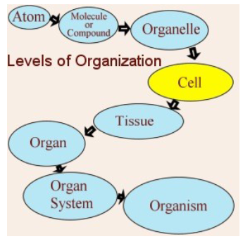
What is the cytoplasm and it’s components?
cytoplasm: non-nuclear portion of the cell
includes:
organelles: little organs
cytoplasmic matrix: mostly water, has ions and organic metabolites
What are the membrane-bound organelles in the cell?
plasma membrane
endosomes
lysosomes
transport vesicles
rough ER
smooth ER
golgi apparatus
mitochondria
peroxisomes
Describe the plasma membrane of the cell.
sets boundaries
fluid mosaic
phospholipid bilayer
compartmentalizes the cell
allows things to be isolated/stored
could have microvilli attached
has peripheral proteins that provides linkage to cytoskeleton
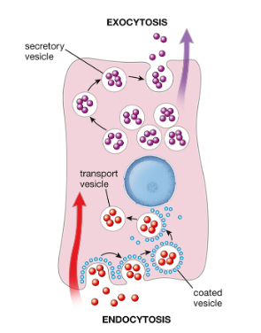
How are endosomes, lysosomes, and transport vesicles formed?
all formed from invagination of cell membrane
Endocytosis and exocytosis need to be __ over time!
balanced

What is an endosome?
membrane bound vesicle formed as a result of endocytosis
protein sorting
What is a lysosome?
forms as a result of maturing endosomes accumulating acid
recycling center
have enzymes that degrade organic molecules
ruptured = causes autolysis
Describe exocytosis.
exocrine glands
pepsinogen is one of the enzymes released
when looking at exocrine glands, they display eosinophilia because they are waiting to release their contents
Describe rough endoplasmic reticulum.
has ribosomes on its surface
mRNA translation
for protein synthesis
basophilic
ex: nissl bodies on a neuron, displays heavy protein synthesis

Describe smooth endoplasmic reticulum.
for lipid synthesis
steroid metabolism
glycogen metabolism
no ribosomes on surface

Describe the golgi appratus.
“cell warehouse”
protein modification
doesn’t stain well

What are peroxisomes?
for oxidation → detoxification in cells (kidney, liver)
degrade fatty acids
Describe the mitochondria.
can use janus green to stain
metabolism
cell that has high metabolic demands have more numerous mitochondria

What are the membrane-free organelles in the cytoplasm?
cytoskeletal elements
microtubules
actin
intermediate filaments
centrioles
ribosomes
Describe actin.
two forms:
fibrous
globular
for muscle contractions

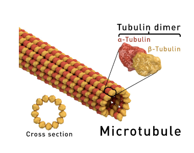
Describe microtubules.
larger in size, quickly assemble and disassemble
important for mitosis and meiosis
cilia beating
highways in cells to move transport vesicles
associated with flagella and sperm
What are centrioles?
microtubule organizing center

What are intermediate filaments?
structural role in cells, no movement
What are the components of the nucleus?
chromatin
nucleolus
nuclear envelope
nucleoplasm
What is chromatin? What are the forms?
DNA-protein complex
two forms:
euchromatin: dispersed, unfolded DNA
unzipped DNA used for replication and transcription
heterochromatin: dense, folded
Describe the nucleus.
membrane-bound organelle
houses DNA (an extremely long molecule)
DNA packed tightly around protein
How can you identify plasma cells?
have a clock-like pattern around nucleus (dot in center, ring around edge because of chromatin)
What are the 4 tissue classes?
epithelium- boundary between outside and inside world; is protecting something
connective tissue- link tissues together
muscle tissue- movement
nerve tissue- specialized, long term communication
Where are the general places you will find epithelium tissue?
surface of body
lines internal cavities (with the exception of joints)
tubes that communicate with outside world
glands/ducts
What are the functions of epithelial tissue?
secretion (ex: mucus)
absorption (ex: sugar, nutrients)
transportation (ex: cilia)
protection (ex: skin)
communication/ receptor function (ex: taste buds)
What are the characteristics of epithelial tissue?
free surface
avascular: no direct blood supply
basement membrane
morphological polarity: unique functions in different parts of the cell
tightly bound cells
What is cell polarity? What are the regions?
unique cellular function in different parts of the cell
regions/domains:
apical
lateral
basal

Describe the apical domain of epithelial cells.
vary among epithelium
can have cilia, microvilli, etc.
if have microvilli = increased surface area
efficient transport
all microvilli filled with actin filaments
pumps / transporters, enzymatic roles
variability in size and density of microvilli
cilia = larger projections than microvilli, microtubules found in cilia provide ability to move entire structure
trachea = pseudostratified
What is Immotile Cilia Syndrome (ICS)?
affects cilia of certain epithelial tissue
abnormal microtubules
dynein arm connects microtubules
affects ~1/ 20,000 individuals
common symptoms:
infertility (both genders but mostly men because it affects sperm swimming)
swelling of ventricles in the brain
respiratory distress = kartageners syndrome
know picture!!

Describe the lateral domain of epithelial cells.
adjacent cells linked to tissue
CAM = cell adhesion molecule
tight junctions: controlling/barriers to paracellular passage
Describe the junctions on the lateral domain of epithelial cells.
tight junctions
link to cytoskeleton by actin filaments
controlling/barriers to paracellular passage
anchoring junctions = desmosomes
maintain tissue integrity
structural role
have intermediate filaments
gap junctions
cell-cell communication
ex: cardiac and smooth muscle cells (Ca2+)

Describe the basal domain of epithelial cells.
epithelial links to underlying connective tissue
PAS = periodic acid schiff
stains basement membrane and carbohydrates
How is epithelium classified?
layers:
simple
stratified
shape
squamous
cuboidal
columnar
special
pseudostratified
transitional
the structure dictates function!

Describe simple squamous epithelium, where its found, and its function.
epithelium = layer of outside tissue
found in blood vessels, kidney (Bowman’s capsule), alveoli in lungs, body cavity, capillaries
on fragile tissues
not a great barrier
function: exchange because thin and one layer thick

What is atherosclerosis?
atherosclerosis = formation of plaques under vessel
buildup of cholesterol, immune cells, fibrotic tissue (= collagen), cellular waste
plaques can break off and block vessels → heart attack
vessels lined with simple squamous epithelium
risk factors:
old age
obesity
smoking
diabetes
sedentary lifestyle
hypertension
hyperlipidemia = excessive LDL’s
Where is simple cuboidal epithelium found, and what is it’s function?
found in kidney tubules, ovary, hormone glands, ducts of exocrine glands
function: absorption or secretion
not really protective
mostly for transport
Where is simple columnar epithelium found, and what is it’s function?
found in small intestine (digestive tract)
function: absorption & secretion
taller than they are wide
one nuclei/ cell
do a lot more transport/ secretion because of large cell size
only one cell thick so easy to move things across without blockage
Where is stratified squamous epithelium found, and what is it’s function?
wet areas = non-keratinized
vagina, esophagus, mouth
dry = keratinized
skin
moisture loss prevention and protection
function: protection
cells at top definitely doesn’t touch basement membrane, so = stratified
divides from bottom up
Where is stratified cuboidal epithelium found, and what is it’s function?
locations: found in sweat gland ducts
function: barrier, conduit, secretion
always only 2 layers thick
difficult to find
Where is stratified columnar epithelium found, and what is it’s function?
locations: largest ducts of exocrine glands
rare, found in eye
function: barrier, conduit
Where is pseduostratified epithelium found, and what is it’s function?
found in trachea
function: absorption, secretion, movement
looks very stratified but isn’t because all cells are touching the basement membrane
changes shape to fit more cells
always ciliated
cells don’t sit straight
Where is transitional epithelium found, and what is it’s function?
found in urinary bladder, ureter, and urethra
function: barrier and stretchability
done by layering of different cell sizes
top layers bigger than underneath (top cells = umbrella cells)
stratified
Describe epithelioid tissue.
“epithelium-like”
lack a free surface
usually associated with endocrine function
ex: Leydig cells of testis
function: hormone (endocrine) production/release
vascularized (has blood vessels)
found in testes, ovaries, thyroid, pancreas (islets), pituitary