2.08 Visual Pathways 2
1/109
There's no tags or description
Looks like no tags are added yet.
Name | Mastery | Learn | Test | Matching | Spaced |
|---|
No study sessions yet.
110 Terms
Layers of the retina from anterior to posterior
Internal limiting membrane
Nerve fiber layer
Ganglion cell layer
Inner plexiform layer
Inner nuclear layer
Outer plexiform layer
Outer nuclear layer
External limiting membrane
Photoreceptor layer
Retinal pigment epithelium
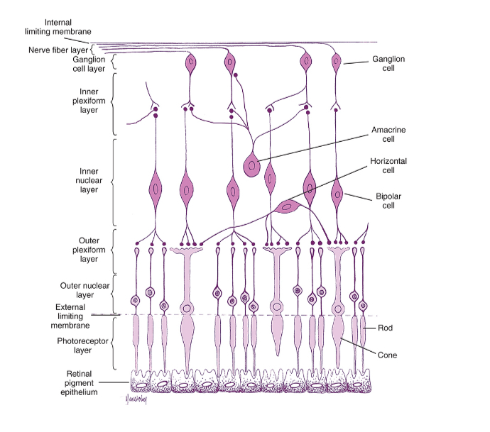
What layer of the retina are glanglion cells found
Ganglion cell layer duhh
What layer of the retina do amacrine cells operate
Inner plexiform layer (where bipolar cells and retinal ganglion cells form synapses)
What layer of the retina are horizonal cells found
Inner nuclear layer
What layer of the retina are bipolar cells found
Inner nuclear layer
What layer of the retina are rods and cones found
Photoreceptor layer
How do photoreceptors, bipolar cells and ganglion cells work together
Rods and cones (photoreceptors) respond to light
The bipolar cells process the signal and send a signal to the ganglion cells
Ganglion cells form the output of the retina
These neurones axons form the optic nerve which leaves the retina via the optic disc
What is the visual receptive field of a neuron
The visual receptive field of a neuron is the region of the visual field in which a stimulus can modulate the firing of the neuron
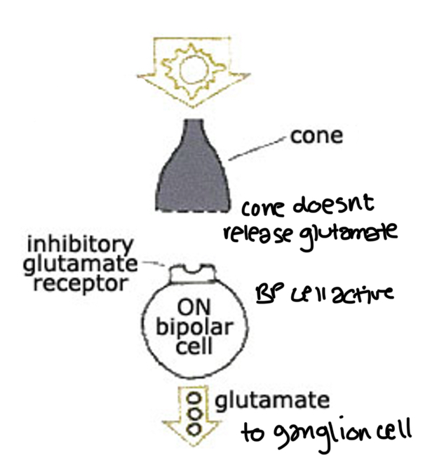
Response of on bipolar cells in the light
On bipolar cells are active in the light
Cones release less/no glutamate (neurotransmitter)
On BP cell has an inhibitory receptor so if theres glutamate it inhibits the BP cell
But since theres no glutamate released, the BP cell is active - its not inhibited so it releases neurotransmitter to ganglion cells
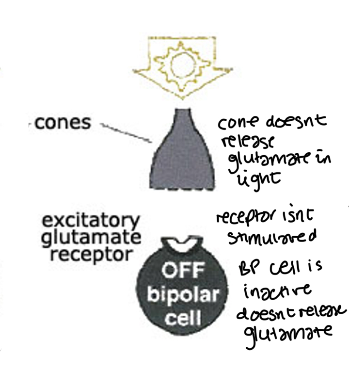
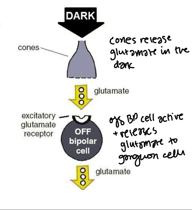
Response of off bipolar cells in light
Off bipolar cells are inactive in light/active in dark
When theres light, cones release less/no glutamate (neurotransmitter)
Off BP cells have an excitatory glutamate receptor
Since no glutamate is released in the light the off bipolar cell receptor is not excited, the BP cell is inacitve and doesnt release any neurotransmitter
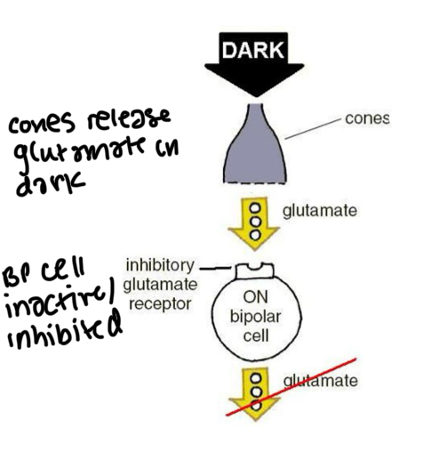
Response of on bipolar cells in the dark
On bipolar cell is inactive in the dark
Cones release glutamate
On BP cells have an inhibitory glutamate receptor so since cones release glutamate to BP cell it inhibits the BP cell
BP cell doesnt release glutamate to ganglion cells
Response of off bipolar cells in the dark
Off BP cells are active in the dark
Cones release glutamate in the dark
Off BP cells have exitatory glutamate receptor so since glutamate is released it excites the off BP cell/ makes it active so BP cell releases glutamate to ganglion cell
Which bipolar cells respond to light images against a dark background
On bipolar cells have inhibitory glutamate receptors so they respond to light images against a dark background
Which bipolar cells respond to dark images on a bright background
Off bipolar cells have excitatory glutamate receptors so respond to dark images on a bright backgrouns
On bipolar cells connect with _
ON bipolar cells connect with ON ganglion cells
(And off BP cells connect with OFF ganglion cells)
What do ON and OFF ganglion cells form
They form the output signals from the retina
What do horizontal cells do
They carry out horizontal processing (at the photoreceptor synapse) which helps refine the responses in the ON and OFF pathways
What do amacrine cells do
Increase the quality of signals
Rods only have _ bipolar cells
On
Which cells in the retina generate action potentials
Only ganglion cells
What are the types of ganglion cells
Magnocellular
Parvocellular
Bistratified/koniocellular
(More detail in eye intro 2)
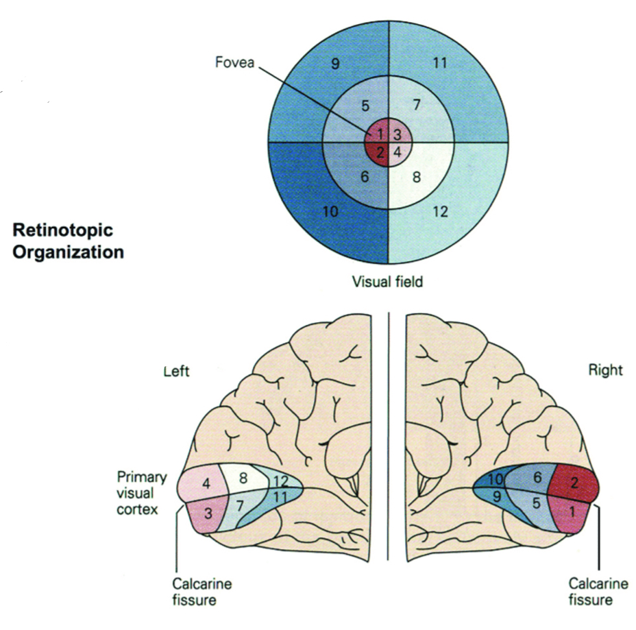
Where does the thalamus transfer sensory information
To the cerebral corticies
What is the main component of the thalamus
The dorsal lateral geniculate nucleus (dLGN/LGN)
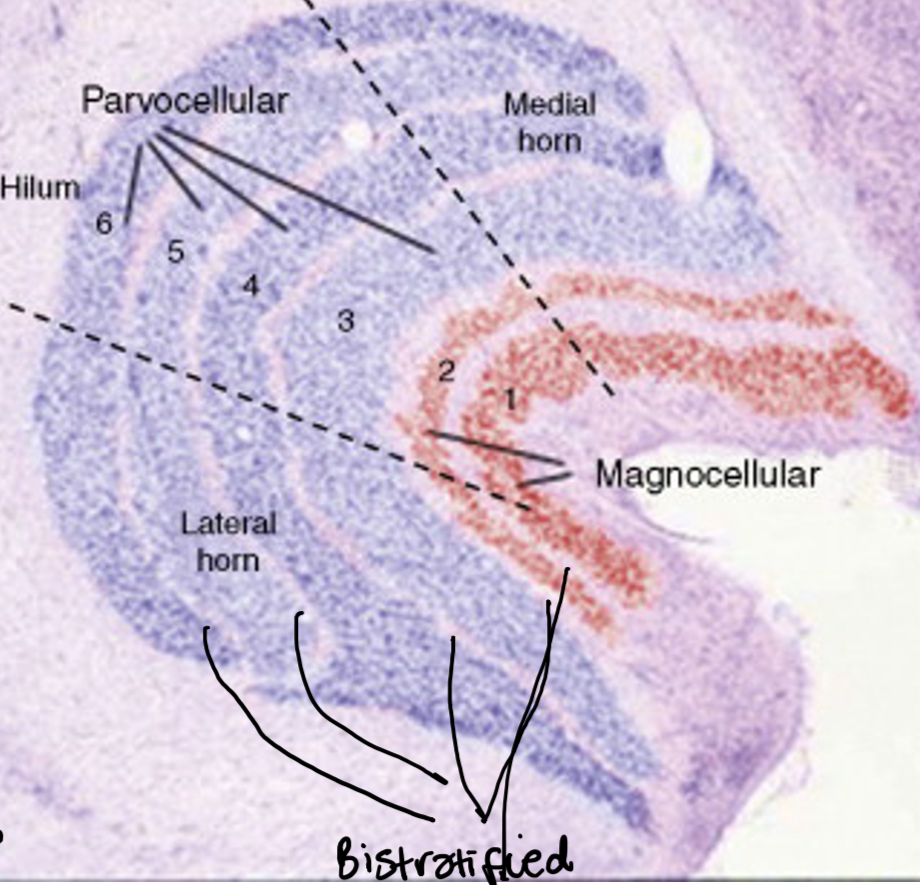
Which ganglion cells project to which layers of the LGN
Layers 1 and 2 = magnocellular cells
Layer 3 to 6 = parvocellular cells
Bistratified cells project to cells between the layers called konicellular
Each layer recieves retinal input from only one eye - This means the same visual space is represented multiple times in each LGN
Right visual field = left LGN
Left visual field = right LGN
What is the retintopic map
Nearby cells in each layer of the LGN represent a nearby location in the visual fiels
Where do 90% of retinal outputs terminate
In the dLGN
Where do 10% of retinal outputs terminate
To non visual functions eg circadian rhythm
What is the magnification factor
Fovea has a larger respresentation than peripheral retina (around half the mass of the LGN represents fovea bc it has the highest density of cones)
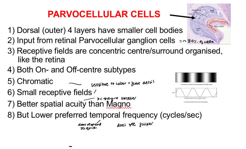
Parvocellular cells (LGN) :
cell body size
Receive input from where
Receptive fields organisation
Subtypes
Chromatic or achromatic
Receptive field size
Good or bad Spacial acuity
Preferred temporal frequency
Magnocellular cells
cell body size
Receive input from where
Receptive fields organisation
Subtypes
Chromatic or achromatic
Receptive field size
Good or bad visual acuity
Preferred temporal frequency

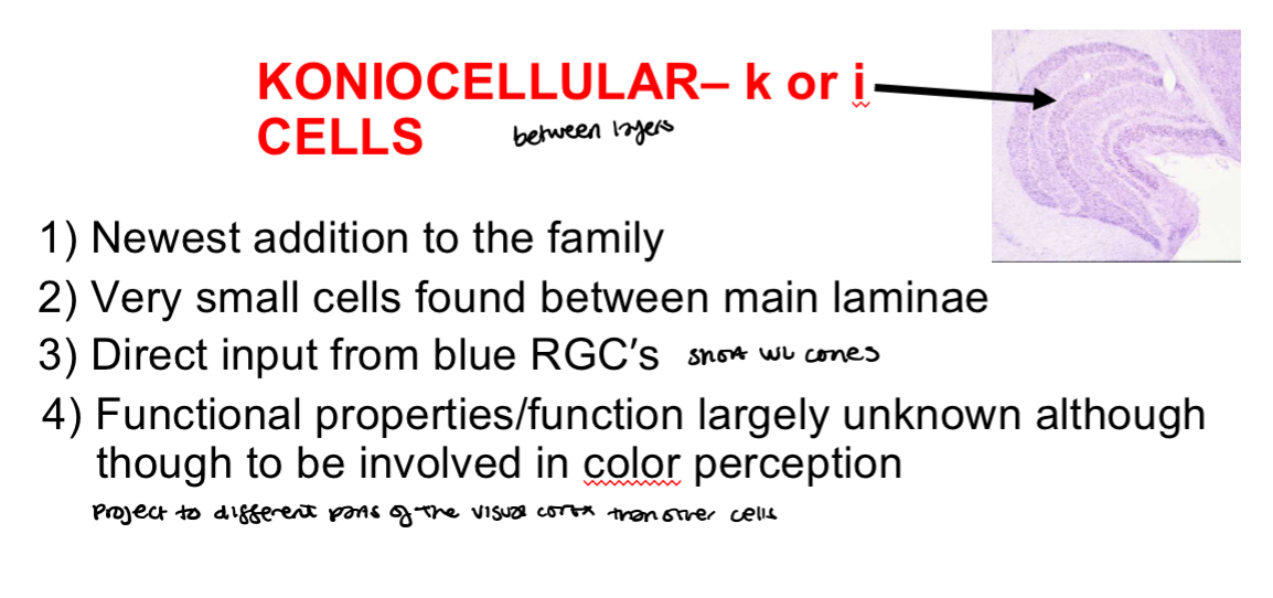
Koniocellular cells (LGN)
cell size
Input from where
Function
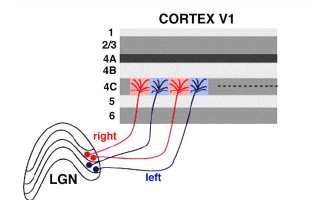
Where do axons leaving the LGN go
They make up the optic radiations that project (mainly) to the primary visual cortex (striate cortex)
What is the neocortex
The outer layer of the brain
How many layers in the neocortex
6 horizontal layers (but can be subdivided)
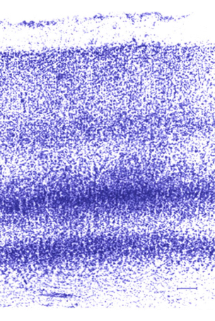
what stain is used to see cell bodies
Nissl staining
Cells in layer 1 of the visual cortex
The outer surface is celll sparse (arent many cells)
The axons go to the next cortical layer
Cells in layer 2 of cortex
The external granule layer
Mainly small spherical cells
Cells in layer 3 of cortex
The external pyramidal layer
Variety of cell tyes, mainly pyramidal cells, cells deeper in the layer are larger than the superficial layers
Cells in layer 4 of cortex
(Internal) granule cell layer
Mainly granule cells
Cells in layer 5 of cortex
The internal pyramindal cell layer
Ppyramidal cells - Cells are typically larger than layer 3
Cells in layer 6 of cortex
Polymorphic or multiform layer
Heterogenous mix. ‘Blends’ into white matter
Which layers of the cortex are supragranular
1, 2 and 3
Which layers of the cortex are infragranular
5 and 6
Which layers of the cortex are granular
4
Function of layer 2 and 3 of the cortex
Project info to extrastriate cortex
Recieves input from konicellular cells
Contains colour tuned cells within the CO blobs and orientation tuned cells outside the blobs (both project to V2 separately)
Function of layer 4A of the cortex
Recieves input from magnocellular and parvocellular cells
Function of layer 4B of cortex
Projects info to extrastriate cortex (processes motion)
Associated with motion perception - Contains direction selective cells
Function of layer 4Ca of cortex
Recieves input from magnocellular cells
Info process in 4B
Function of layer 4Cb of cortex
Receives input from parvocellular cells
Info processed in 2/3
Function of layer 5B of cortex
Projects/output to superior colliculus
Function of layer 6 of cortex
Projects/Feedbacks to LGN - Helps modulate responses eg helps you pay more attention to something or enhance response to stimulus
Receives secondary inputs from magnocellular and parcocellular cells
Projects/outputs info to extrastriate cortex
What is retinotopy
the way visual information from the retina is mapped onto areas of the brain, especially the visual cortex. Neighboring parts of the retina connect to neighboring neurons in the brain, preserving the spatial layout
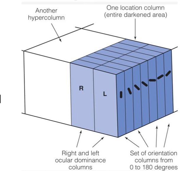
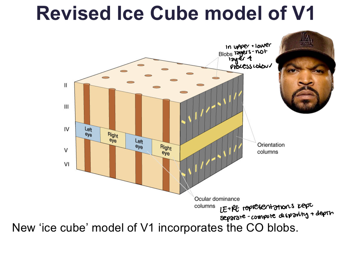
What is ocular dominance
Left eye info is kept separate from right eye info in the LGN
Axons from the different layers of the LGN (each layer only responds to one eye) go to separate ‘zones’ in the cortex, forming ocular dominance columns. Theres equal space devoted to each eye, each about 500 micrometres in width forming a zebra like pattern
Left and right eye info project next to eachother allowing you to compare visual representation from LE+RE for the same location in space - important in depth perception
What is orientation selectivity
Cells in the cortex are most excited when stimulus is at its orientation. Other cells nearby will also prefer a similar orientation
The orientation is the same as you penetrate the cortex vertically (apart from layer 4) but gradually shifts as you walk along horizontally
This property is not found in the LGN or retina
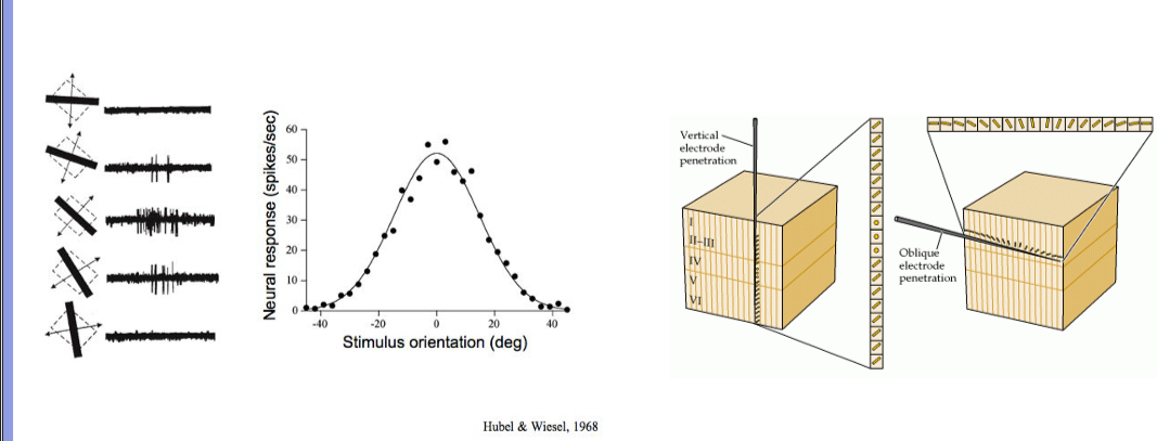
Ice cube model
Orientation columns run perpendicular to ocular dominance columns and orientation columns vary smoothly from location to location

What is a hypercolumn
One block of tissue in the visual cortex will represent both eyes and represent all orientations for one location in the visual space
How do neurons become orientation selective
Concentric receptive fields (CRFs) from several neurons can be combined in a line. When a bar of light with the right orientation hits them, they all activate together. This creates a new neuron with an elongated receptive field that responds best to that bar angle — giving it orientation selectivity. ON and OFF zones in the combined field help it respond to light-dark patterns
The new cell is called a simple cell
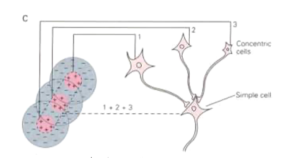
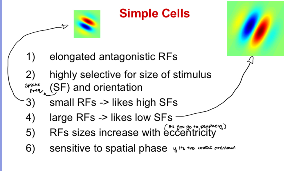
What is a simple cell
Cells which have distinct regions that like bright/dark bars
Sensitive to spacial phase
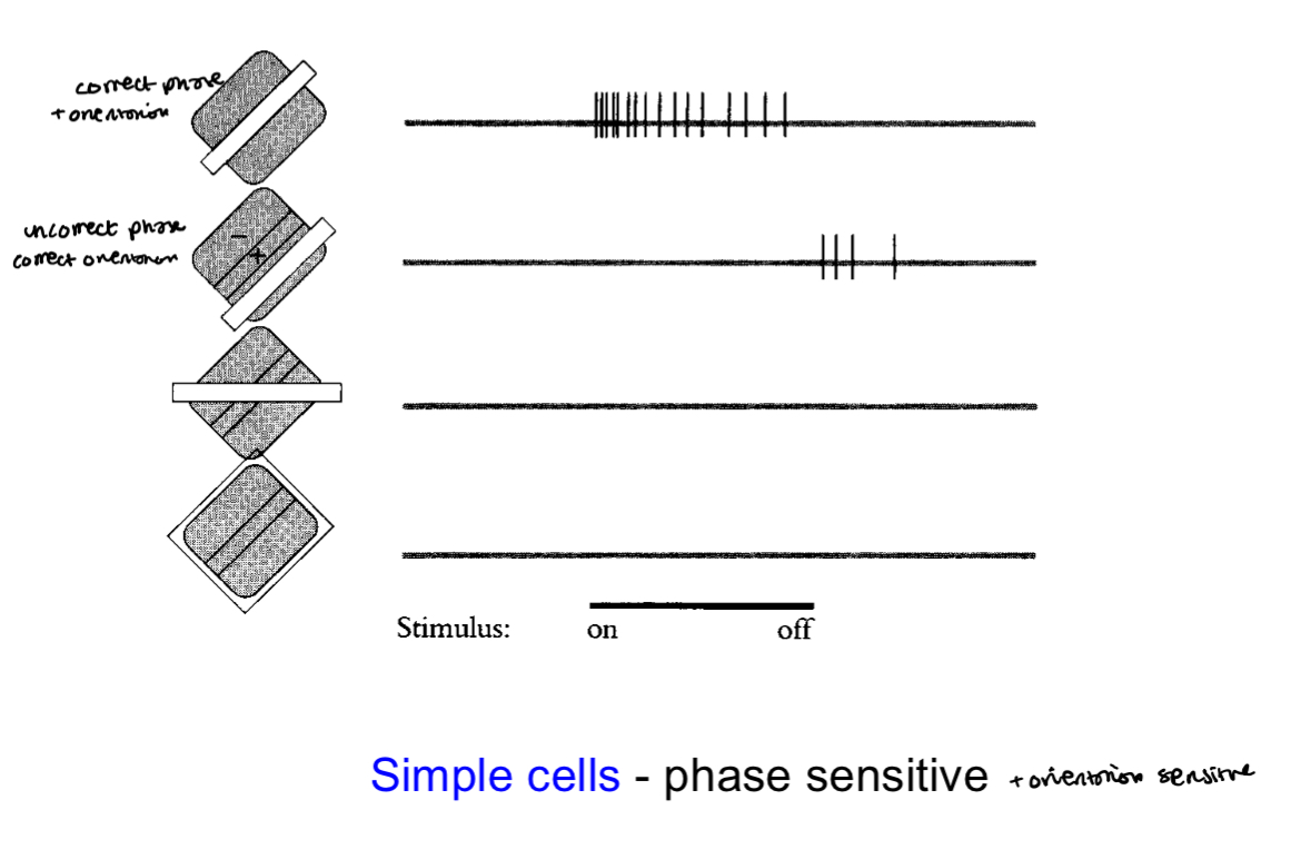
Simple cell properties
receptive fields
What are they selective for
Small RFs → like __ SFs
Large RFs → like __ SFs
RFs sizes increase with __
Sensitive to __
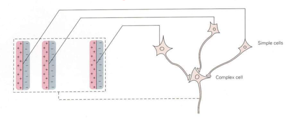
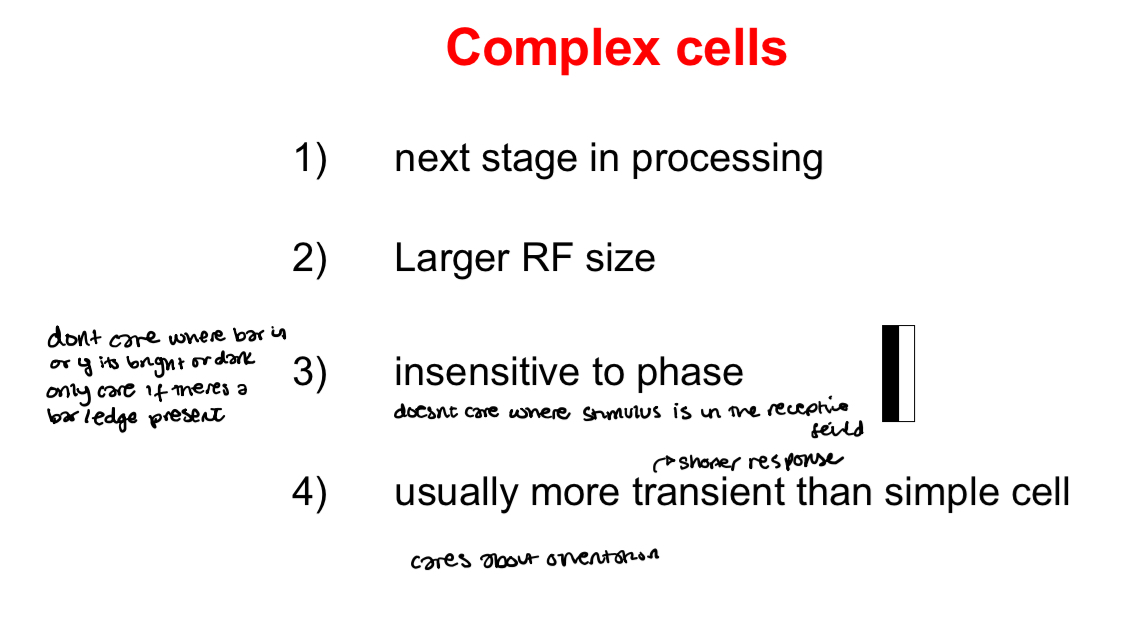
What is a complex cell
Cells that sum up multiple simple cells to produce a receptive fields that has overlapping on and off responses
They can also be produced by summing lots of concentric (LGN-like) RFs together
Complex cell properties
receptive field size
Insensitive to what
Response time
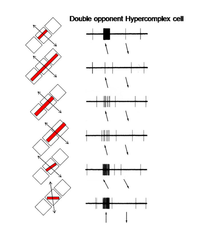
What are hypercomplex cells
Cells that are length tuned - they respond maximally to a defined length and are inhibited if the stimulus is longer than this
Thought to be involved in processing corners
Dont care where bar is or if its black/white
Care about length of bar therefore only respons when the central region is activated at the right orientation
Phase insensitive mixed on and off subregions
Why do we need oriented recepetive fields
Edges define when objects start and end
What is direction selectivity
Cells tuned to the direction the stimulus is moving at
What layer of the cortex is direction selectivity found And where does it project to
Direction selectivity is found in layer 4B
4B projects out of V1 to V2 and also directly to MT (these regions are involved in motion processing)
What is disparity selectivity And wheres it found (depth selectivity)
Selectivity for the difference between the two eyes views
Found mainly in the upper layers
What are interblob cells selective to
Orientation
What are blob cells selective to
Wavelength
They respond the same to all orientations (not orientation selective)
Konicellular cells (blue/yellow) project directly to these blobs
Revised ice cube model
How is information processed in V1
Info is inputted to layer 4
Vertical and horizontal connections between neurons
V: Its then projected upwards and downwards to layers 2/3 and 5 and other sublaminar
Filled pyramindal cells in layer 2/3 - axon collarerals branch off to neighbouring cells
Lateral connections in visual cortex
Lateral connections are like to like. Cells in close layers are connected. Cells connect to cells that are similar eg same orientation
These connections may be involved in grouping responses together
Or they mat play a role in sharpening responses to the orientaiton of a stimulus or to the length of a stimulus or all of the above
What is the dorsal stream
The where pathway
Its involved with processing the objects spatial location relatitve to the viewer (motion and location)
Goes to the prefrontal cortex
What is the ventral stream
The what pathway
Involved with object and visual identification and recognition
Recognising objects, form, colour
Goes to the inferotemporal cortex
Full Dorsal pathway
V1 → V2 → V3 → MT → MST → prefrontal cortex
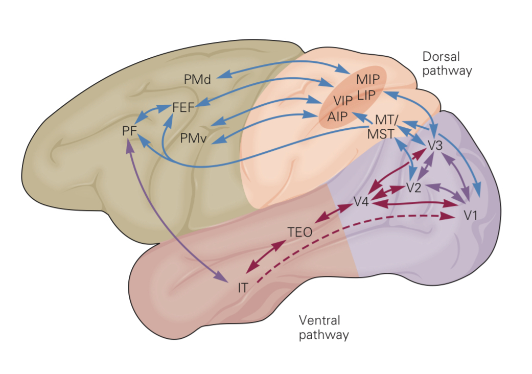
Full ventral pathway
V1 → V2 → V4 → inferotemporal cortex

What do connections between the ventral and dorsal pathways lead to
Object identification and location
Damage to the ventral pathway can lead to what
Prosopagnosia - cant recgonise people
Damage to the dorsal pathway can lead to what
Optical ataxia - dont know where things are in space
Where do parallel streams from V1 originate from
From layers 2/3 and layer 4B (project info out of V1)
Wher is V2
Surrounds V1
Thick stripes in V2 are selective for
Movement direction
Disparity
Thick stripes in V2 receive input from which layer of V1
4B
Thin stripes in V2 are selective to
Colour
Contrast
Thin stripes in V2 receive input from where in V1
Blobs (layer 2/3)
Interstripes in V2 are selective for
Orientation
Contrast
Interstripes in V2 recieve input from where in V1
Interblobs - layer 2/3
Full V2 thick stripes stream
4B → thick stripes → V3 (depth perception)
Full V2 thin stripes stream
Blobs → thin stripes → V4 (colour and form)
Full V2 interstripes stream
Interblobs → interstripes → V4 (colour and form)
Where does V3 receive input from
Magnocellular cells
Receives inputs from layer 4B in V1 and thick stripes in V2
What is V3 selective for
Has a large number of cells for orientation, direction and disparity
Where does V3 make connections to
V1, V2, V4 and MT
What is V4 associated with
Processing colour vision and form vision
First area to show strong modulation by attention
Where does V4 receive input from
V2 (thin stripes and pale stripes) and direct connections from central V1)
Where does V4 send outputs to
Strong outputs to Posterior inferotemporal cortex (PIT)
Also weaker outputs to MT/V5
What is V4 selective for
Orientation, colour and object features of intermediate complexity
Inferotemporal cortex Properties
Very large receptive fields
All include fovea
May include both hemifields ie input from opposite cortex
Cells tuned to specific shapes
Inferotemporal cortex (IT) selective for
Dont care about the position or size of the object within the recpetive fields
Cells respond to faces
What are face cells
Cells that are responsive to faces and hands
Cells may be more or less responsive according to familiarity
What is MT And where does it get input from
Part of V5 is involved in motion processing and receives mainly magnocellular inputs from V1 directly and indirectly from V2, V3 and V4