Chapter 12 - REPRODUCTION PRODUCES OFFSPRING
 Call Kai
 Learn
Learn Practice Test
 Spaced Repetition
 Match
Match Flashcards
 Knowt Play
1/110

There's no tags or description
Looks like no tags are added yet.
Name | Mastery | Learn | Test | Matching | Spaced | Call with Kai | Chat |
|---|
No analytics yet
Send a link to your students to track their progress
111 Terms
insemination
During sexual intercourse, an erect penis releases semen into the vagina through an ejaculation.
oocyte
an immature egg cell in the ovary
Gene
The factor that determines a hereditary characteristic; part of a chromosome
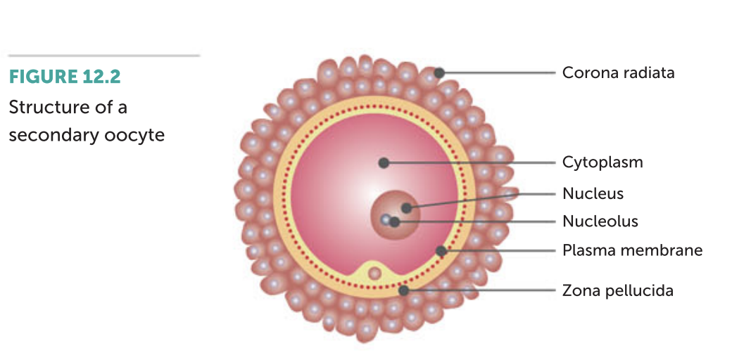
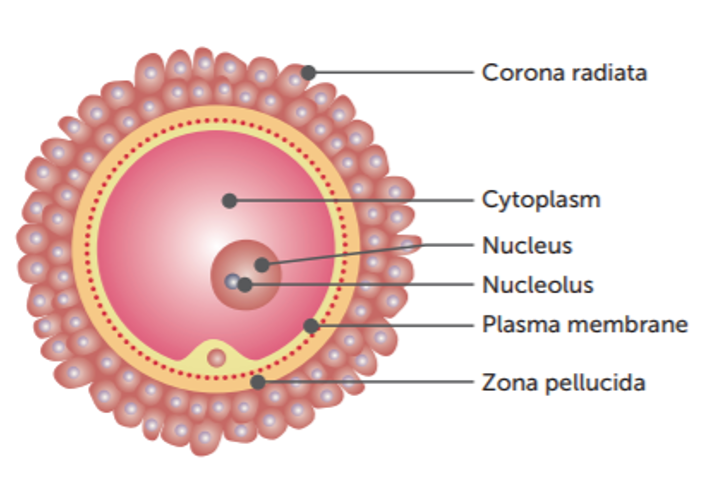
The secondary oocyte that is released at ovulation is at metaphase II. It is surrounded by two layers.
• The outer corona radiata consists of follicle cells held together by cementing materials that
contain acid.
• The inner zona pellucida is a glycoprotein matrix surrounding the plasma membrane of the
oocyte.
implantation
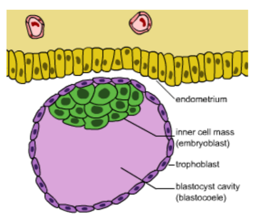
The blastocyst remains free within the cavity of the uterus for two to three days, and then sinks into the soft endometrium (uterine lining) to become firmly attached to the wall of the uterus. This process is called implantation, and enables the blastocyst to gain nourishment for growth and development by absorbing nutrients from the glands and blood vessels of the uterine lining.
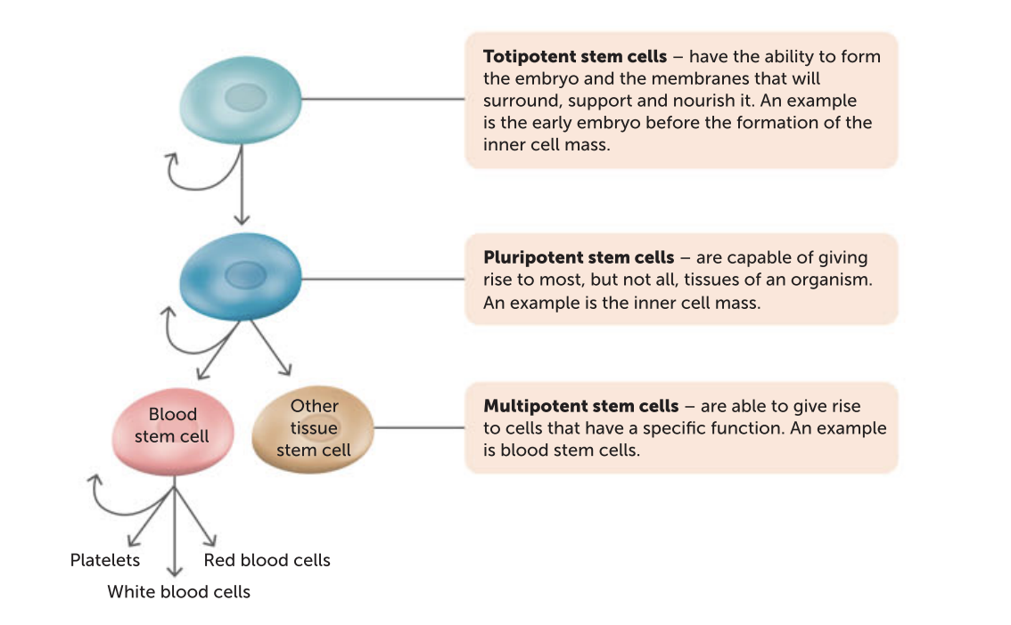
stem cells are very different from other cells because:
• they are not specialised for any particular role
• they are capable of repeated division by mitosis – a process called proliferation
• given the right conditions, they can differentiate into specialised cells. All the 200 or more
types of cells that make up a mature human body develop from the stem cells of the inner
cell mass.
zygote life journey
A zygote develops from an unspecialised totipotent cell to a blastocyst, and then to an embryo and its membranes.
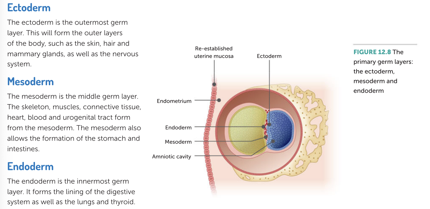
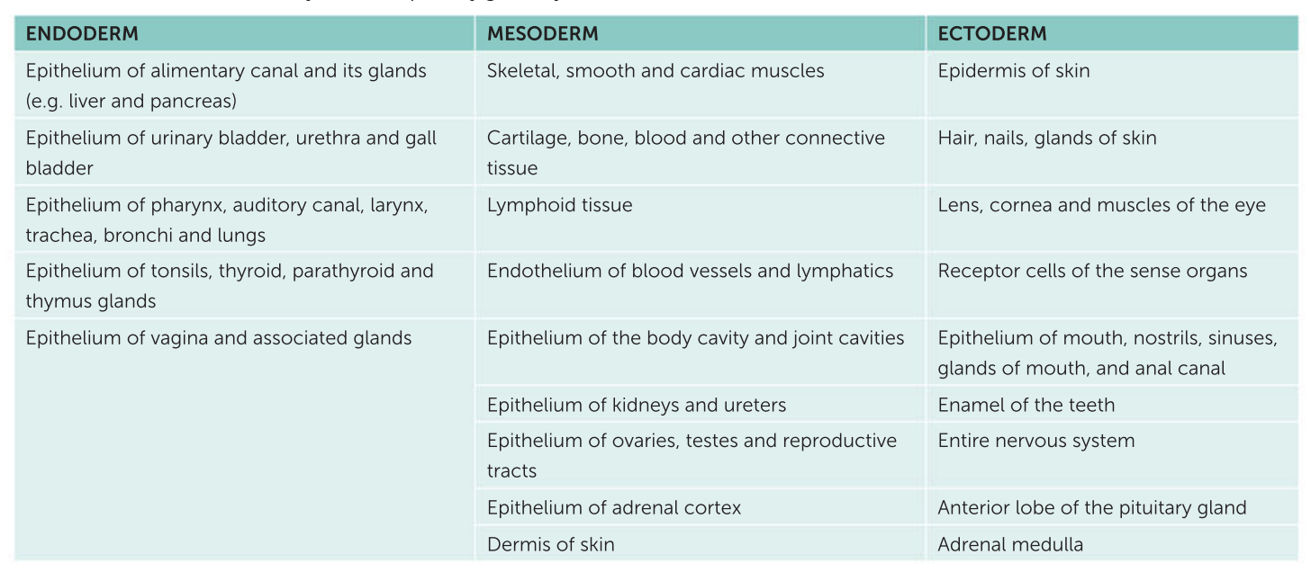
The primary germ layers differentiate to form the specialised structures of the embryo.
Structures formed by the three primary germ layers
what is an embryo
When a fertilized egg has implanted in the uterus, the group of cells that will become a baby
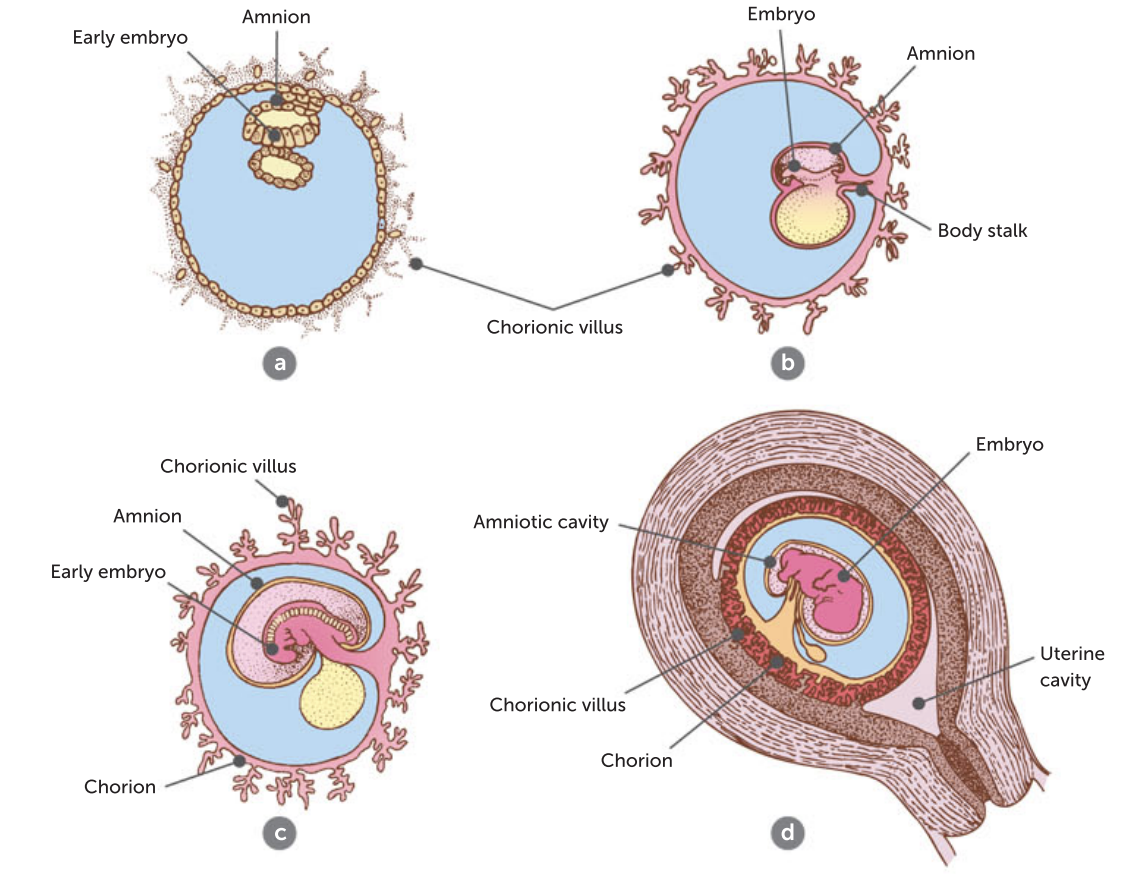
Amnion
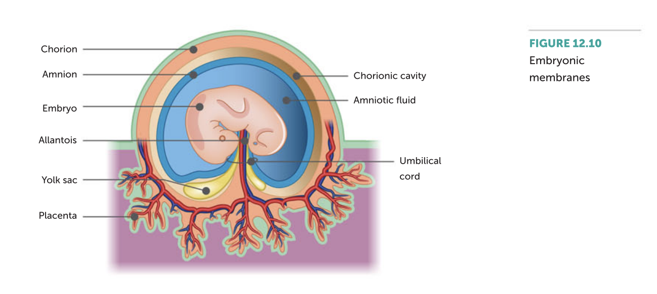
The amnion is the first membrane to develop. By the eighth day after fertilisation, it surrounds the embryo, enclosing a cavity into which it secretes amniotic fluid. This fluid serves to protect the embryo against physical injury by acting as a shock absorber. It also helps to maintain a constant temperature and allows the developing embryo, and later the foetus, to move freely. The amnion expands as growth takes place. It usually ruptures just before childbirth, releasing the amniotic fluid, an event commonly referred to as ‘breaking of the waters’.
Chorion
Another embryonic membrane is the chorion. It is formed from the outer cells of the blastocyst together with a layer of mesodermal cells. The chorion surrounds the embryo and the other three embryonic membranes. As the amnion enlarges, it fuses with the inner layer of the chorion. Eventually, the chorion becomes the main part of the foetal portion of the placenta.
Yolk sac and allantois
In addition to the chorion and the amnion, there are two other membranes – the yolk sac and the allantois. These are not as important in humans as they are in the development of many other animals; however, they do form the outer structure of the umbilical cord.
Functions of the placenta
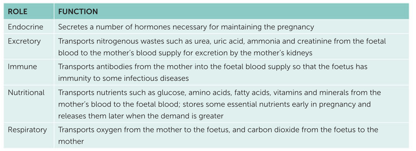
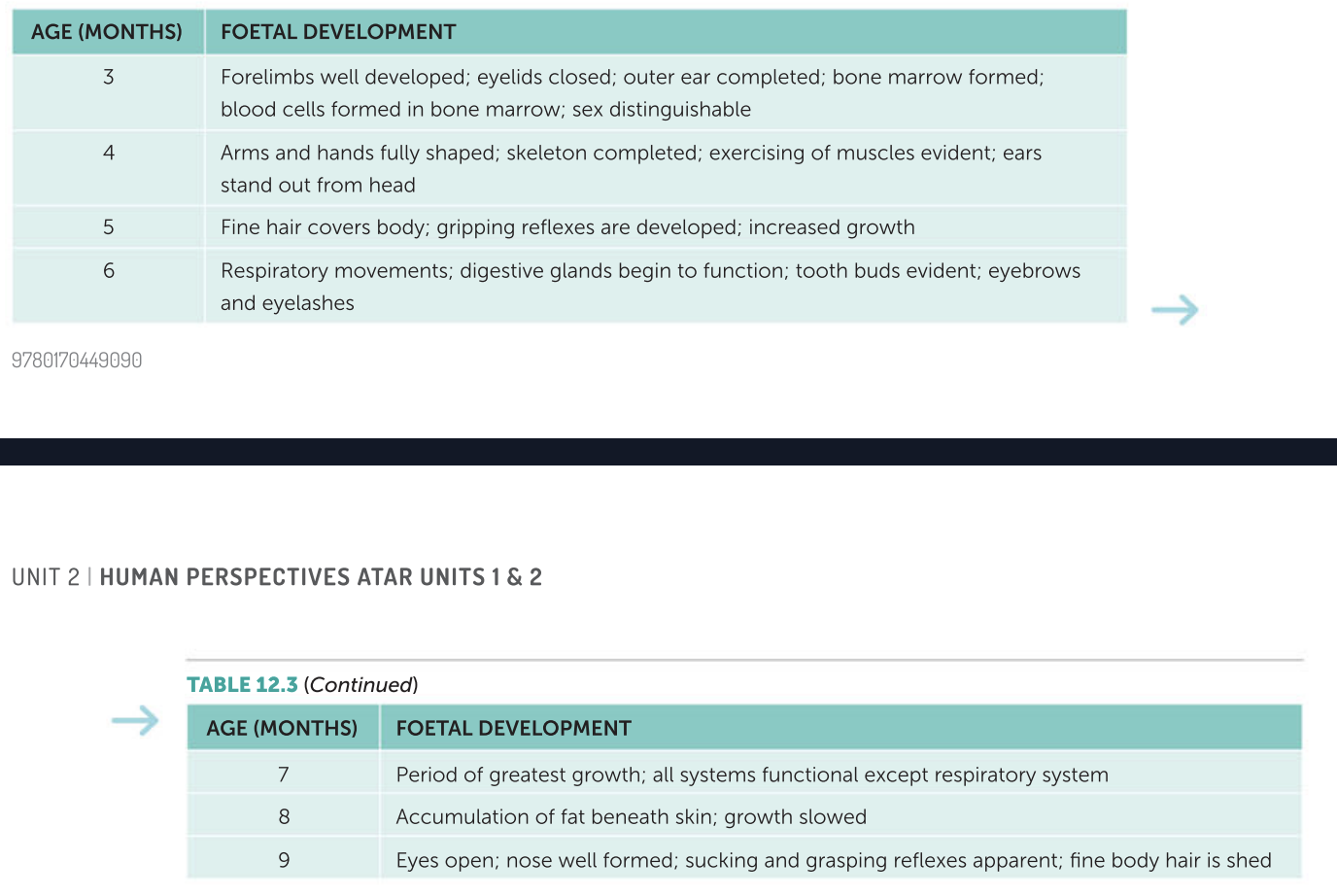
Stages of foetal development
Pregnancy also affects the mother in less obvious ways.
• There is an increase in the size of the heart and in blood volume. This is to cater for the extra blood that is flowing through the placenta.
• The greater blood volume results in an increased blood flow to the kidneys and, therefore, increased urine production. Additionally, during the first three months of pregnancy, the expanding uterus presses on the bladder so that it feels as if it is filled with urine. As the uterus grows, it moves up the pelvic cavity, releasing this pressure. Then, during the last stages of pregnancy, the foetus presses on the bladder again.
• The emotional state of the mother may be affected due to the changes in hormonal balance and as a result of natural fears accompanying pregnancy. The mother may be concerned about her child’s development, the problems that may occur at the time of birth, and the effect the newborn child will have on the rest of the family. Many of these factors are beyond the control of the pregnant woman and so support and reassurance from family and friends are very important in maintaining a positive outlook.
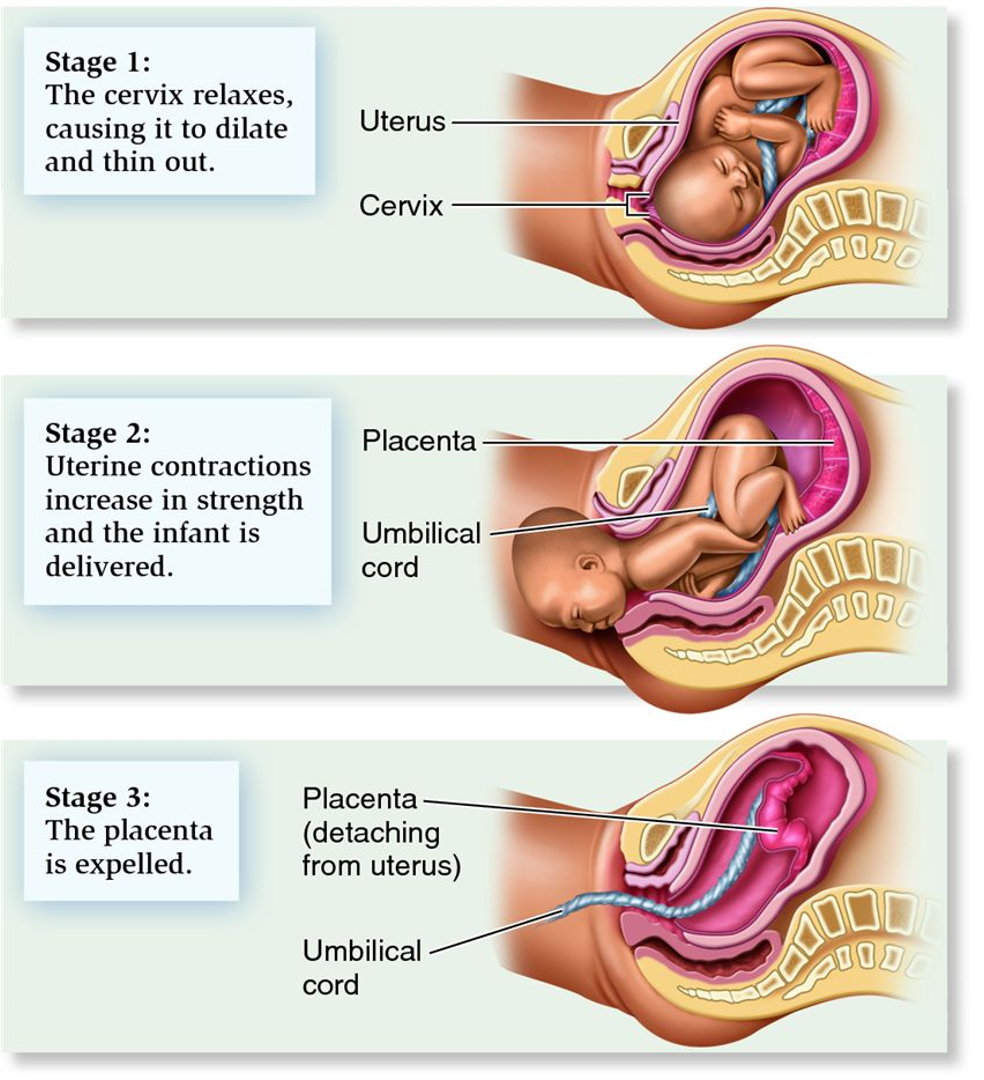
During the first stage of labour,
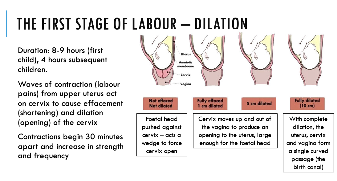
During the first stage of labour, uterine contractions gradually increase in strength and the cervix dilates until it forms the birth canal with the uterus, cervix and vagina
During the second stage
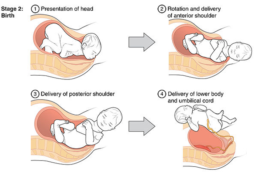
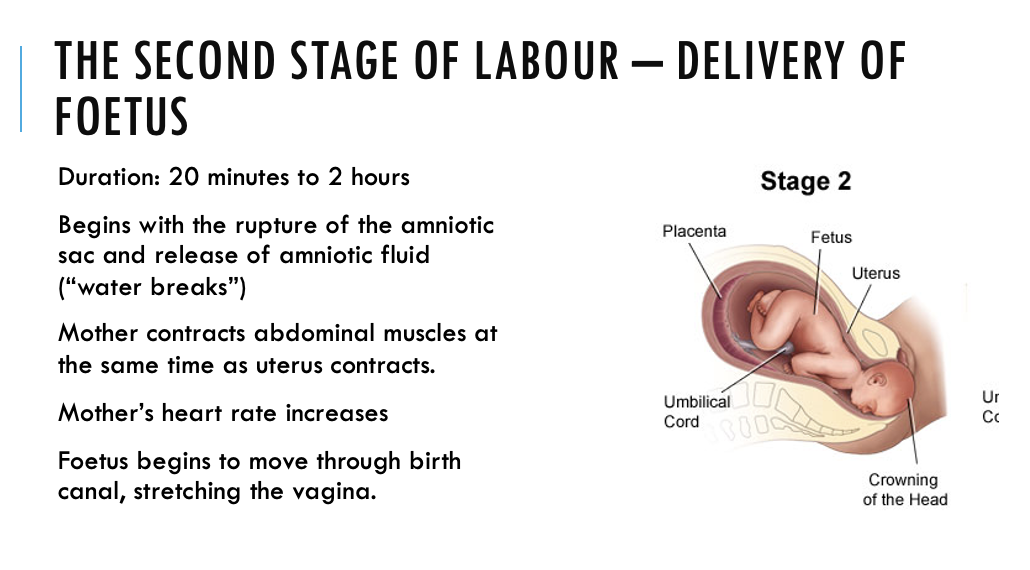
During the second stage of labour, the membrane bursts and contractions push the foetus through the birth canal.
In the final stage
In the final stage, the placenta, other membranes and the remains of the umbilical cord are expelled.
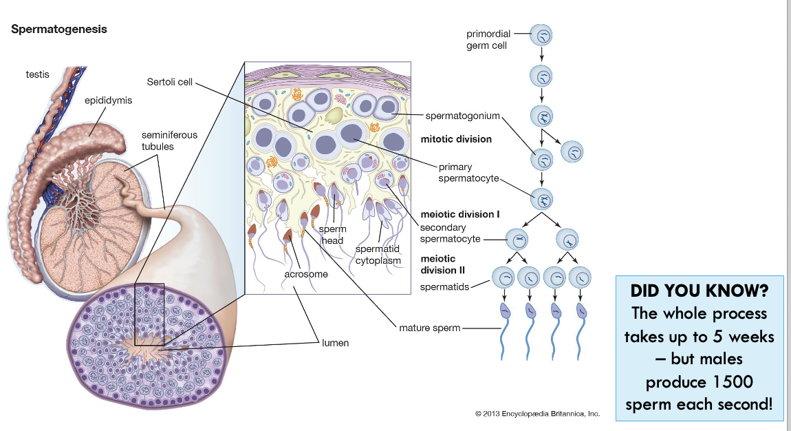
SPERMATOGENESIS
Seminiferous tubules are lined with spermatogonia (immature diploid cells)
At puberty spermatogonia begin dividing by mitosis to provide a continuous source of cells for sperm production.
Some new cells are pushed into the centre of the tubule where they enlarge to form primary spermatocytes
Each primary spermatocyte undergoes the first stage of meiosis to become two haploid cells called secondary spermatocytes.
The second meiotic division occurs and two secondary spermatocytes form four haploid spermatids.
One diploid spermatocyte produces four haploid spermatids
MATURATION OF SPERM – THE EPIDIDYMIS
Sperm travel from the testes to the epididymis by muscular contractions (peristalsis)
During maturation they:
§Gain mitochondria
§Lose cytoplasm
Swimming ability is gained at ejaculation when sperm are mixed with other secretions.
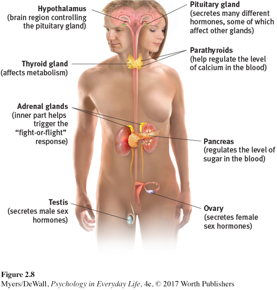
The endocrine system
The endocrine system consists of glands found throughout the body, which produce hormones.
Hormones are chemical messengers that travel through the bloodstream and communicate with specific target cells.
The effects of the endocrine system are slow, but long-lasting.
HORMAL CASCADE
Regulation of spermatogenesis starts in the hypothalamus in the brain!
At puberty, the hypothalamus begins releasing gonadotropin releasing hormone (GnRH).
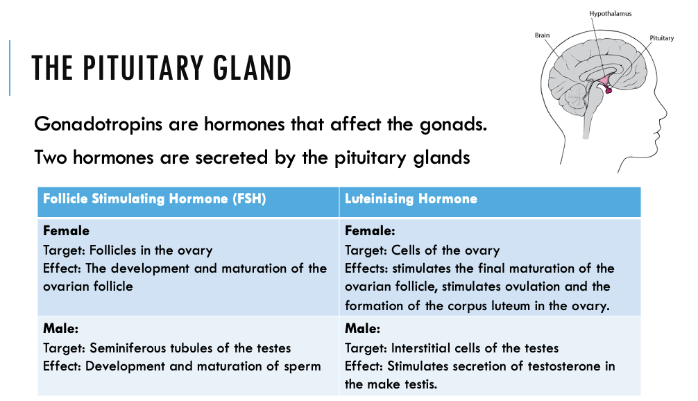
This communicates with the anterior pituitary gland – to release Follicle Stimulating hormone FSH & Luteinising hormone (LH) into the bloodstream.
LH communicates with cells in the testes to begin producing testosterone.
FSH causes testosterone to accumulate in the testes – triggering the production of sperm.
Ovary (Female Gonad)
Produces female gamete (ovum).
One on each side of lower abdomen.
Mass of connective tissue (stroma) surrounded by a layer of cells including germ cells. Germ cells develop into eggs.
Each germ cell is enclosed in a follicle. Follicles mature, move to surface of ovary & rupture.
Egg is expelled into funnel-shaped opening of uterine tube.
Oogenesis – the production of ova
Occurs in the ovary. Involves meiosis and maturation of ova.
Ovaries of the female foetus contain diploid oogonia that divide by mitosis and then enlarge to form hundreds of thousands of primary oocytes.
At birth the primary oocytes are in prophase of the first meiotic division.
They are surrounded by a single layer of cells, forming a primary follicle.
Development now ceases until puberty.
oogenesis
From puberty, one primary follicle develops per month, usually in alternative ovaries.
As the follicle matures, the primary oocyte completes the first stage of meiosis, forming 2 haploid cells.
Each has 23 chromosomes, but one is much larger and contains almost all the cytoplasm (secondary oocyte)
The smaller cell is called the first polar body.
Ovulation occurs: the follicle ruptures and the secondary oocyte and polar body enter the uterine tube.
If penetrated by a spermatozoan, the secondary oocyte completes meiosis II, forming two haploid cells of unequal size.
The larger cell develops into an ovum, the smaller cell develops into the second polar body.
The first polar body may also complete meiosis
All polar bodies disintegrate.
One primary oocyte produces only one ovum.
The presence of cytoplasm prolongs the life of the ovum.
If fertilised, the zygote can survive until implantation in the uterus.
Fertilisation occurs only if the nuclei of the ovum and sperm fuse.
Uterine tube
Extends from each ovary to the uterus.
End of tube near ovary has funnel-like opening fringed with finger-like fimbriae that guide the egg into the tube.
Ciliated cells line the tube.
Beating cilia aided by contractions of smooth muscle in walls of uterine tube carry egg along to uterus.
Fertilisation occurs in uterine tube.
Uterus
Single, hollow, pear-shaped organ held by ligaments in pelvic cavity between rectum (behind) & urinary bladder (in front).
Wall of uterus is of smooth muscle with soft mucous membrane lining (endometrium).
Protects & nourishes developing foetus during pregnancy.
Cervix
Neck of uterus.
Protrudes into vagina.
Sealed with plug of mucus during pregnancy.
Vagina
Muscular canal 10 cm long leading to exterior.
Lined with mucous membranes.
Capable of stretching & widening to become the birth canal.
Receives penis during sexual intercourse.
Vulva:
External genitalia.
Labia majora & labia minora: oily secretions, pubic hair & fleshy folds protect the vagina & provide lubrication for penis during intercourse.
Clitoris
sexual stimulation
what are hormones?
Hormones are chemical messengers that are produced in the endocrine glands.
They are transported in the blood and act upon specific target cells or organs.
The ovarian cycle
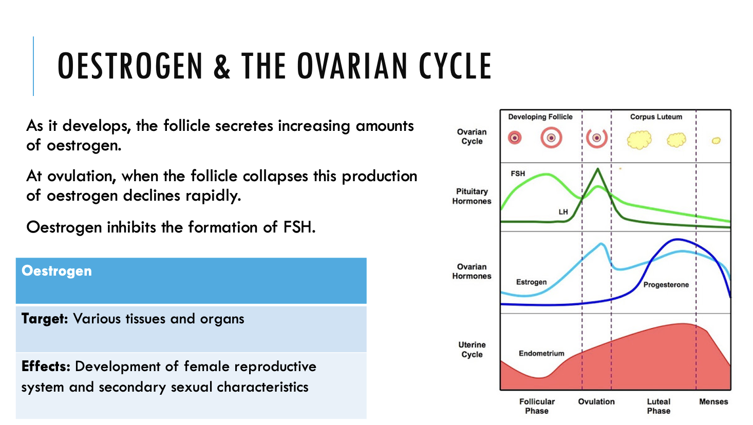
At the start of the cycle, high levels of FSH start the development of the follicle.
Follicles are glandular and they secrete oestrogen – which travels in the blood to the uterus and causes the endometrium to thicken.
LH hormone is released, causing the mature follicle to burst.
The ovum is released (ovulation) and the ovum is swept into the fallopian tube.
High levels of LH causes the empty follicle to develop into a glandular structure, known as the corpus luteum.
The corpus luteum secretes progesterone (and small amounts of oestrogen) – maintaining the endometrium in preparation for fertilisation.
Oestrogen & the ovarian cycle
Hormones and fertilisation
The embryo implants into the wall of the uterus.
The placenta begins to develop.
The developing placenta secretes human chorionic gonadotropic hormone (HCG).
HCG maintains the corpus luteum until the placenta develops enough to secrete its own oestrogen and progesterone.
The endometrium does not degenerate (no menstruation)
The ovarian cycle
If the egg is not fertilised, the rising levels of progesterone and this results in the pituitary gland to reduce its production of LH.
Lower levels of LH causes the corpus luteum to degenerate – triggering menstruation.
Menstruation
occurs 14 days after ovulation. Blood, mucous and cell debris from the endometrium are shed through the vagina.
Menarche
is the first menstrual period
Menopause
is the gradual decline in frequency and eventual cessation of menstruation –the end of the reproductive life.
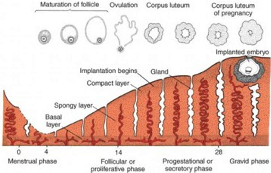
The menstrual cycle
Occurs in the uterus.
Involves changes in the endometrium lining the wall of the uterus.
Closely associated with the ovarian cycle
If the ovum is not fertilised, then the endometrium must be shed.
The menstrual cycle is said to begin on the first day of menstruation as a period can easily be detected.
Stages of the menstrual cycle
Relating the ovarian and menstrual cycles
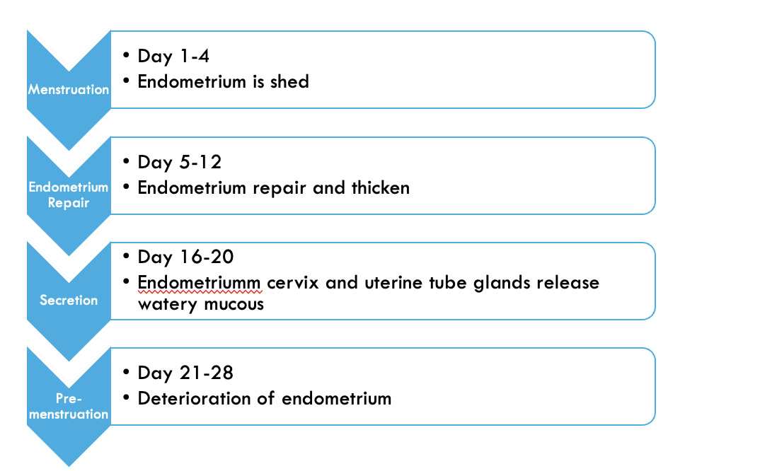
While the follicle is maturing, the endometrium is repairing and thickening, with an increase in blood vessels and mucous glands
After ovulation, the endometrium continues to thicken and secrete watery mucous rich in glycogen
If fertilisation does not occur, the corpus luteum in the ovary degenerates, causing the endometrium of the uterus to break down.
The endometrium, blood, mucous and cell debris are shed through the vagina.
ovulation
Ovulation usually happens around day 14 of the menstrual cycle.
Luteinizing hormone (LH) surges, triggering the release of the secondary oocyte (at metaphase II).
At the same time, cervical mucus becomes more slippery to help sperm make their way to the egg.
The structure of the oocyte
A layer of follicle cells surrounding the mature egg (corona radiata) is held together by an acid.
The zona pellucida is a glycoprotein matrix surrounding the plasma membrane
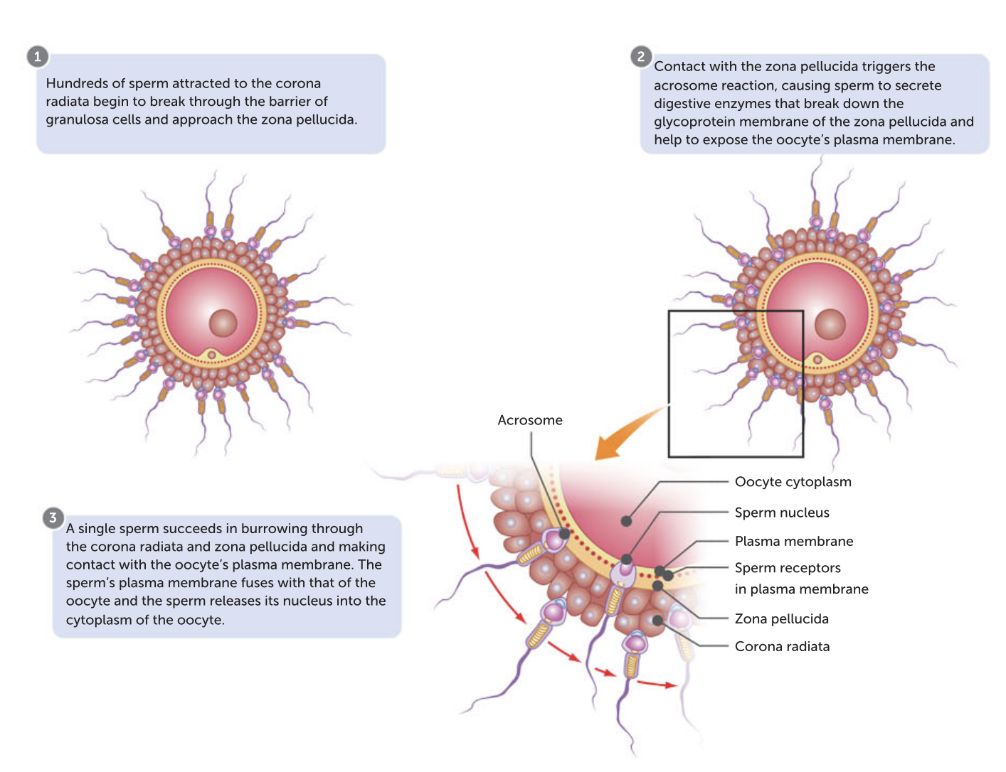
Only one sperm penetrates the egg.
ejaculation
250-350 million sperm are released into the vagina.
Only a few thousand enter the uterine tube
Fertilisation takes place in the uterine tube
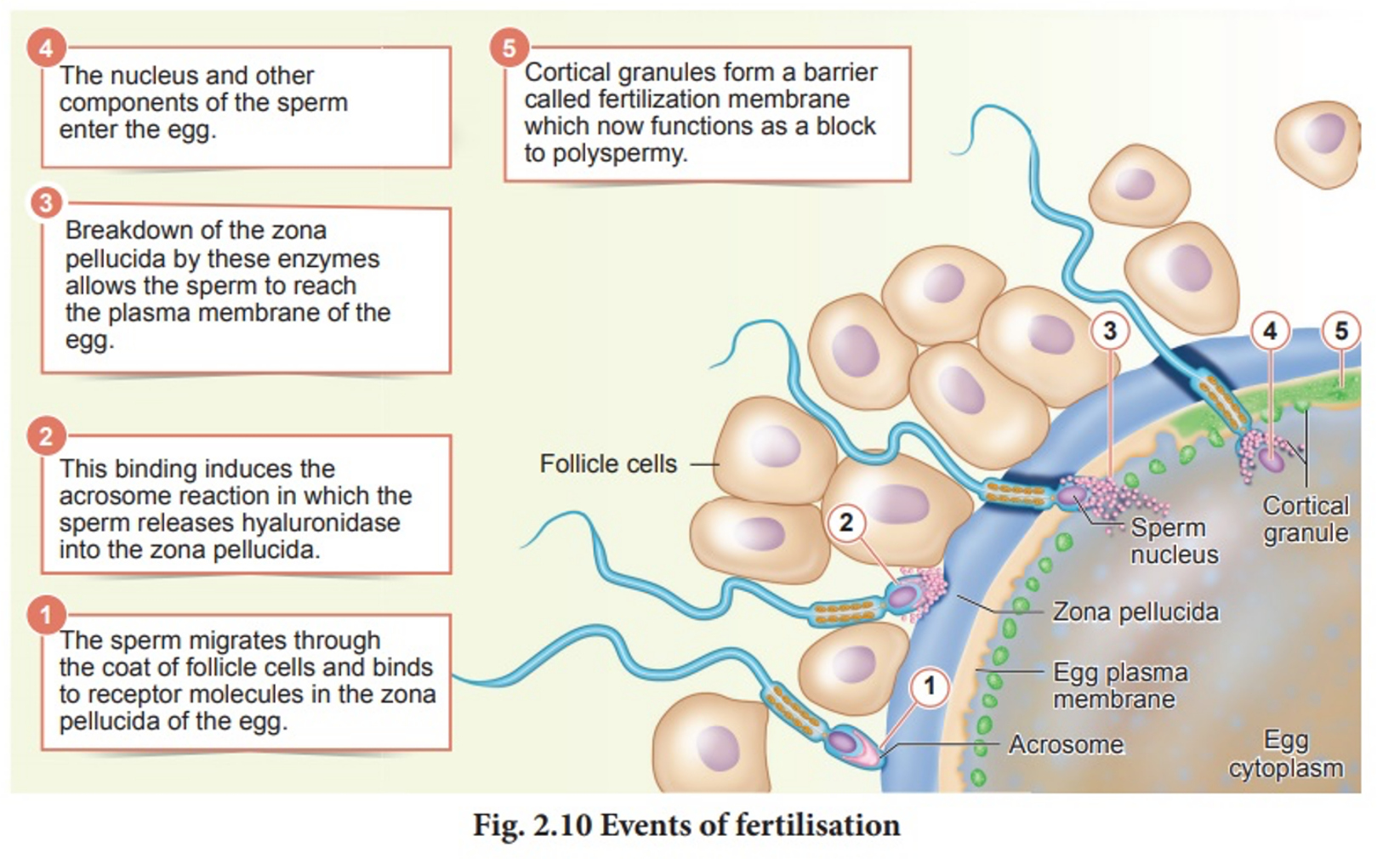
Enzymes in the vesicle of sperm tip can break down the acid, but thousands of sperm are needed to supply enough enzyme.
During fertilisation, the sperm and egg unite in one of the uterine tubes, forming a zygote.
Formation of the zygote
A fertilisation membrane forms (hard shell- si no chromosomes enter) around the egg, preventing the entrance of any ore sperm.
Inside the egg, the tail of the sperm is absorbed.
Head moves through the cytoplasm of egg as the male pronucleus.
Entrance of sperm stimulates the egg to complete meiosis II.
Egg nucleus develops into a female pronucleus
Male and female pronuclei fuse to form diploid zygote.
From zygote to blastocyst
From zygote to blastocyst
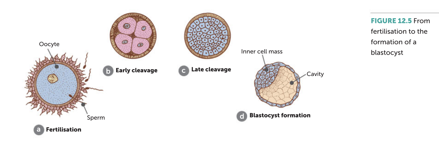
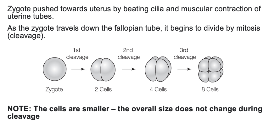
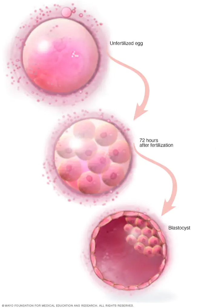
3-4 days after fertilisation, a solid ball of cells called a morula is formed.
The morula contains 16 totipotent cells
Day 5: A blastocyst forms (sphere of cells surrounded by a layer of cells) and reaches the uterus.
The inner cell mass develops into the embryo.
Implantation of blastocyst
The blastocyst is free in the uterine cavity for 2-3 days.
Blastocyst sinks into the endometrium and becomes firmly attached to the uterine wall (implantation)
Implantation enables blastocyst to absorb nutrients from the glands and blood vessels of the endometrium
Development of the placenta
Immediately after implantation, small, finger-like projections (chorionic villi) develop from the outer layer of blastocyst cells.
Chorionic villi penetrate endometrium and will become the foetal part of the placenta.
Secretion of the gonadotropin hcg
The developing placenta secretes HCG (human chorionic gonadotropic hormone)
HCG targets the ovary and prevents degeneration of the corpus luteum.
The corpus luteum secretes oestrogen and progesterone to maintain the endometrium
Menstruation does not occur
Diagnosis of foetal health
First trimester
Foetal Ultrasound and maternal blood testing
Process can help determine the risk of certain birth defects
Second trimester
Blood tests to provide further information about genetic conditions
Further ultrasounds check for foetal growth, estimate due dates and look for structural abnormalities
Additional testing includes – amniocentesis or chorionic villus sampling
imaging the foetus
Ultrasound
Foetoscopy
analysing biochemicals or chromosomes
Amniocentesis
Chorionic villus sampling
Maternal & foetal blood sampling
DNA probes
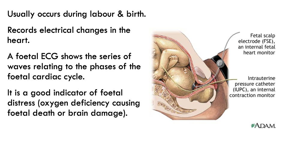
Foetal monitoring
Electrocardiogram
Maternal contractions (labour)
FOETAL IMAGING TECHNIQUES: ULTRASOUND
Inaudible, high-frequency sound waves are used to image the foetus.
Ultrasound uses the principle of reflection of sound waves as echoes.
The echoes are collated as an image by a computer.
Visible defects can be detected.
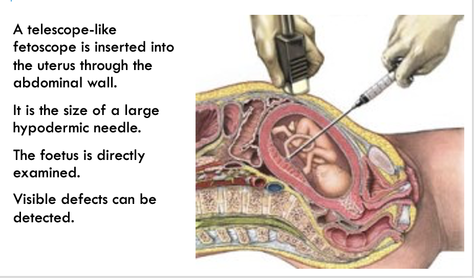
FOETAL IMAGING TECHNIQUES: FETOSCOPY
BIOCHEMICAL AND CHROMOSOME ANALYSIS:
AMNIOCENTESIS
A needle is inserted into the amniotic fluid surrounding the foetus during weeks 16-20.
10-20 mL of amniotic fluid
is extracted.
Tests can be performed
on the foetal cells floating in the fluid to determine a large number of chromosomal or biochemical abnormalities.
Performed only when there is a risk of a birth defect.
BIOCHEMICAL AND CHROMOSOME ANALYSIS:
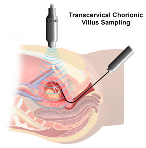
CHORIONIC VILLUS SAMPLING
A catheter (flexible hollow tube) is inserted into the chorion through the maternal vagina during weeks 9 - 19.
A specimen of foetal cells is obtained from the chorion (one of the foetal membranes) and analysed as in amniocentesis.
Analysis can be done quicker than with amniocentesis but the risk of miscarriage is 1%.
Spina bifida cannot be detected by CVS.
BIOCHEMICAL AND CHROMOSOME ANALYSIS:
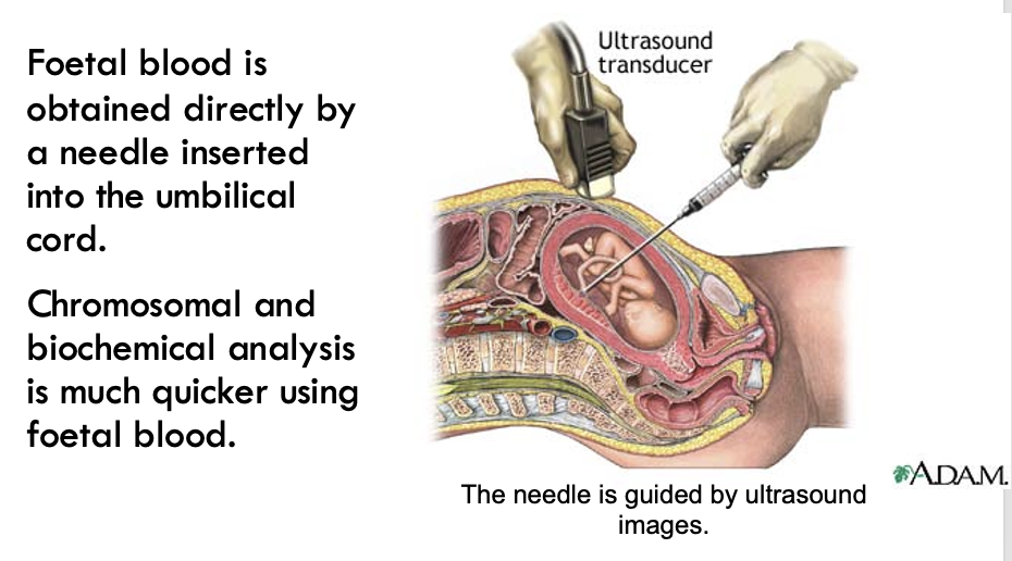
MATERNAL & FOETAL BLOOD TESTS
Searching for DNA sequences
Gene probes can be used to search for a specific, small DNA sequence, such as the mutated allele that leads to Huntington’s disease.
FOETAL MONITORING:
ELECTROCARDIOGRAPHY (ECG)
BIOCHEMICAL ANALYSIS ON THE NEWBORN:
ASSESSMENT OF MARKER PROTEINS
Heel-prick blood test for the presence of abnormal amounts of certain substances which are markers for certain diseases. e.g.
large quantities of phenylalanine indicate PKU.
WHAT IS PKU (Yr 12)
Phenylketonuria
Autosomal recessive condition
Phenylalanine is an essential amino acid, your body must have it to grow and function
Too much can be harmful, so the body uses the enzyme (phenylalanine hydroxylase) to convert it into tyrosine.
In PKU, levels of this enzyme are low. As a result, phenylalanine builds up in the blood and other tissues, particularly the brain.
Causes severe and irreversible brain damage.
Down syndrome
Identifying Down Syndrome: Chorionic villus sampling (CVS).
In CVS, cells are taken from the placenta and used to analyse the foetal chromosomes. This test is typically performed in the first trimester, between 10 and 13 weeks of pregnancy.
Cystic fibrosis
Cystic fibrosis can be diagnosed during pregnancy by obtaining genetic material from the foetus through chorionic villus sampling or amniocentesis.
For couples who are carriers of cystic fibrosis, testing during pregnancy allows them to plan or make decisions about termination.
Muscular dystrophy
There are two main ways of performing a prenatal diagnosis.
1. Chorionic villus sampling (CVS), which involves removing tissue from the placenta for analysis, usually after 11 weeks into the pregnancy.
2. Amniocentesis, which isn't usually carried out until 15 to 16 weeks of pregnancy.
Huntington's disease
Huntington's disease is a progressive brain disorder caused by a single defective gene on chromosome 4.
This defect is "dominant," meaning that anyone who inherits it from a parent with Huntington's will eventually develop the disease.
Pre-implantation genetic diagnosis (PGD) is a way to test an embryo before it’s implanted in a woman’s uterus.
Using IVF (In Vitro Fertilization) techniques, the egg and sperm are combined outside the body.
Once the embryo reaches a certain level of development it can be tested to see if it carries the gene causing HD.
Only embryos that are unaffected with HD are implanted in the woman’s uterus
Gestation/Pregnancy
Period of embryonic and foetal development in utero.
In utero:
inside the uterus
Parturition/Birth:
Process by which the foetus is expelled from the maternal body
Labour:
Processes resulting in parturition and expulsion of the placenta from the maternal body.
Changes in the mother during pregnancy
There is an increase in the size of the heart and in blood volume – this is to allow for the additional blood that is flowing through the placenta.
During pregnancy, the blood volume increases by 30 to 50 percent, the heart pumps more blood each minute and the heart rate increases.
The heart is tipped on an angle – forced to occupy a smaller space due to the expanding uterus.
Changes in the mother during pregnancy
Greater blood volume causes an increase in blood flow to the kidneys, resulting in increased urine production.
In the first 3 months, the uterus presses on the bladder, creating the feeling that the bladder is full of urine.
In the final stages of pregnancy, the foetus presses on the bladder again
Emotions
Changes in hormonal balance impact the regulation of emotions
Natural fears about the pregnancy and pending birth also increase stress on the mother.
Preparing for labour
Hormone (Relaxin)
§Cause ligaments of the pelvis to soften and become more pliable.
§Increased response of the uterus to stimuli
§Strengthen uterine muscle contractions
§Cervix softens and shortens
Foetus
§Head of the foetus engages in maternal pelvis, facing hips
The first stage of labour – dilation
The second stage of labour – delivery of foetus
Contraction and retraction repeated until head reaches entrance to vagina
Foetal head turns to face mother’s back and passes through the vagina
Head turns sideways again to face hips so shoulders and body pass out easily.
Shape of head may distort, but joints/bones of skull are pliable and shape remoulds quickly
The first breath
Umbilical cord is clamped and cut, severing the child’s oxygen supply.
Carbon dioxide builds up in infant’s blood, stimulating the child’s first breath through the lungs.
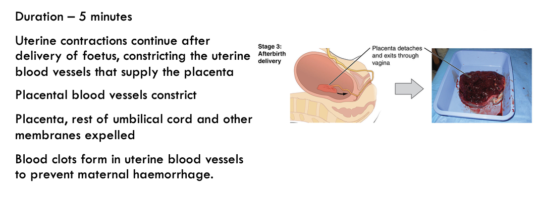
Third stage of labour – after birth
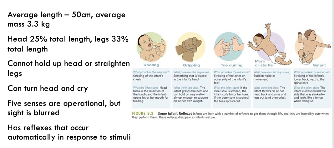
Characteristics of the newborn
FOETAL CIRCULATION BEFORE BIRTH
Deoxygenated foetal blood low in nutrients & high in wastes is carried to the placenta from the foetus through the 2 umbilical arteries.
Oxygenated foetal blood high in nutrients is carried from the placenta to the foetus through the umbilical vein.
The liver is not functional & receives only enough blood to grow & develop. The rest passes through the ductus venosus into the inferior vena cava & then through the heart.
The lungs do not function & receive only enough blood to grow & develop.
Blood bypasses the lungs to reach the foetal tissues more quickly. The 2 bypasses are
from the pulmonary artery through the ductus arteriosus into the aorta & through the body.
from the right atrium into
the left atrium through a hole, the foramen ovale, in the septum.
Recall:
The septum is the wall separating the right and left sides of the heart.
The pulmonary artery carries blood from the right ventricle of the heart to the lungs.
AFTER BIRTH THE BABY STARTS BREATHING
The baby starts to
breathe due to
1.The shock of birth.
2.Clamping of the umbilical cord with the result that:
The baby is cut off from placental oxygen.
The level of carbon dioxide rises in the baby’s blood.
The respiratory centre in the brain is stimulated.
FOETAL CIRCULATION AFTER BIRTH:
THE DUCTUS ARTERIOSUS
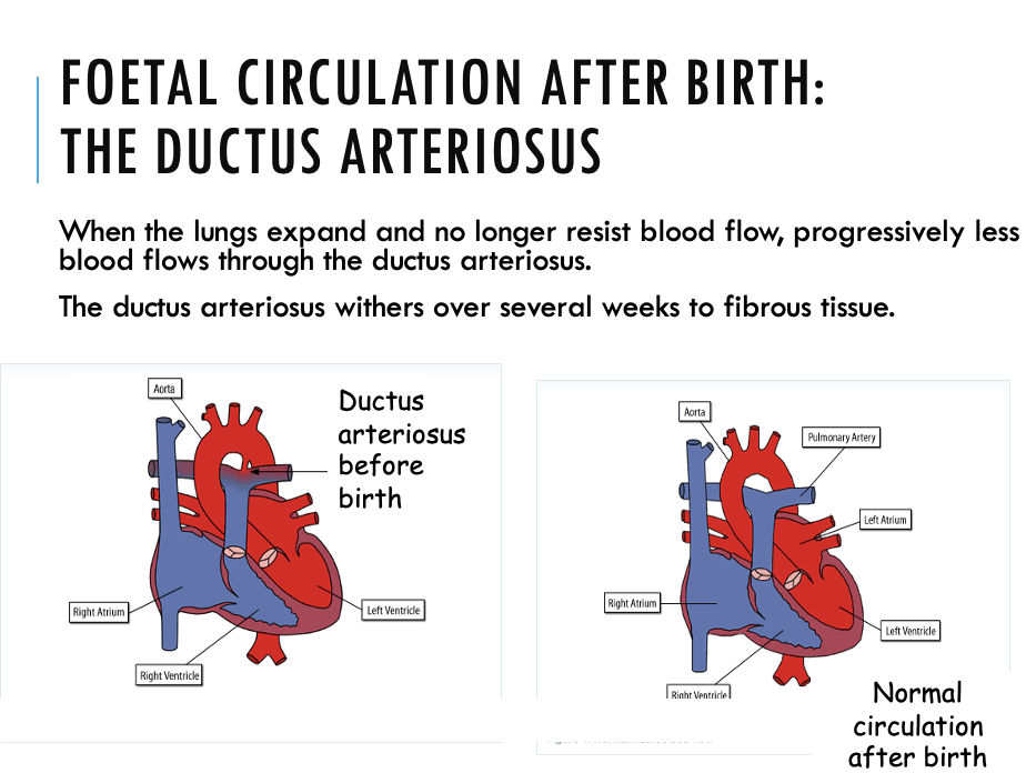
FOETAL CIRCULATION AFTER BIRTH: THE FORAMEN OVALE
Progressively more blood returns to the left atrium of the heart from the lungs once the lungs expand.
The blood pressure in the left atrium increases and forces the flap of the foramen ovale against the septum.
The hole fuses closed.
If it does not, (‘hole in the heart’) surgical correction is needed to stop oxygenated & deoxygenated blood mixing.
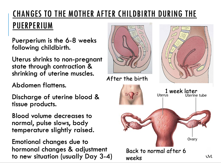
CHANGES TO THE MOTHER AFTER CHILDBIRTH DURING THE PUERPERIUM
GAMETOGENESIS
Gametes are the sex cells.
•Male: spermatozoon (sperm)
•Female: ovum (egg).
Gametogenesis is gamete development, from meiosis to the development of a mature gamete.
Spermatogenesis is the development of spermatozoa.
Oogenesis is the development of ova.
Gametes are produced by meiosis and cell development.
Gametes
Gametes are haploid (having half the normal human diploid number of 46 chromosomes)
• There are 23 chromosomes in each gamete.
Sexual reproduction involves
• fusion of the nucleus of a haploid male gamete (sperm) with the nucleus of a haploid female gamete (ovum) to form a diploid zygote.
• Development of the zygote and growth by mitosis into a new human.
Spermatogenesis

Maturation of spermatozoa
Spermatids mature into spermatozoa in the epididymis
Much of the cytoplasm is lost
Cells are nourished by specialised cells extending from the outer lining of the seminiferous tubules to the central lumen.
Spermatozoa develop a tail containing contractile material