PITUITARY GLAND HISTOLOGY
1/28
There's no tags or description
Looks like no tags are added yet.
Name | Mastery | Learn | Test | Matching | Spaced |
|---|
No study sessions yet.
29 Terms
What are endocrine glands composed of?
What do secretory cells do?
Describe the cells of endocrine glands?
composed of islands of secretory epithelial cells with intervening connective tissue, rich in blood and lymphatic capillaries.
discharge hormones into the interstitial spaces, and it is rapidly absorbed into the circulatory system.
Cells of endocrine glands have prominent nuclei and abundant mitochondria, endoplasmic reticulum, Golgi bodies, and secretory vesicles.
Describe the four main groups of hormones?
Four main groups of hormones:
Protein and glycoprotein molecules
insulin, GH, PTH
Small peptide molecules
vasopressin (ADH), and products of enteroendocrine cells.
Amino acid derivatives
thyroxine, EPI & NE
Steroids derived from cholesterol
adrenal cortical hormones, ovarian and testicular hormones.
Where are hormone receptors located and describe which hormone uses each type of hormone receptors
Hormone receptors are located on:
cell membrane
protein, peptide, and catecholamines hormones (nonsteroid hormones)
Cytoplasm
Steroid hormone
Nucleus
Thyroid hormone
What are the three parts of the endocrine system?
3 Parts:
The major endocrine organs
Major function of the organ = synthesis, storage, and secretion of hormones
possess an extensive vascular supply that is rich in fenestrated capillaries
Endocrine components within other solid organs,
Ex: the endocrine components of the pancreas, ovary, testis, and kidneys, are found in the form of clusters of endocrine cells within other tissues.
The diffuse endocrine system
scattered individual cells or small clumps of cells usually within an extensive epithelium e.g GI & Respiratory tracts
function = paracrine
Differentitae between endocrine, paracrine, and autocrine
Endo: hormones are discharged from a cell into the bloodstream and transported to effector cells
Para: hormones act on target cells in their immediate vicinity
Auto: hormones act on the same cells that produce them
Describe the Pituitary Gland (AKA? Location?)
How is it divided?
How is it controlled?
AKA: hypophysis,
@ base of brain beneath the 3rd ventricle,
sitting in a bony cavity in the base of the skull (sella turcica).
………………………………………………………………………………………………………………………
divided into anterior and posterior parts, which have different embryological origins, functions, and control mechanisms.
………………………………………………………………………………………………………………………
Control:
Hypothalamus Hormone Secretion
Negative Feedback Mechanism
What are the two functional groups of Pituitary Hormones?
List out the pituitary-dependent endocrine glands?
Direct Hormones:
Hormones which act directly on non-endocrine tissues
GH, prolactin, ADH, oxytocin, and melanocyte-stimulating hormone (MSH).
Trophic Hormones:
Hormones which modulate the secretory activity of other endocrine glands
TSH, ACTH, FSH, LH.
………………………………………………………………………………………………………………………
thyroid gland, adrenal cortex, and gonads are pituitary–
dependent endocrine glands
What is the anterior pituitary gland also known as?
Where did it arise from?
What divides the anterior pituitary and what are the three parts?
Origin/Name:
AKA: Adenohypophysis
arises as an epithelial outgrowth from the roof of the primitive oral cavity known as the Rathke’s pouch. This specialized glandular epithelium, wrapped around the anterior aspect of the posterior pituitary,
…………………………………………………………………………………………………………………
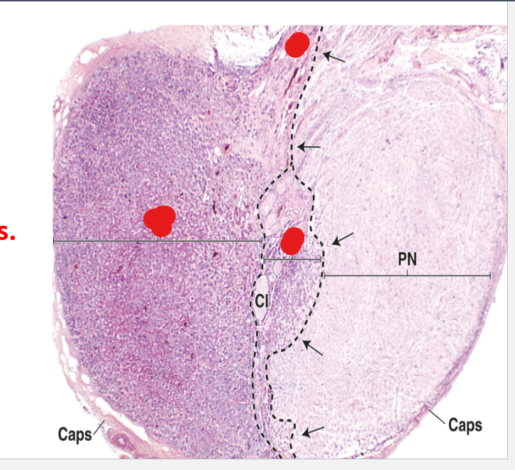
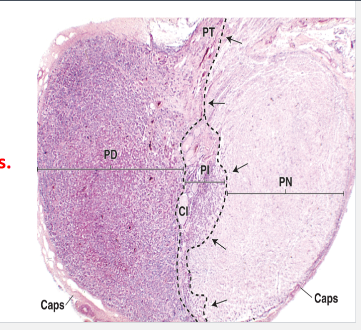
The remnants of Rathke’s pouch divides the anterior pituitary into 3 parts:
Pars distalis
Pars tuberalis
Pars intermedia
Describe the Pars Distalis
Components?
Desribe the different types of cells
Chromophils? Acidophils? Basophils Chromophobes?
~75% of the adenohypophysis has a thin fibrous capsule.
components = cords of well-stained endocrine cells interspersed with fenestrated capillaries and supporting reticular connective tissue.
Two different types of cells with different staining affinities are seen :
Chromophils:
Acidophils (the DIRECT hormones):
SOMATOTROPHS (somatotropic cells): secrete GH
LACTOTROPHS (lactotropic cells): secrete prolactin
Basophils (basically the trophic hormones):
CORTICOTROPHS: ACTH
GONADOTROPHS: FSH/LH
THYROTROPHS: TSH
Chromophobes:
stain weakly, have a few secretory granules, represent a heterogeneous group of cells including stem cells & undifferentiated progenitor cells.
Differentiate between Gonadotrophs and Corticotrophs
Gonadotrophs: secrete 2 different glycoproteins, follicle-stimulating hormone (FSH) and luteinizing hormone (LH).
Corticotrophs: secrete adrenocortical trophic hormone (ACTH) and β-lipoprotein (β-LPH).
*In the Pars Distalis*
What is the pars tuberalis? What types of cells comprised the majority of this place?
It is a smaller funnel-shaped region surrounding the infundibulum of the neurohypophysis.
Most of the cells of the pars tuberalis are gonadotrophs.
Where is the Pars Intermedia?
What does it contain?
When is it best developed/active?
What does it secrete?
Location: A narrow zone lying between the pars distalis and the pars nervosa (posterior pituitary).
Contains:
basophils (corticotrophs),
chromophobes
small colloid-filled cysts derived from the embryonic hypophyseal pouch.
Best developed and active during fetal life.
Corticotrophs of Pars Intermedia secrete:
smaller peptide hormones (ex: melanocyte-stimulating hormone (MSH))
γ-LPH
β-endorphin
***Overall functional significance of the pars intermedia remains uncertain.***
Describe the cells of the Anterior Pituitary.
cells are round to polygonal in shape with a prominent nucleus that has one or more nucleoli.
…………………………………………………………………………………………………………………
These cells synthesize and secrete protein hormone >> have a prominent Golgi complex, many mitochondria, an extensive rough endoplasmic reticulum, and secretory granules (vesicles).
…………………………………………………………………………………………………………………
Secretory vesicles scattered in the cytoplasm or near the cell surface are discharged by exocytosis close to thin- walled and highly permeable fenestrated capillaries.
Describe the control of hormone secretion in the Anterior Pituitary
Controlled by? Secreted by? Released from? Transported by?
Function of these hormones?
Which hormones are inhibitory?
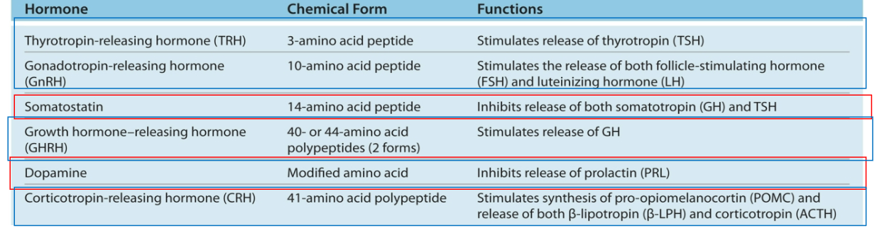
controlled by specific hypothalamic releasing hormones (peptides):
secreted by small neurons near the 3rd ventricle,
released from the axons near the median eminence
transported by capillaries of the portal system throughout the anterior pituitary.
…………………………………………………………………………………………………………………
Most of these releasing hormones are stimulatory, which stimulate the secretion by specific anterior pituitary cells.
…………………………………………………………………………………………………………………
Two of the hypothalamic releasing hormones are inhibitory; (Somatostatin/Dopamin)
Describe the 6 hypothalmic releaseing hormones and their functions
Beside the hypothalamus, what other factors controls the secretions of the pituitary gland?
By negative feedback
by other hormones,
Ex: ghrelin secreted by the stomach mucosa → growth hormone secretion,
oxytocin from the posterior pituitary → prolactin during breastfeeding.
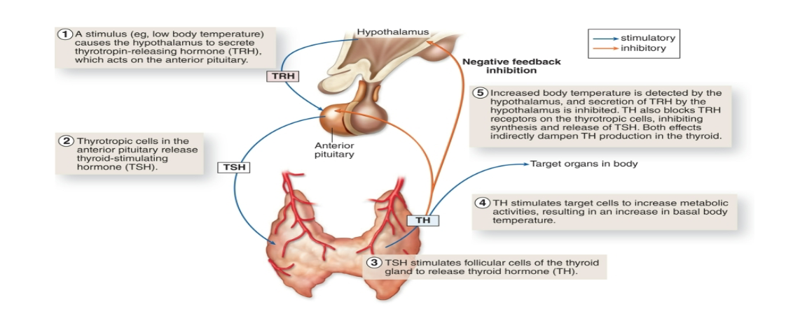
Describe the example of temperature and negative feedback on the hypothalamus/pituitary gland
What is a general consequence of Benign pituitary adenomas? What can happen if there is an adenoma involving the somatotropic cells?
Benign pituitary adenomas often produce excessive numbers of functional acidophils and basophils.
Adenomas involving the somatotropic cells → gigantism in children or acromegaly in adults
with associated musculoskeletal, neurologic, and other medical consequences.
Describe the Composition and function posterior pituitary.
Consists of?
Describe its neurons?
What does it not contain????? ****
What is the most abundent cells in the posterior pit.
Function?
Composition:
Consists of pars nervosa & infundibular stalk.
composed of neural tissue, containing some 100,000 unmyelinated axons of large neurons with cell bodies in the supraoptic and paraventricular nuclei of the hypothalamus.
does not contain the cells that synthesize the hormones
Glial cells called pituicytes are also present, which resemble astrocytes, and are the most abundant cells in the posterior pituitary.
Function:
Not an endocrine gland.
It is a storage site for neurosecretions of the neurons of the supraoptic and paraventricular nuclei of the hypothalamus.
Describe how the posterior pituitary can secrete ADH and oxytocin despite not producing it themselves?
Production:
secretory neurons synthesize ADH and oxytocin
accumulate in axonal dilatations called neurosecretory bodies or Herring bodies
can be visible in the light microscope as lightly eosinophilic structures.
Herring bodies contain a carrier protein called neurophysin
Secretion:
Nerve impulses along the axons trigger the release of peptides from neurosecretory bodies, taken up by fenestrated capillaries of the pars nervosa, and the hormones are then released into the general circulation
Describe the ultrastructure of the posterior pituitary
Axonal swellings have prominent mitochondria and many microtubules for axoplasmic transport.
…………………………………………………………………………………………………………………
Abundant sinusoidal capillaries with fenestrations are also seen. These capillaries are near the axonal terminal, filled with secretory vesicles, hence facilitating the axonal discharge of hormones and the rapid distribution of their contents into the circulation.
Describe the conditions for which ADH is released and its effect? AKA?
Condition of release:
increased blood tonicity, sensed by osmoreceptor cells in the hypothalamus.
…………………………………………………………………………………………………………………
Effect:
increases permeability of renal collecting ducts to water → water reabsorption is increased → osmotic balance of body fluids restored.
…………………………………………………………………………………………………………………
ADH is also called arginine vasopressin.
What can happen if a head trauma affects the posterior pituitary?
Any head trauma affecting the posterior pituitary can lower the production of ADH → Diabetes Insipidus → inability to concentrate urine:
frequent urination (polyuria)
excessive thirst (polydipsia).
What are the effects of oxytocin? Conditions for release of oxytocin?
Effect:
stimulates contraction of uterine smooth muscle during childbirth
acts on the myoepithelial cells in the mammary gland alveoli.
produces psychological effects, such as promoting pair-bonding behavior
…………………………………………………………………………………………………………………
Condition of Release:
nursing infant induces oxytocin secretion by stimulating sensory tracts that act on the hypothalamus (supraventricular nuclei), causing rapid ejection of milk.
Describe the connection between the hypothalamus and the adenohypophysis
Connection between the hypothalamus and the adenohypophysis is vascular.
The vascular portal system carries the small regulatory peptides from the hypothalamus to the adenohypophysis.
Describe the blood supply of the pituitary gland
Pituitary gland receives its blood supply from the right and left superior hypophyseal arteries (branches of the internal carotid arteries), serving:
median eminence,
pars tuberalis
infundibulum.
Pars nervosa receives its blood supply from the right and left inferior hypophyseal arteries.
Describe the hypothalamic–hypophyseal portal system
hypothalamic–hypophyseal portal system:
The 2 superior hypophyseal arteries give rise to the
hypothalamic–hypophyseal portal systemcomposed of two fenestrated capillary beds that are connected to each other by a set of portal veins.
……………………………………………………………………………………………………………………
Function:
crucial for the control of the adenohypophysis by neurosecretions from hypothalamic neurons, which convey releasing and inhibiting hormones to the primary plexus.
……………………………………………………………………………………………………………………
Primary Capillary Plexus:
Superior hypophyseal arteries give rise to the primary capillary plexus located in the median eminence
receive the releasing hormones produced by the hypothalamic neurons
Hypophyseal portal veins drain the primary capillary plexus and deliver the blood containing these releasing hormones into the secondary capillary plexus.
……………………………………………………………………………………………………………………
Secondary Capillary Plexus:
in the pars anterior, where these releasing hormones regulate the hormone secretion by the parenchymal cells.
The hormones released by the cells of the adenohypophysis are carried away by the blood from the secondary capillary plexus for systemic distribution
……………………………………………………………………………………………………………………
***Important general concept: this portal system is how the hypothalamus and ant. pituitary communicate w/ each other***
Describe how the posterior pituitary communicate w/ the hypothalamus
Hypothalamic–Hypophyseal Tract (H-H Tract):
Posterior pituitary has a direct neural connection to the
hypothalamus via a bundle of axons called the hypothalamic–
hypophyseal tract, which courses into the neurohypophysis from two important hypothalamic nuclei.
Mechanism of tract:
ADH; synthesized in supraoptic; Oxytocin: synthesized in paraventricular nuclei
Both hormones → axonal transport → accumulate (temporarily) in axons of (H-H Tract) → release and uptake by large capillaries branching from the inferior hypophyseal arteries.