Electrophysiology Methods
 Call Kai
 Learn
Learn Practice Test
 Spaced Repetition
 Match
Match Flashcards
 Knowt Play
1/58

There's no tags or description
Looks like no tags are added yet.
Name | Mastery | Learn | Test | Matching | Spaced | Call with Kai | Chat |
|---|
No analytics yet
Send a link to your students to track their progress
59 Terms
Electrophysiological Methods
single cell recordings
electroencephalography (EEG)
event-related potentials (ERP)
Magnetoencephalography (MEG)
Brain Imaging Methods
MRI
fMRI
Brain Stimulation Techniques
Transcranial Magnetic Stimulation (TMS)
Transcranial Direct Current Stimulation (tDCS)
Intercranial Stimulation
Single Cell Recordings
electrode(s) placed in or near a neuron (invasive)
Measure number of action potentials per second
after establishing a baseline firing rate for a given cell, researchers then determine what properties of a stimulus make the cell fire maximally above that baseline (e.g., light, movement, etc.)
Event-Related Potentials (ERP)
electrode(s) placed on the skull
measures change in voltage at the scalp
summed electrical potentials from thousands of neurons (sensitive to dendritic currents)
Single Cell Recordings Example: Fields
a specific region of sensory space in which an appropriate stimulus can drive an electrical response in a neuron

Single neuron recordings in humans
gnostic cells/grandmother cells —> neurons that respond only to a highly complex, specific, and meaningful stimulus, such as the image of one’s grandmother or other specific object categories
discoveries through single cell recording in epileptic patients (e.g., Quian Quiroga et al., 2008)
Jennifer Aniston neuron
Multimodal invariance
Friends neuron, Star Wars neuron
Reflects person’s knowledge, not world knowledge
Probably not just one neuron, rather gnostic fields (sparse coding)
Single-cell recording - how good?
Advantages:
great spatial resolution
great temporal resolution
Disadvantages:
samples only a very small fraction of a functional neural system
invasive (mostly restricted to animal studies)
What signals do single cell recordings capture?
A. action potentials
B. neurotransmitter release
C. postsynaptic potentials
Single-cell recordings have
A. poor temporal, but excellent spatial resolution
B. excellent temporal and spatial resolution
C. excellent temporal but poor spatial resolution
Electroencephalography (EEG) - break down the word
Electro - electrical
encephalo - brain
graphy - picture
Electroencephalography (EEG)
records electrical activity of the brain via metal electrodes positioned on the scalp
each electrode acts as its own recording site
electrical potential is recorded at the scalp as a waveform having
a particular voltage/amplitude (a measure of its size)
a particular frequency (measured in Hertz or cycles per second)
Origin of signal: summed postsynaptic potentials of similarly aligned neurons
Neurophysiological Basis of EEG
the EEG signal reflects the summation of synchronous synaptic activity around a population of post-synaptic neurons with similar spatial orientations
Local EPSP
as depolarizing current (positive charge) flows into the neuron, the extracellular space becomes more negatively charged
Pyramidal Neurons
apical dendrites contribute strongest signals measurable with EEG
perpendicular to surface
uniform, columnar organization
long enough to form dipoles

Formation of EEG Signals from EPSP and IPSP

Factors that Affect Magnitude and Direction of EEG Signal
(all occurring in post-synaptic neurons)
magnitude and direction of dipole effects in individual neurons
density of parallel dendrites (typical signal from 500k - 1M neurons)
Synchrony of dipole effect in neighboring neurons
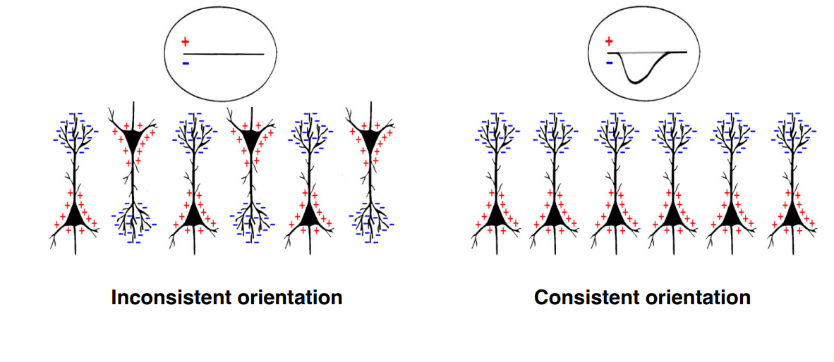
Orientation relative to scalp and other neurons
Magnitude and Direction of Dipole Effects in Individual Neurons
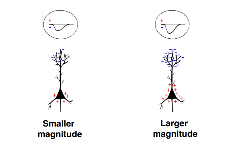
Density of Parallel Dendrites

Synchrony of Dipole Effect in neighboring Neurons
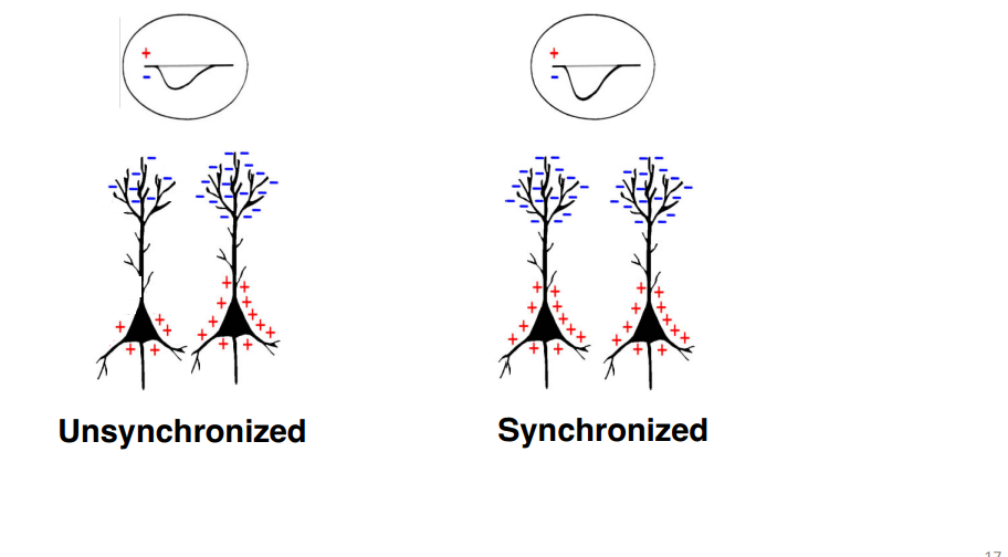
Dipole Orientation Relative to Scalp

Dipole Orientation in Neighboring Neurons
Summary of EEG Signal
To measure the electrical activity of the brain from scalp recordings, it must be of sufficient strength and duration
Action potentials
No
The EEG signal (from scalp recordings) are mainly post- synaptic potentials (PSPs) of cell body and large dendrites of pyramidal neurons.
Weaker than action potentials
Longer duration
The column structure (summation rather than cancellation)
Extracellular current
EEG profiles
EEG can detect different normal brain states
These states vary by frequency and pattern of oscillations

EEG: Abnormal Activity
EEG can detect abnormal brain states, such as epilepsy
in epilepsy, large quantities of neurons fire randomly at once
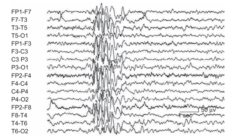
Event-Related potentials (ERPs)
EEG recordings provide a continuous measure of brain activity
ERPs are recorded in reference to a specific event: voltage changes that are related to brain’s response to a stimulus
Low signal to noise ratio!
Size of variation in EEG signal induced by event small compared to ongoing EEG signal.
Detecting ERPs requires signal averaging, which averages waveforms from multiple trials so they can be seen against variations in EEG from trial to trial.
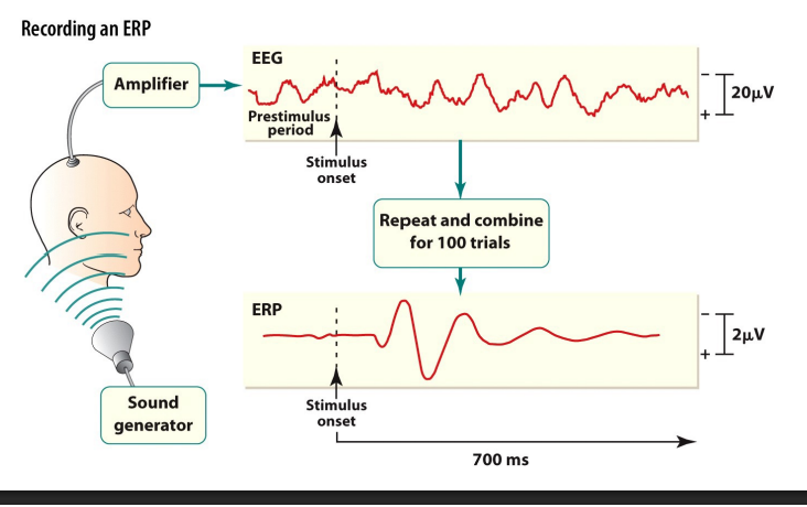
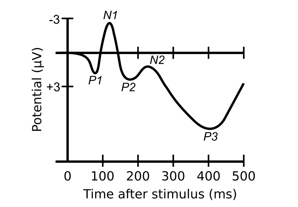
ERP: Wavelength Components
Waveforms can be divided into components, characteristic portions of the wave linked to certain psychological process.
Components: a letter & a subscript number.
The letter is a P or an N to denote whether the deflection of the electrical signal is positive or negative.
The number shows, the order of events or how many milliseconds after the stimulus the component appears.
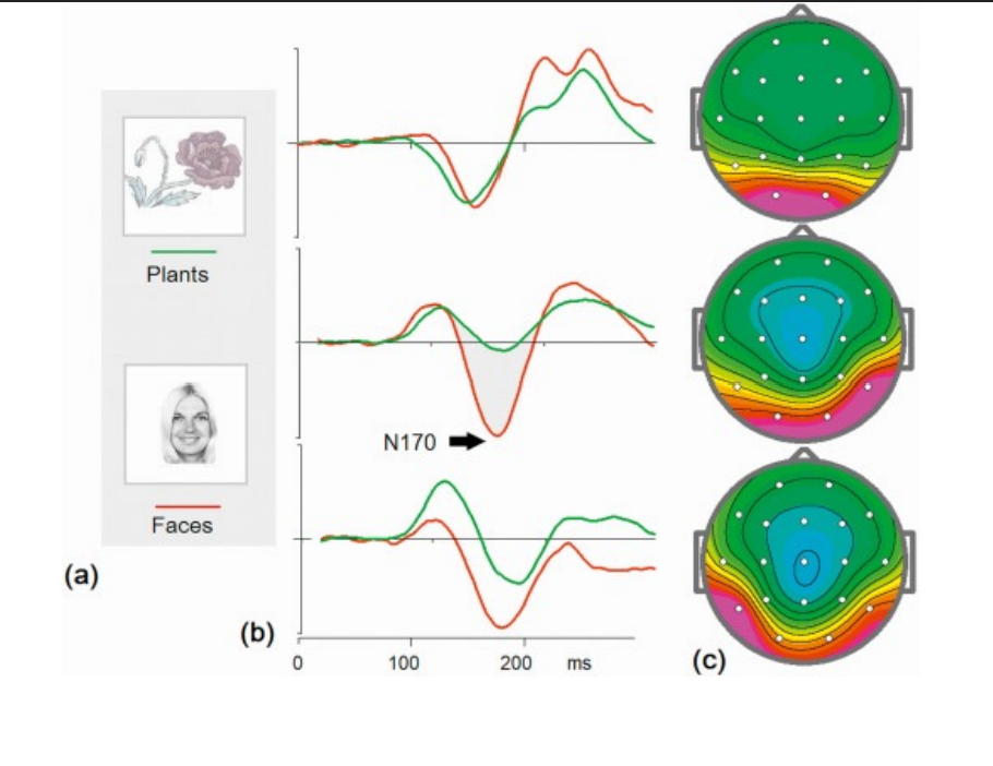
ERP: Plants and Faces
ERP: How Good?
ERP has an excellent temporal resolution
ERP signal is directly related to neural activity and this electrical activity is conducted instantaneously from the scalp
ERP has a poor spatial resolution
The ERP signal is derived from different sources in the brain and it is not possible to infer exactly where these sources are from the scalp
What does the ERP component N170 signify?
a. a negative peak at 170s after stimulus onset
b. a negative peak at 170ms after stimulus onset
c. a negative peak with a magnitude of 170mV
d. a negative peak caused by activity of 170 neurons
EEG can be used to measure/diagnose which of the following? (select all that apply)
a. subcortical brain lesions
b. epilepsy
c. brain death
d. sleep stages
EEG has excellent temporal resolution
• True
• False
EEG has excellent spatial resolution
• True
• False
Magnetoencephalogram (MEG)
Magnetic equivalent of EEG
Electricity and magnetism are intrinsically linked
Sources of MEG Signal
right hand rule
The MEG signal is also mainly post-synaptic potentials (PSPs) of cell body and large dendrites of pyramidal
Mainly from sulci of cortex
vs EEG: gyri and sulci
EEG signal is stronger than MEG signal
MEG has relatively better spatial resolution than EEG

Why MEG?
Advantages:
High temporal resolution: milliseconds
Direct link to neural activity: post-synaptic potentials
Little effect of skull/scalp
Slightly better spatial resolution than EEG
Disadvantages
High cost (still cheaper than MRI scanner, but much more expensive than EEG)
Poor portability
Weaker signal than EEG
MEG Summary
MEG signal also mainly comes from postsynaptic potentials of pyramidal neurons (like EEG)
High temporal resolution
Slightly better spatial resolution than EEG (inverse problem)
Weaker signal than EEG
MEG signal mainly from the sulci of cortex than gyri
More expensive than EEG
Clinical applications: epilepsy and surgical planning
Transcranial Stimulation
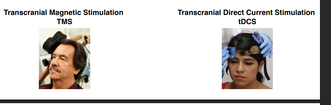
TMS Mechanism
Induces electric current in brain
Disrupts neural circuits with rapid changes to electric currents:
Facilitation/Excitation – high frequency stimulation (~20 Hz)
Suppression/Inhibition – low frequency stimulation (~1 Hz)
“Virtual” lesions
Mostly non-invasive and safe, minimal pain

TDCS Mechanism
Applies electric current to specific brain areas
Current flows between electrodes
Placement of electrodes determines type of disruption
Facilitation/Excitation – positive anodal depolarizes
Suppression/Inhibition – negative cathodal hyperpolarizes
Relatively non-invasive, painless

From Correlation to Causation
MEG measures neural correlates of cognitive phenomenon
TMS drives neuronal activity, causing changes in behavior
Brain Imaging: Structural & Functional
Structural
looking at brain structure independent of function, based on different physical properties of different tissues
Computed Tomography (CT)
Magnetic Resonance Imaging (MRI)
Functional
Revealing brain structures that are active during specific tasks
Positron Emission Tomography (PET)
Functional Magnetic Resonance Imaging (fMRI)
Structural Magnetic Resonance Imaging (MRI)
Body contains 70% water. MRI signal picks up electromagnetic energy released from hydrogen atoms.
MRI scanner = powerful electro-magnet (a field strength of 3 teslas is about 50,000 times greater than the Earth’s magnetic field).
Magnetic field inside the scanner affects the magnetic nuclei of atoms. Normally atomic nuclei are randomly oriented but under the influence of a magnetic field the nuclei become aligned with the direction of the field.
Orientation of nuclei is disturbed by applying RF pulses. When pulse is turned off, hydrogen atoms realign with magnetic field while releasing electromagnetic energy.
The signal from hydrogen nuclei varies in strength depending on the surroundings. This provides a means of discriminating between grey matter, white matter and cerebral spinal fluid in structural images of the brain.

Neuroplasticity - London Taxi Drivers
Maguire et al. (2206)
“Thus, we have found that London taxi drivers have greater gray matter volume in the mid-posterior hippocampi and are better at identifying London landmarks and knowing their proximal relations than London bus drivers. By contrast, taxi drivers have less gray matter volume in their anterior hippocampi and are worse at acquiring or retrieving new visuospatial information.” (Maguire et al., 2006, p. 1095)

Functional Magnetic Resonance Imaging (fMRI)
Neural activity consumes oxygen and glucose
About 20% of the oxygen and 25% of the glucose consumed by the human body are dedicated to cerebral functions, yet the brain represents only 2% of the total body mass.
To compensate for increased oxygen consumption, more blood is pumped into the active region
The time taken for this response is slow (several seconds) - functional imaging has poor temporal resolution, but good spatial resolution

fMRI exploits the need of increased blood flow to active regions
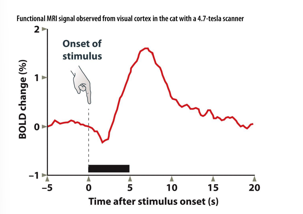
fMRI signal based on ratio between deoxygenated/oxygenated hemoglobin: BOLD response (Blood Oxygen Level Dependent effect)
The change in BOLD response over time is called the hemodynamic response function
The Hemodynamic Response Function peaks 6–8 seconds after stimulus onset
limits the temporal resolution of fMRI

Hemoglobin
Hemoglobin carries oxygen
Oxygenated hemoglobin is diamagnetic (repelled by magnet)
Deoxygenated hemoglobin is paramagnetic (attracted to magnet)
Hemodynamic Response Function
Advantages and Disadvantages of fMRI
Advantage
best spatial resolution
Noninvasive
broader spatial coverage than single-cell recordings
Disadvantage
poor temporal resolution
expensive
What Does it Mean to Say a Brain Region is "Active"?
The brain has a constant supply of blood and oxygen; if it didn’t, it would die
To infer functional specialization, one needs to compare RELATIVE differences in brain activity between two or more conditions
This involves selecting a baseline or comparison condition
A region is "active" if it shows a greater response in one condition relative to another
Subtraction method: compare activity in experimental and control task that differ only in the process of interest.
fMRI Study Designs: Block Design
Participant is presented with a stimulus or engages in a task during a block of time (block 1) and a control stimulus/process during another block of time (block 2)
collapsing across many trials attains adequate signal-to-noise ratio • good for finding regions of interest
good for continuous tasks (i.e., reading of a narrative or continuous finger-tapping task)
Blocked designs cannot be used if we want to consider trials that depend on subject’s performance (e.g., correct or wrong; chooses among different alternatives) or need to present trials in a non- blocked fashion (e.g., the oddball paradigm).

fMRI Study Designs: Event-related design
Read out BOLD response to specific stimuli
allows for greater flexibility and randomization
conditions can be defined by participant behavior (e.g. correct/incorrect trials)
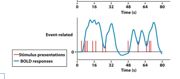
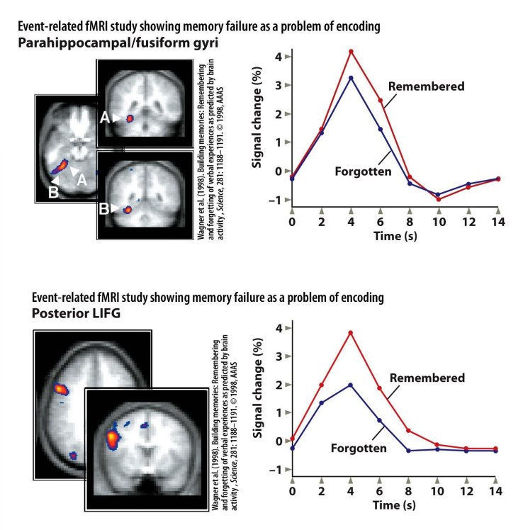
Event-Related Design: Is memory failure due to encoding or retrieval failure?
Wagner et al. 1998
Block Design: Faces and Objects

Subtraction Method Exercises
compare brain activity between two conditions that differ only in the specific variable of interest
Subtraction Method: You want to know what brain areas are specifically active when a person views photos of a romantic partner.
comparison condition?
Guess what brain regions would be commonly activated across both conditions, and what would be uniquely activated in the experimental condition.

Subtraction Method: what brain areas are specifically active when a person uses their visual imagination. You have people listen to a series of words (e.g., horse, apple, etc). For each word they are told to imagine the object.
comparison condition?
Guess what brain regions would be commonly activated across both conditions, and what would be uniquely activated in the experimental condition.

fMRI study on lying

You want to know what brain area is specifically active when a person looks at emotionally charged images.
comparison condition?
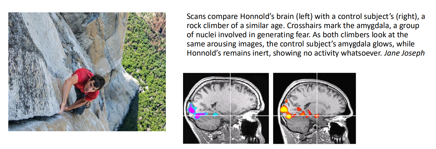
Guess what brain regions would be commonly activated across both conditions, and what would be uniquely activated in the experimental condition.