BSCI222
1/73
There's no tags or description
Looks like no tags are added yet.
Name | Mastery | Learn | Test | Matching | Spaced | Call with Kai |
|---|
No analytics yet
Send a link to your students to track their progress
74 Terms
Genetics
The study of heredity and unders; how genetic information (variable traits) is passed down (transmitted) through generations
Heritable variation
Differences in phenotype/traits
Ex. siblings look similar, but not exactly
Gene
A unit of heritable information for a trait; genes encode information for a heritable trait
Alleles
variants of a single gene
Ex. Gene: hair
Allele: white, black, brown coat color
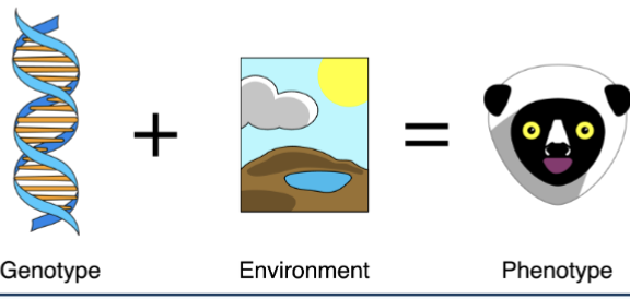
Genotype
The combination of alleles an organism has
Phenotype
The trait(s) an organism displays
What is phenotype influenced by?
Phenotype is determined by both genotype AND the environment
Ex. Genotype: A person may inherit genes that predispose them to be tall.
Environment: Nutrition and overall health during childhood.
Outcome (Phenotype):
Two people with similar “tall” genes can end up different heights if one had good nutrition growing up and the other did not.
What is genetic information coded by?
All genomes are encoded in DNA (deoxyribonucleic acids) and RNA (ribonucleic acids); nucleic acids are polymers of nucleotides (a sugar, a phosphate group, and a nitrogenous base (A, G, C, T, or U)
Nucleotides link together to form long strands of nucleic acids, which carry genetic instructions
DNA - stores long-term genetic information
RNA - helps use that information to make proteins
The sequence of nucleotides code genetic information
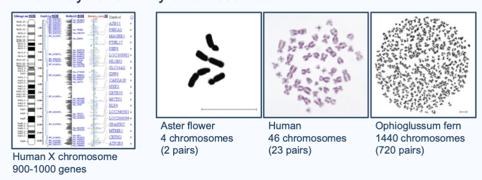
Where are genes found?
On chromosomes; many genes are on one chromosome, and a cell may have many chromosomes
Ex. The human X chromosome has 900-1000 genes
Aster flowers have 4 chromosomes (2 pairs), Humans have 46 chromosomes (23 pairs), and Ophioglussum fern have 1440 chromosomes (720 pairs)
How many chromosomes to humans have?
46 chromosomes (23 pairs)
What does cell division do?
Chromosomes are copied by DNA replication
copies of chromosomes separate during cell division
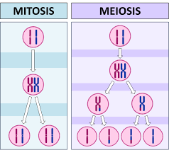
Mitosis - copies of each pair of chromosome go to daughter cells
Meiosis - half of each chromosome pair goes to each cell
Information flows from…
DNA to RNA to protein
DNA replication duplicates information prior to cell division
Transcription copies information from DNA to RNA
Translation uses information in RNA to build polypeptides
Mutations
changes in DNA resulting in changes to protein
Quantitative traits
measurable phenotypes, such as height, weight, or blood pressure, that exhibit continuous variation within a population rather than discrete differences
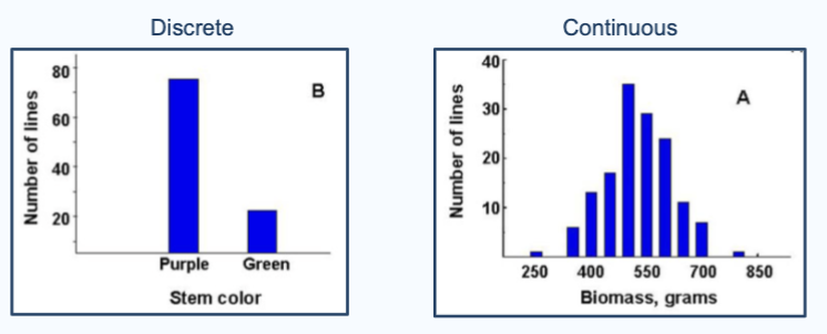
What are quantitative traits influenced by?
Many genes and environment
How do traits evolve?
Through changes in allele frequency; mutations create new alleles, and alleles can change in frequency in a population over time through natural selection, sexual selection, and drift
Cell division
Make duplications of genetic material (copy of chromosomes) and then separate and move them into daughter cells
a parent cell divides into two (mitosis) or four (meiosis) daughter cells
One cell —> 2 cells (2 copies of DNA then splits up between 2 cells)
What happens during cellular reproduction (cell division)?
One cell —> 2 cells (2 copies of DNA then splits up between 2 cells)
Before division: a cell must grow, chromosomes must be replicated
During division: 1) cell contents (ex. mitochondria) must be split between daughter cells, and 2) chromosome copies (DNA) must be split between daughter cells
Cell division in prokaryotes
Binary fission - process when a prokaryotic cell reproduces, and its singular circular chromosome replicates, and the cell divides
Cell growth and DNA replication are COUPLED (replication of dna, movement of dna, and growth of the cell are all connected)
What does it mean by cell growth and RNA replication are coupled?
They happen at the same time and are directly linked
In bacteria (binary fission):
There is no nucleus
There are no distinct cell-cycle phases like G1, S, G2, M
DNA replication starts early and continues as the cell grows
So instead of:
Grow
Replicate DNA
Divide
…it’s more like:
Grow + replicate DNA simultaneously
Segregate DNA
Divide

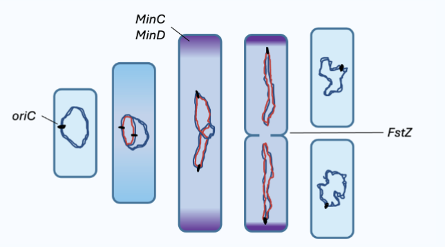
Steps of binary fission
1) DNA REPLICATION: DNA replication begins at the origin of replication (oriC), where proteins bind to the origin and connect it to the cell wall
The chromosome replicates, creating 1 additional new origin of replication
Other proteins called Min proteins interact with the DNA as it’s being replicated
2) COPY SEPARATION: The origin of the two newly replicated chromosomes move away from each other and toward opposite ends of the cell (Min proteins move to the 2 poles of the cell as the cell grows, and drag the DNA with them)
3) DIVISION OF CYTOPLASM: A new cell wall forms between the two chromosomes, producing two cells, each with an identical copy of the chromosome (FstZ proteins establish where the cell physically divides, and tehy accumulate in the center)
End result: 2 identical cells, each with 1 of the chromosomes
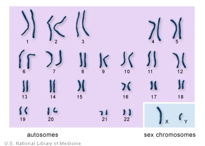
Chromosomes in eukaryotes
Most eukaryotes are sexually reproducing, and chromosomes come in homologous pairs (one from mom, one from dad)
Humans have 46 chromosomes in 23 pairs
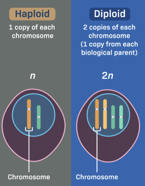
Diploid: 2N = 46; Haploid: N = 23
Diploid
Diploid (2N) means two sets of chromosomes
In humans:
46 total chromosomes
Organized as 23 pairs
One set of 23 comes from your mother
One set of 23 comes from your father
Most body (somatic) cells are diploid.
What does 2N = 46 mean?
2N → 2 sets of chromosomes and diploid
In humans, there are 46 total chromosomes
Haploid
Haploid (N) means one set of chromosomes
In humans:
23 total chromosomes
No pairs—just one copy of each chromosome
Gametes (sperm and egg) are haploid.
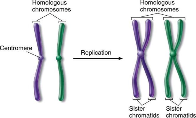
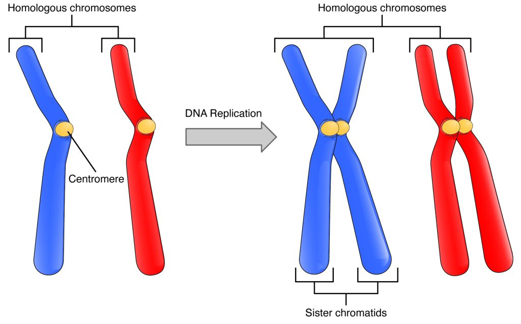
Homologous chromosomes
Pairs of chromosomes in a diploid organism that have the same content and order of genes but may contain different gene variants, or alleles
pairs of matching chromosomes in a diploid organism, with one set inherited from the mother (maternal) and one from the father (paternal). They contain the same genes in the same order, but may have different versions (alleles).
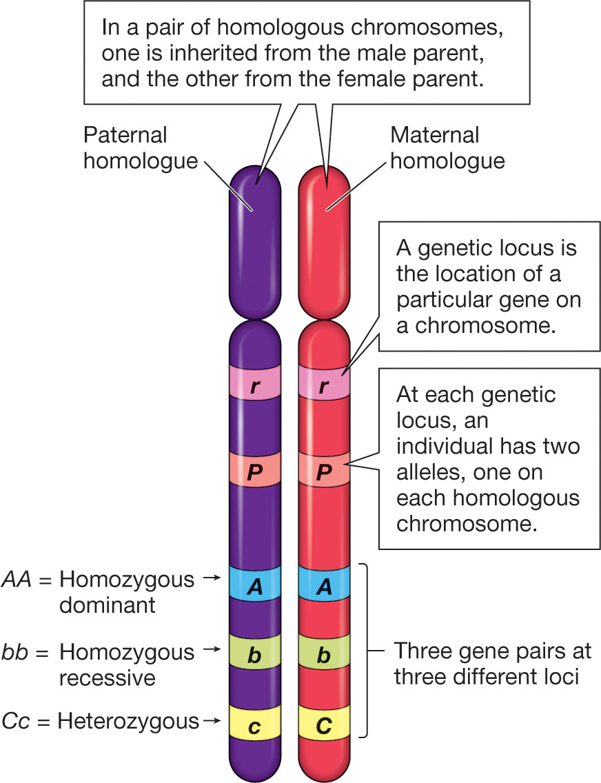
Homologous chromosomes have..
the same genes in the same order along the length of the chromosome, but may have different alleles
Ex. gene: eye color
1 from mom could be for black eyes, 1 from dad could be for blue eyes
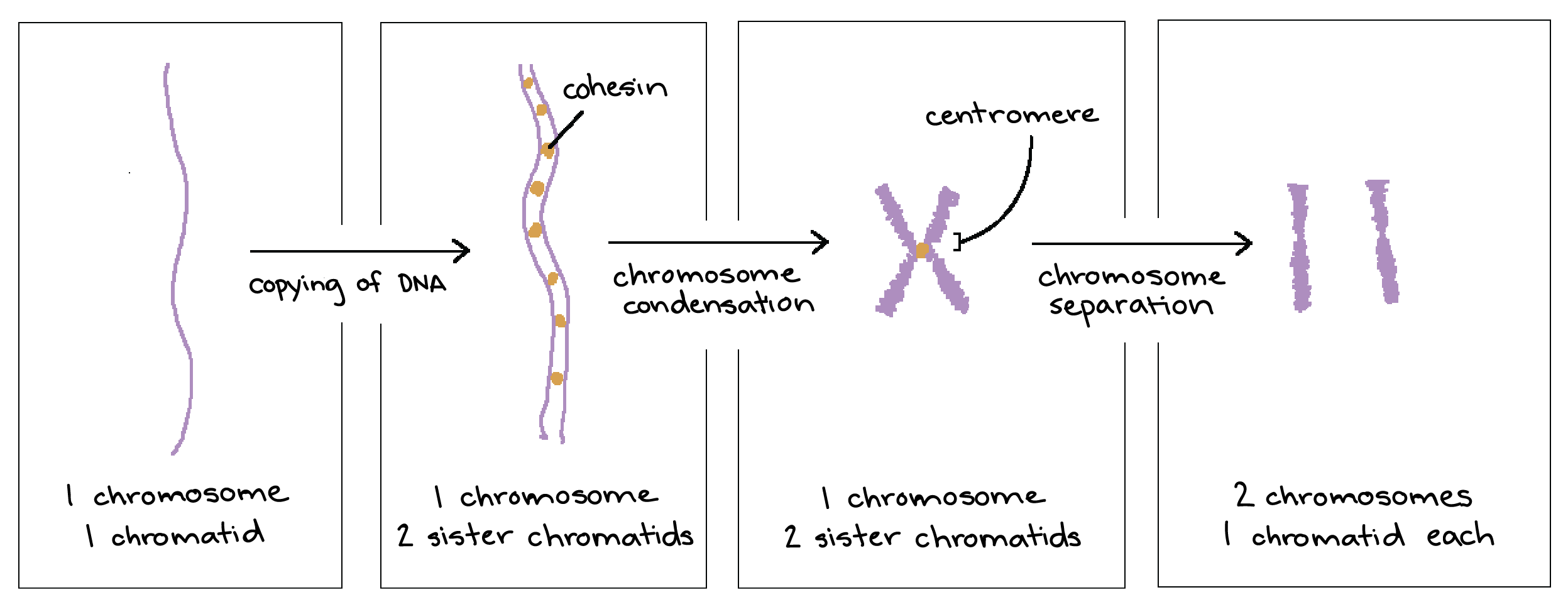
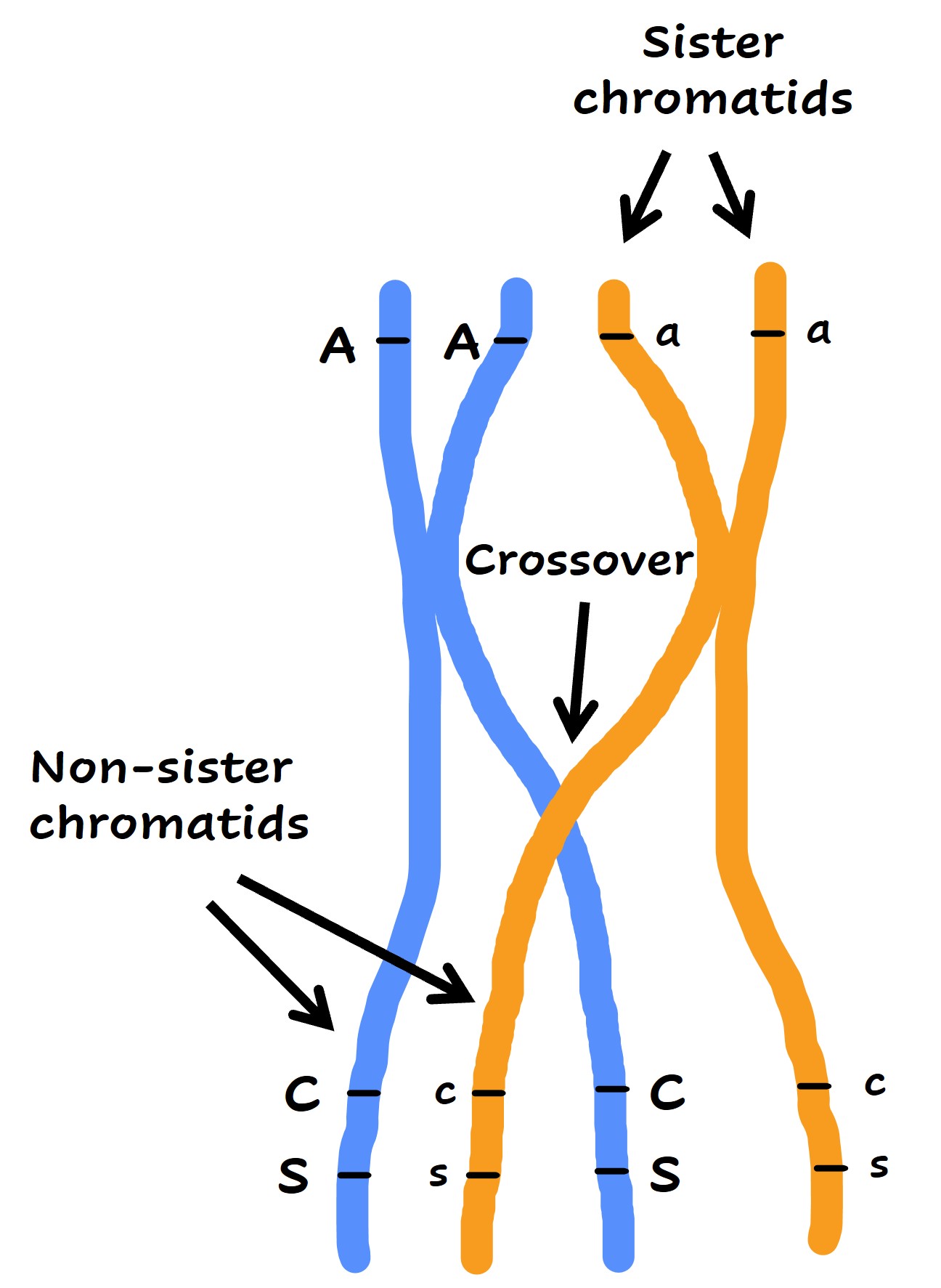
After DNA replication, each chromosome has..
two sister chromatids (two duplicated pieces of DNA)
sister chromatids
two identical copies of a single replicated chromosome, formed during the S-phase of interphase and held together by a common centromere. They are essential for cell division (mitosis and meiosis), ensuring that each daughter cell receives an exact copy of the genetic material.

Chromatin
Long, linear, loose complex of DNA and histone proteins; DNA + Proteins
DNA is wrapped around histone proteins = loose, thread-like substance
Chromatin vs. Chromosomes
Chromatin: DNA is wrapped around histone proteins and is long, linear, and loose, like a thread (relaxed state)
Chromosome: DNA is tightly coiled/condensed and organized in structures like spools; formed from chromatin during cell division (meiosis/mitosis) to ensure accurate DNA packaging and segregation into daughter cells (HIGHER LEVEL OF ORGAINZATION)
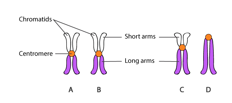
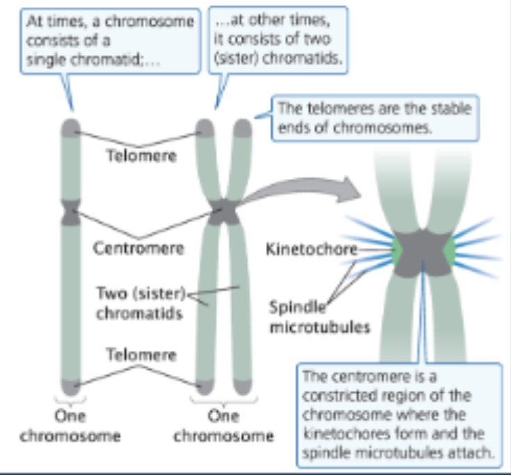
Chromosome structure
Chromatin = DNA + proteins (DNA wrapped around histone proteins) —> make long pieces of DNA
Components of a chromosome
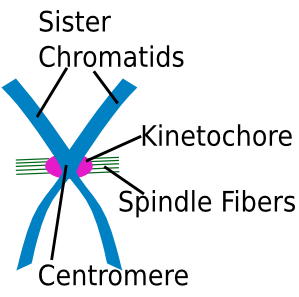
1) Centromere - region of the chromosome that joins sister chromatids together and forms the kinetochore
2) Telomeres - the tips/ends of eukaryotic chromosomes; required for complete DNA replication
3) Origins of replication - initiation site for DNA replication; multiple per chromosome
Centromere
A constricted region of the chromosome where the kinetochores form and the spindle microtubules attach
During cell division, it’s where the cell grabs the chromosome and pulls it apart (the attachment point for spindle microtubules, the filaments responsible for moving chromosomes in cell division)
Special multiprotein complex gathers there (the kinetochore), and tiny fibers (spindle microtubules) attach to the kinetochore to move the chromosome
Kinetochore
A protein complex that assembles on the centromere to bind spindle microtubules to the chromosome (anchor chromosomes to microtubules)
Telomeres
The specific DNA sequences and associated proteins located on the tips (ends) of whole linear chromosomes
Telomeres protect and stabilize the chromosome ends, like plastic tips protect the ends of a shoelace (chromosome stability)
REQUIRED FOR COMPLETE DNA REPLICATION.
Why are telomeres required for DNA replication?
They act as protective caps at the ends of linear chromosomes, preventing the loss of crucial genetic information during cell division
Origins of replication
Initiation site for DNA replication; multiple per chromosome
Mitosis
Clonal reproduction of cells, producing genetically identical daughter cells (ex. healing wounds)
Involves cell growth, replication of organelles (cytokinesis - division of cytoplasm and all of its contents, such as the mitochondria)
Separation of sister chromatids
Meiosis
Production of reproductive (sex) cells (eggs, sperm)
Reduction division: each cell gets half the chromosome
Produces genetically diverse cells
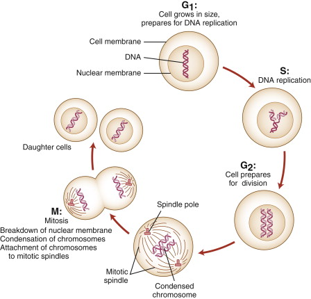
Cell cycle
Series of stages through which a cell passes from one division to the next, and genetic information for all characteristics are accurately passed from parent to daughter cells
Takes place in cells that are actively dividing; a new cycle begins after a cell has divided and produced 2 new cells
Cell cycle steps
1) Interphase
A) G1
B) S
C) G2
2) Mitosis - cell actively dividing
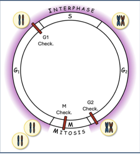
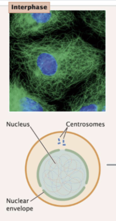
Interphase
First stage of cell cycle; extended period of growth and development between cell divisions (cell usually spends most of its time in interphase)
DNA is being synthesized, RNA and proteins are being produced, and hundreds of biochemical reactions necessary for cellular functions are taking place
Growth and development
Several checkpoints
3 stages of interphase:
1) G1 (gap 1)
2) S (DNA synthesis)
3) G2 (gap 2)
Stages of interphase
During interphase, the chromosomes are in a relaxed state but not uncoiled; individual chromosomes cannot be seen under a microscope
G1 (gap 1): the cell grows and proteins necessary for cell division are synthesized, lasting several hours
A. Critical checkpoint G1/S: holds the cell in G1 until the cell has all the enzymes and proteins necessary for the replication of DNA; the cell can divide once passing this checkpoint
B. G0: however, some cells can exit the cell cycle in response to regulatory signals and pass into the non-dividing phase G0, a stable state during which cells maintain a constant size
S (DNA synthesis): each chromosome is duplicated and is composed of 2 identical sister chromatids
G2 (gap 2): two chromatids per chromosome
G2/M checkpoint: cell passes if its DNA is completely replicated and undamaged
G1
gap 1; first stage of interphase; the cell grows and proteins necessary for cell division are synthesized
one chromatid per chromosome
S
DNA synthesis; second stage of interphase; DNA replication occurs (copy all double helix —> 2 of each; copies stay connected at the centromere X)
G2
gap 2; third stage of interphase; two chromosomes, each with 2 sister chromatids
several additional biochemical events necessary for cell division take place
Progression through the cell cycle is controlled by..
checkpoints
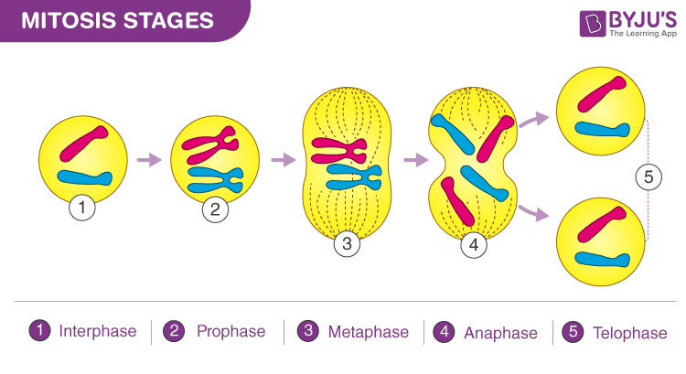
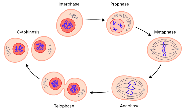
Mitosis
Second phase of the cell cycle; cell separates sister chromatids, creating two daughter cells
5 stages of mitosis:
1) Prophase
2) Prometaphase
3) Metaphase
4) Anaphase
5) Telophase
Before mitosis occurs…
Interphase occurs; DNA is replicated during the S phase of interphase and centrosomes are duplicated (organelles that organize the microtubules into the mitotic spindle for separating chromosomes during mitosis)
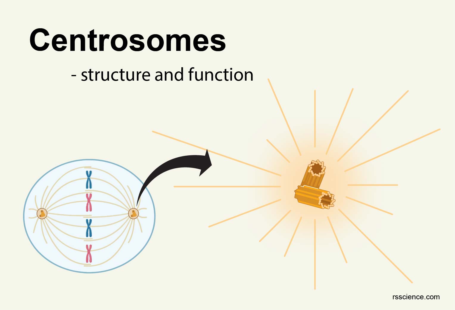
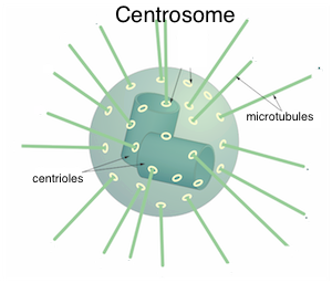
Centrosome
Organelles that organize the microtubules into the mitotic spindle for separating chromosomes during mitosis
2 centrosomes are required to divide a cell so that two spindle poles can form, allowing sister chromatids to be pulled to opposite sides of the cell
Centrosomes migrate to opposite sides
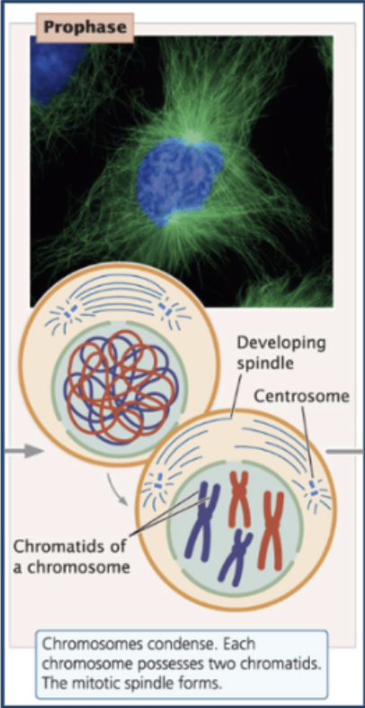
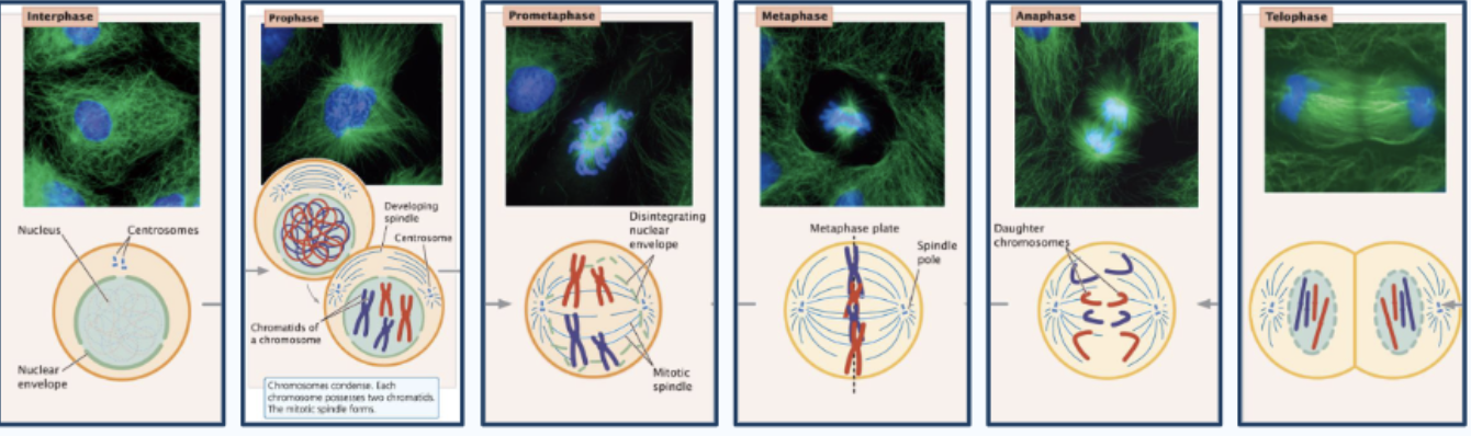
Prophase
First stage of mitosis
Chromosome condensation (DNA gets tightly packed and compact, chromosomes can be seen under the microscope)
Centriole migration to poles - drive formation of spindle
Assembly of spindle - distinct, microtubule-based structure responsible for physically pulling the chromosomes apart
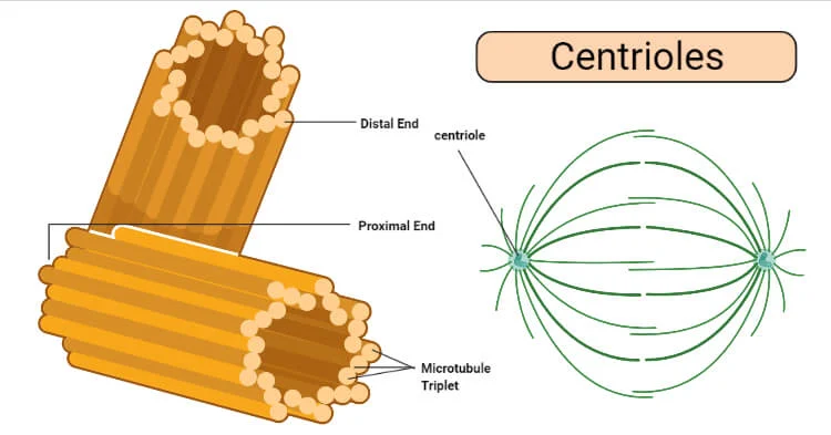
What is centriole migration to poles?
The movement of the two duplicated centrioles (within centrosomes) to opposite ends of the cell during prophase, establishing the two spindle poles that pull chromosomes apart
Centriole - barrel-shaped structure made of microtubules that helps organize the mitotic spindle during cell division
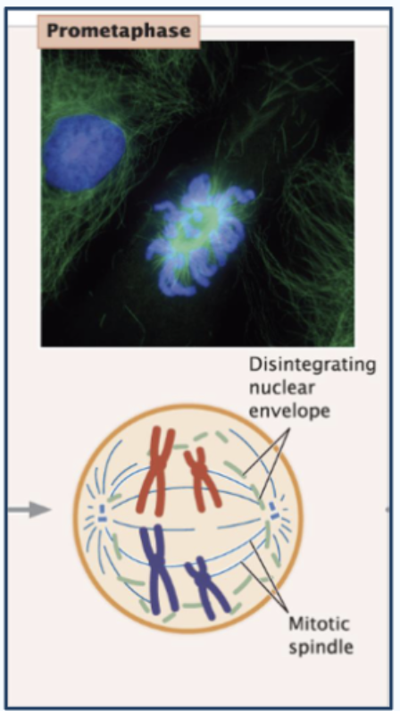
Prometaphase
Second stage of mitosis; when chromosomes gain access to the spindle and get physically connected to it so they can be moved
Nuclear envelope disintegrates (chromosomes can spill out in the cytoplasm; chromosomes now attach to spindle microtubules)
This removes the barrier between:
Chromosomes (formerly inside the nucleus)
Spindle microtubules (in the cytoplasm)
The spindle can now reach and interact with chromosomes.
Spindle microtubules (built by centrioles) attach to kinetochores (structure built around the centromere where the 2 chromosomes are connected together)
spindle microtubules do the actual pulling
Kinetochores are the handles spindle fibers grab; each sister chromatid has its own kinetochore
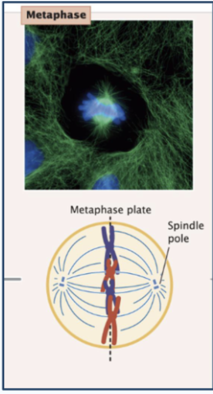
Metaphase
Third stage of mitosis; chromosomes are perfectly aligned and the cell checks that everything is ready before separation
Chromosomes line up at the metaphase plate
Spindle microtubules arrange chromosomes (make sure they are lined up correctly; every chromosome attached to opposite microtubules so the chromatids can be pulled apart) - Microtubules from opposite centrosomes attach to opposite kinetochores
M checkpoint prevents next phase, ensures:
Both sister chromatids are attached to spindle fibers (No unattached kinetochore is allowed)
Lined up at metaphase plate
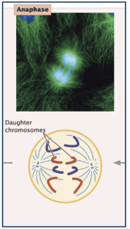
Anaphase
Fourth stage of mitosis; when sister chromatids separate and move to opposite poles of the cell
Centrosomes split (one goes to each cell)
Chromosomes pulled to poles
Sister chromatids go in opposite directions
Telophase and Cytokinesis
Fifth stage of mitosis; marked by the arrival of the chromosomes at the spindle poles
Chromosomes decondense
Nuclear envelopes reform
Spindle disassembly
Cell membrane formation
Two daughter cells
Return to interphase, repeat cycle
After the sister chromatids have separated, each is considered a separate chromosome!
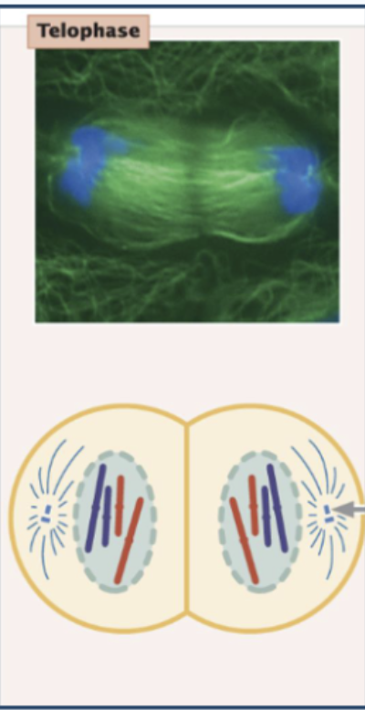
Telophase vs. Cytokinesis
Telophase: the nucleus divides, reforming the nuclear envelope around two sets of separated chromosomes
Cytokinesis: the separate physical division of the cytoplasm, organelles, and cell membrane into two distinct daughter cells
Mitosis consequences
Genetically identical cells
Clonal - do not contribute to genetic variation in a population
Mitosis
“I Picked My Apples Today” IPMAT
I: interphase
P: prophase
M: metaphase
A: anaphase
T: telophase (and cytokinesis)
Meiosis
sexual reproduction
Makes HAPLOID specialized reproductive (sex) cells (eggs, sperm/pollen)
Combines genetic material from two parents (1/2 from mom, ½ from dad)
Recombination - alleles from both parents
Meiosis starts with..
Meiosis does not start with sex cells (sperm or eggs)
it starts with diploid germ cells (2N = diploid)
N = one complete set of chromosomes
2N = two complete sets
!!!! fix later
Haploid cells
Cells containing only a single set of chromosomes (n); half the genetic material of a diploid (2n) organism
Diploid cells have 2 sets (one from each parent)
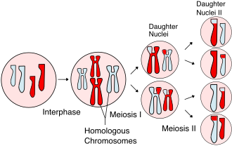
Stages of meiosis
1) Meiosis I
Interphase
Prophase I
Metaphase I
Anaphase I
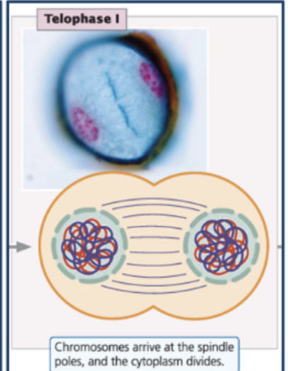
Telophase I
2) Meiosis II:
Prophase II
Metaphase II
Anaphase II
Telophase II
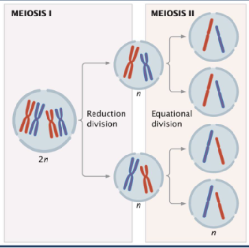
Why are there two stages of meiosis?
1) Meiosis I: results in two cells that each have two sets of chromosomes, like in mitosis; introduces genetic diversity through crossing over
2) Meiosis II: creates four haploid cells (gametes) that each contain one set of chromosomes, because the genetic info isn’t copied a second time
!!
Meiosis I
1) Interphase
2) Prophase I
3) Metaphase I
4) Anaphase I
5) Telophase I
Meiosis II
1) Prophase II
2) Metaphase II
3) Anaphase II
4) Telophase II
Interphase
Prophase I
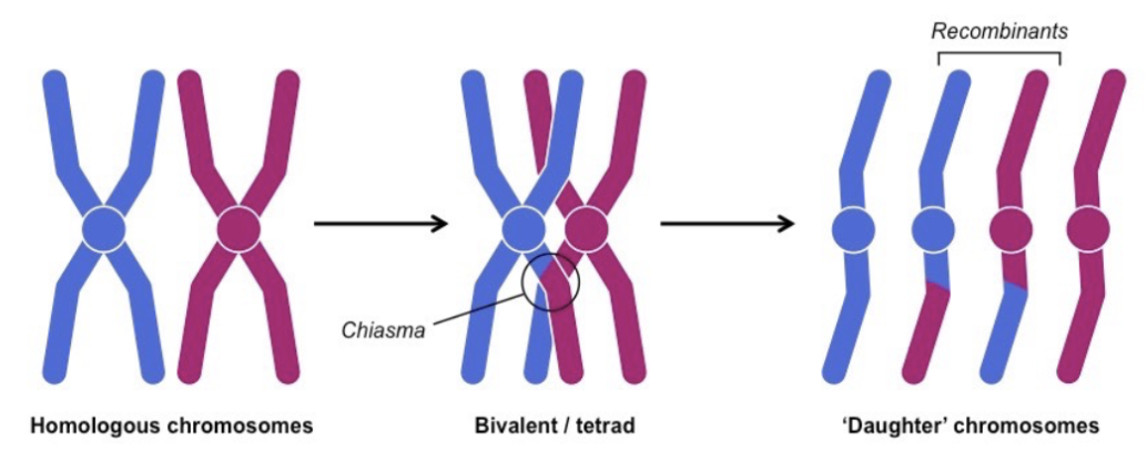
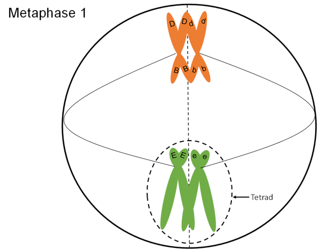
Synapsis of homologous chromosomes - homologous chromosomes (one from each parent) pair up and align lengthwise, forming a structure called a bivalent or tetrad, which enables genetic recombination through crossing-over, increasing genetic diversity in offspring
Crossing over between homologues occurs at chiasmata - X-shaped, physical points of contact between non-sister chromatids of homologous chromosomes (one from mom, one from dad; they carry the same genes, but may have different alleles)
5 substages (Leptotene, zygotene, pachytene, diplotene, diakinesis)
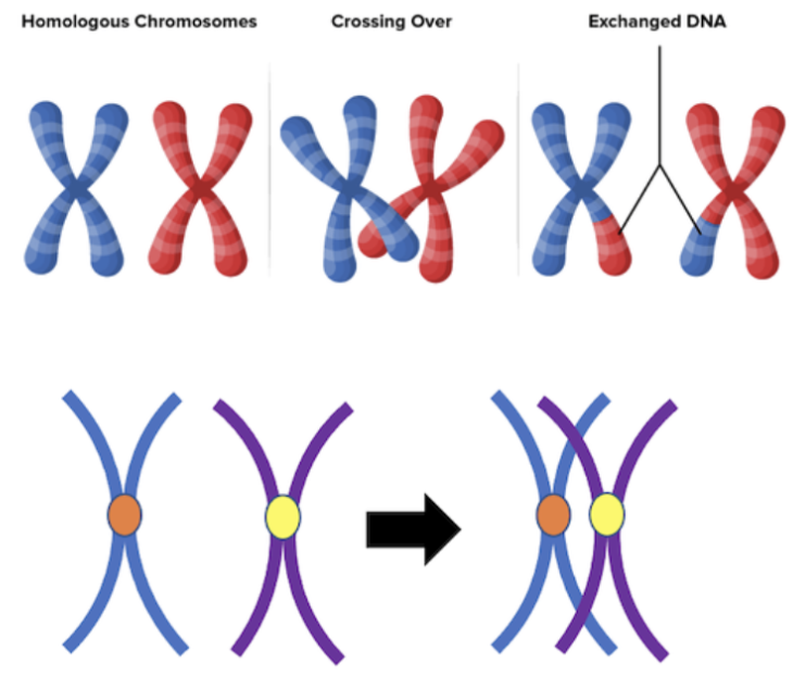
Crossing over
The exchange of genetic material in synapsis (close pairing) between homologous chromosomes during prophase I of meiosis, creating new allele combinations in gamete
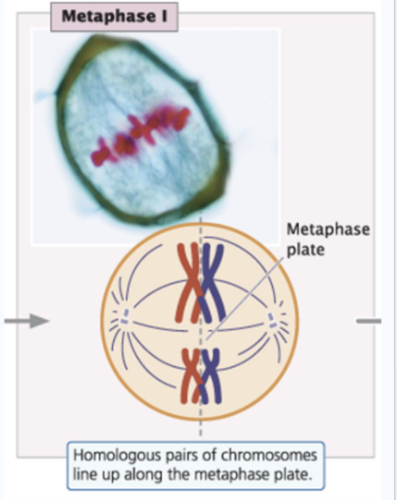
Metaphase I
Homologous pairs of chromosomes (one from mom, one from dad) align along the metaphase plate; a microtubule from one spindle pole attaches to one chromosome of a homologous pair, and a microtubule from the other pole attaches to the other member of the pair
Microtubules from spindle poles attach to different homologues in a pair
Bivalent line up at a metaphase plate
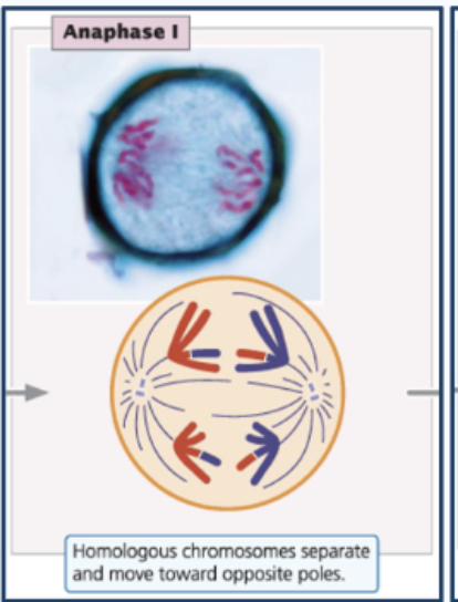
Anaphase I
Bivalents split
Homologues go to poles (but the sister chromatids remain attached and travel together)
Telophase I
Chromosomes decondense
Nuclei reform
Cytokinesis divides cytoplasm into two separate cells
The resulting two daughter cells are HAPLOID
Result of meiosis I
Formation of two haploid (n) daughter cells