Osteology of the Skull - First Lecture
 Call Kai
 Learn
Learn Practice Test
 Spaced Repetition
 Match
Match Flashcards
 Knowt Play
1/42

There's no tags or description
Looks like no tags are added yet.
Name | Mastery | Learn | Test | Matching | Spaced | Call with Kai | Chat |
|---|
No analytics yet
Send a link to your students to track their progress
43 Terms
What do bones do (4)?
support tissue and vital structures (heart/lungs)
mechanical basis for movement
regulate calcium/salt levels
provide continuous supply of new blood cells
What is the axial skeleton made up of?
the head, neck, and trunk
What is the appendicular skeleton made up of?
limb bones
What are osteoblasts?
bone building cells that secrete extracellular/bone matrix
What are osteocytes?
mature bone cells derived from osteoblasts surrounded by bone matrix
What are osteoclasts?
bone reabsorbing cells, important during bone remodeling and repair, derived from cells in the bone marrow
What are long bones?
tubular bones, longer in one dimension (humerus, tibia, metacarpals)
What are short bones?
bones appearing cuboidal due to being nearly equal in length and diameter (tarsus and carpus bones)
What are flat bones?
thin or plate-like bones serving protective function (skull bones, sternum)
spongy bone
cancellous bone that forms the mesh work of interior bone, consisting of trabeculae
Trabeculae
thin tubule network of spongy bone
compact bone
dense bone forming the outer portion of bones
What is irregular bone?
bones of various shapes, bones of the face
What are sesamoid bones?
unique classification of bones where tendons cross the ends of long bones in the limbs, protecting tendons from wear (patella)
What is intramembranous ossification?
bone formation without a cartilage precursor, formed directly from mesenchyme; bone forms from a membranous sheet of connective tissue where membranous models of bone form and ossify in the embryo during the fetal period
What are some bones developed by intramembranous ossification?
flat bones of the skull, mandible, maxilla
What does this picture show?
intramembranous ossification

What does this image show?
endochondral ossification
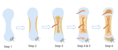
What is endochondral ossification?
cartilagenous bone formation where cartilage models of bones form from the mesenchyme during the fetal period, and bone replaces most of the cartilage
What are some bones developed by endochondral ossification?
bones of the extremities, vertebrae, or cranial base
What is cranial synchondroses?
cartilaginous joints of the base of the skull that act as growth centers until puberty where they begin to ossify
when is calcification usually complete?
25 years old
What are the 5 zones of the growth plate?
zone of reserve cartilage, zone of proliferation, zone of hypertrophy, zone of calcified cartilage, and zone of resorption
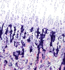
zone of reserve cartilage
no cellular proliferation or matrix production

zone of proliferation
chondrocytes divide and organize into columns, actively producing collagen and matrix proteins

zone of hypertrophy
enlarged chondrocytes, metabolically active
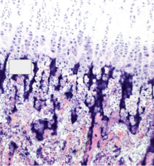
zone of calcified cartilage
matrix is being calcified and serves as a scaffold for new bone
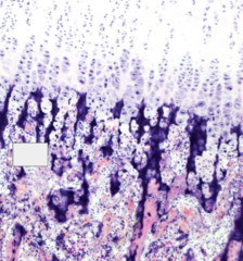
zone of resorption
osteoprogenitor cells are invading this area
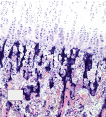
Bone modeling
bone size and shape can change
Bone remodeling
selective bone resorption by osteoclasts and bone apposition by osteoblasts and size and shape remain the same
True or False: Orthodontics involve both modeling and remodeling
True
Elements of cleidocranial dysplasia
birth defect causing failure to shed primary teeth, delayed eruption of permanent teeth, more teeth than normal (supernumerary), lack of collarbones, lack of Runx2 gene
osseointegration
direct structural and functional connection between ordered living bone and the surface of a load bearing implant
prerequisites to osseointegration (4)
immobilization, adequate cells, adequate nutrition of cells, adequate stimulus for bone repair
immobilization (dental implant)
minimal movement of the implant
adequate cells (dental implant)
mesenchymal cells that can differentiate into osteoblasts, osteoclasts should arise at the bone implant interphase through the bloodstream
adequate nutrition of cells (dental implant)
diffusion of nutrients that will facilitate the process will occur locally until microcirculation is reestablished
adequate stimulus for bone repair (dental implant)
trauma from surgery and inflammatory response will trigger healing
phases of bone healing (3)
inflammatory response, regeneration, remodeling
inflammatory response (bone healing)
48 hours after surgery, chemotaxis of mesenchymal cells and phagocytosis of tissue debris, blood clotting and formation of fiber network attaching to implant surface, osteogenic cells migrate to the implant surface and form osteoids, followed by bone directly on the implant surface
regeneration (bone healing)
microcirculation restored in 10 days following surgery, osteogenesis occurs between 4-6 weeks, immature/woven bone is first formed and replaced by dense lamellar bone
remodeling (bone healing)
18 months post op, occurs in response to functional loading, low strain stimulates regeneration while high strain will prevent it
bisphosphonates
drug suppressing osteoclast function and increasing bone density and mineralization, increasing jaw osteonecrosis that may delay or inhibit healing