Biopsychology
1/50
There's no tags or description
Looks like no tags are added yet.
Name | Mastery | Learn | Test | Matching | Spaced | Call with Kai |
|---|
No analytics yet
Send a link to your students to track their progress
51 Terms
Nervous System
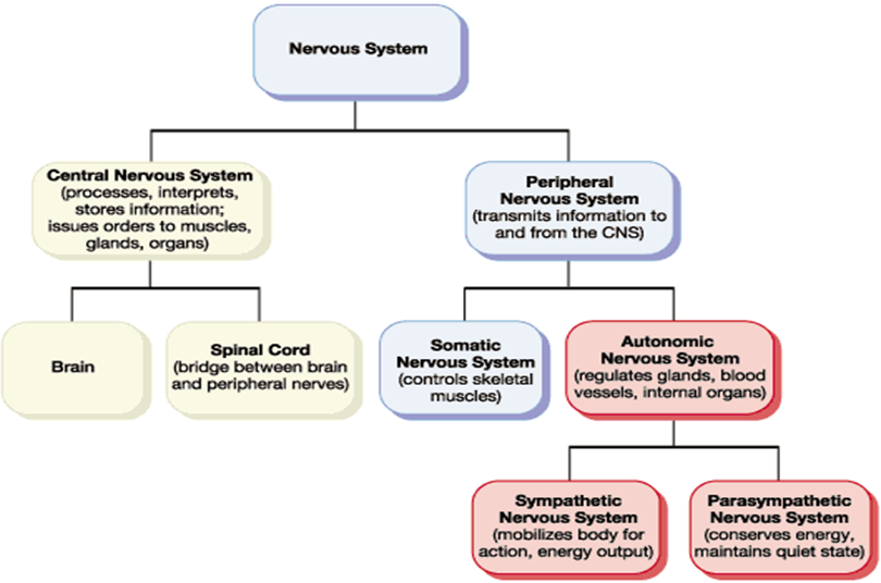
Central Nervous System (CNS)
Consists of the brain and spinal cord. The spinal cord passes messages to and from the brain and connects nerves to the PNS. The CNS processes, interprets and stores information and issues orders to muscles, glands and organs.
Peripheral Nervous System (PNS)
The PNS sends and receives messages to and from the central nervous system. The peripheral nervous system is further sub-divided into the Autonomic Nervous System and Somatic Nervous System
Autonomic Nervous System (ANS)
governs vital functions in the body such as
breathing,
heart rate,
digestion,
sexual arousal
and stress response.
The organs controlled by the autonomic nervous system include the heart, lungs, eyes, stomach, and the blood vessels of the internal organs. It can be divided into the sympathetic and parasympathetic nervous system.
Somatic Nervous System (SNS)
Consists of nerves carrying sensory signals from e.g. eyes, ears, skeletal muscles, and the skin to the CNS. It also contains the nerves that carry the messages from the brain to the other areas of the body. It controls muscle movement and receives information from sensory receptors.
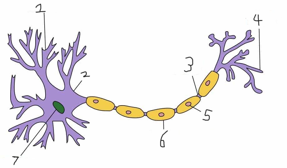
1- Dendrite
2- Soma
3- Node of Ranvier
4- Axon Terminal
5- Schwann Cell
6- Myelin Sheath
7- Nucleus
Motor Neurons
Connect the CNS to muscles and glands
Carry messages AWAY from the brain
Have short dendrites and long axons
Cause movement
Sensory Neurons
Carry messages such as sight, sound and sensations from the PNS to the CNS
Have long dendrites and short axons
Relay Neurons
Connect sensory neurons to motor neurons (or to other relay neurons)
Make up 97% of the CNS
Have short dendrites and short axons
Neurons and the Reflex Arc
The sensory neuron detects the heat and sends a signal to a relay neuron in the CNS (in the spinal cord) which in turn sends a signal to the motor neuron to move our hand away.
Excitatory Neurotransmitters
increase the chance of the next neurons firing
Inhibitory Neurotransmitters
decrease the chance of adjacent neurons firing
Function of the Endocrine System (Glands and Hormones)
To provide a chemical system of communication via the blood stream - occurs through the glands secreting hormones into the bloodstream
To secrete the hormones which are required to regulate many bodily functions
Pituitary gland
Controls the release of hormones from all other endocrine glands in the body.
Parasympathetic Nervous System
Stimulates flow of saliva
Slows heartbeat
Contracts bladder
Allows for digestion
Sympathetic Nervous System
Dilates pupil
Inhibits flow of saliva
Accelerates heart rate
Inhibits bladder contraction
Evaluation of fight or flight
Likely to have evolved as a response to threats posed for our early ancestors (e.g. wild animals). However, it is an inappropriate response to modern day stressors (e.g. exams) and is likely to create panic and anxiety.
Taylor (2000) argues that females are more likely to ‘tend and befriend’. This involves protecting themselves and their young through nurturing behaviours (tending) and forming protective alliances with other women (befriending). Women may have a completely different way of coping with stress as their responses evolved in the context of primary care giver. Fleeing too readily would have put their offspring at risk.
Von Dawans et al (2012) challenge the classic view that under stress men respond with fight and flight, whereas women tend and befriend. They found that acute stress can lead to greater co-operative and friendly behaviour, even in men. This could explain the human connection that happens during times of crises such as the 9/11 terrorist attacks.
Localisation
the theory that specific areas of the brain are associated with particular physical and psychological functions
Motor Cortex
This is located at the back of the frontal lobe and is responsible for the generation of voluntary motor movements.
Somatosensory Cortex
This is located in the parietal lobe, close to the motor area. This produces sensations of touch, pressure, pain and temperature, which it then localises to specific body regions.
Visual Centre
The primary visual centre is the visual cortex in the occipital lobe. The visual cortex contains different areas for colour, shape etc.
Auditory Centre
Is concerned with hearing and is located in the temporal lobe in both hemispheres.
Broca’s Area
This area is located in the left frontal lobe and is essential for speech production.
Wernicke’s Area
Located in the left temporal lobe and is responsible for the recognition and processing of language.
Evaluation of Localisation
+Petersen et al (1988) used brain scans to demonstrate how Wernicke’s area was active during a listening task and Broca’s area was active during a reading task.
+Broca’s aphasia is an impaired ability to produce language. In most cases, this is produced by damage to Broca’s area. Wernicke’s aphasia is an impaired ability to understand language. This is usually the result of damage to Wernicke’s area (e.g. from a stroke).
-Lashley (1950) claimed that intact areas of the cortex could take over cognitive functions following injury to the area normally responsible for that function. He investigated rats ability to learn a maze and found that the basic motor and sensory functions were localised, but that higher mental functions were not. He found the effects of deliberate damage to the rat’s cortex was determined by the extent, rather than the location, of the damage.
Hemispheric Lateralisation
The dominance of one hemisphere of the brain for particular physical and psychological functions.
One difference in the hemispheres is the presence of the language areas (Broca’s and Wernicke’s areas) which are mainly found on the left hand side.
Research has also found that the right hemisphere excels at visual attention tasks and face recognition. Therefore research indicates that these functions (and maybe others) are hemispherically lateralised.
Evaluation of Lateralisation
Lateralisation changes with age –lateralisation of function appears not to stay exactly the same throughout an individual’s lifetime, but changes with normal ageing. Across many types of tasks and many brain areas, lateralised patterns found in younger individuals tend to switch to bilateral patterns in healthy older adults
There are individual differences - Brain function lateralization is evident in the phenomena of right- or left-handedness, but a person's preferred hand is not a clear indication of the location of brain function. Although 95% of right-handed people have left-hemisphere dominance for language, 20 % of left-handed people have right-hemisphere dominance for language function. Additionally, 20 % of left-handed people have bilateral language functions.
Brain Plasticity
To the brain’s ability to change and adapt as a result of experience.
Axonal Sprouting (method of Brain Plasticity)
The growth of new nerve endings which connect with other undamaged nerve cells to from new neuronal pathways.
Neuronal Unmasking (method of Brain Plasticity)
‘Dormant synapses’ in the brain are reactivated.
Recruitment of similar areas on the opposite side of the brain to perform specific tasks. (method of brain plasticity)
An example would be if Broca’s area was damaged on the left side of the brain, the right-sided equivalent would carry out its functions.
Maguire et al (2000) - Evidence for Brain Plasticity
Studied the brains of London taxi drivers and found significantly more volume of grey matter in the hippocampus than in a matched control group of novice drivers.
Ballantyne et al (2008)
Found there is more plasticity for recovery after a stroke in infancy and childhood than in adulthood.
Functional Magnetic Resonance Imaging (fMRI)
It measures changes in blood flow in particular areas of the brain which indicates increased neural activity in those areas.
Evaluation of fMRIs
+It is non-invasive. It does not expose the individual to harmful radiation unlike CT scans and PET scans (which involve injecting a person with radioactive glucose).
-overlooks the networked nature of brain activity, as it focuses on localisation.
Electroencephalogram (EEG)
Measures electrical activity in the brain via electrodes that are fixed to an individual’s scalp either individually or using a skull cap.
Evaluation of EEGs
+Has high temporal resolution.
-Generalised nature of the information received (that of many thousands of neurons).
Event-related potential (ERP)
Very small voltage changes in the brain that are triggered by specific events or stimuli (e.g. a word).
Evaluation of ERPs
+have excellent temporal resolution
-It may be difficult to eliminate background ‘noise’
Post-mortem Examinations
This is a technique involving the analysis of a person’s brain following their death.
Evaluation of PMEs
+allow for a more detailed examination of neuroanatomical and neurochemical aspects of the brain than would be possible with the sole use of non-invasive techniques
-Causation is an issue with these investigations as the evidence is correlational.
Circadian rhythms
Changes in biological activity that show regular variation over 24hrs e.g. sleep-waking cycle.
Infradian rhythms
Changes in biological activity that show regular variation over a time period greater than 24 hours. e.g. the menstrual cycle.
Ultradian rhythms
Changes in biological activity that show regular variation over a time period of less than 24 hours e.g. the stages of sleep follow a pattern of cycles lasting about 90 minutes.
Endogenous pacemakers
Internal body clocks that maintain biological rhythms e.g. SCN/Pineal gland.
Exogenous Zeitgebers
External cues from outside the body which are also important in maintaining bodily rhythms. Light and dark is an important Zeitgeber for both animals and plants and Social cues.
Siffre (1975) - Circadian Rhythms
Spent 6 months inside a cave in Texas. There were no zeitgebers such as natural light or time. He had contact with the outside world via telephone, but had no idea what time it was.
His behaviour e.g. when he slept/woke and when he ate his meals was monitored.
When he was awake, researchers put the lights on and when he went to bed, they turned the lights off.
His sleep/waking cycle was erratic at first but then settled into a fairly regular cycle of about 25 hours i.e. slightly longer than the 24 hour day. When he emerged on the 179th day, it was only his 151st ‘day’!
Russel et al - Infradian Rhythms
Applied the pheromones of one woman to a group of 5 women.
A swab was taken from the donor's armpits daily and the pads were rubbed on to the upper lips of the women.
It was found that 4/5 of the women had menstrual cycles that had synchronized to within a day of the odour donor. This confirmed the role of pheromones in regulating the menstrual cycle
Dement et al - Ultradian Rhythms
Participant is wired up to a Polysomnograph. i.e. Electro Encephalograph, Electro Oculograph, Electro Myograph and sensors to detect pulse, body temp, Blood pressure etc.
The participant then goes to sleep in the lab, and all these functions are recorded. Usually the participant spends several nights in the sleep lab.
In the Dement study, 9 participants were studied for up to 61 nights in a laboratory. The mean time of a NREM/REM cycle was 92 minutes.
Examples of Endogenous Pacemakers
THE SUPRACHIASMATIC NUCLEUS - a pair of tiny clusters of nerve cells in the HYPOTHALAMUS which have an inbuilt circadian rhythmic firing pattern. The SCN gets information on light from the optic nerve, even when our eyes are shut as special photoreceptors in the eyes pick up light signals and carry them to the SCN.
THE PINEAL GLAND AND MELATONIN - converts the neurotransmitter serotonin into the hormone melatonin. It contains light sensitive cells. When the level of light falls, melatonin is produced by the pineal gland. When light levels are high, the production of melatonin in the pineal gland is inhibited and we wake up.
Morgan (1995) - Endogenous Pacemakers
Established the presence of circadian rhythms in a group of hamsters by recording their sleep/wake cycle over a period of time.
They then removed their SCN. They still ate, slept etc, but their circadian rhythms disappeared.
Later they transplanted SCN tissue from hamster foetuses, and found that after a few days the circadian rhythms re-established.