Topic 7B (Muscles, skeleton, heart and breathing)
 Call Kai
 Learn
Learn Practice Test
 Spaced Repetition
 Match
Match Flashcards
 Knowt Play
1/4

There's no tags or description
Looks like no tags are added yet.
Name | Mastery | Learn | Test | Matching | Spaced | Call with Kai | Chat |
|---|
No analytics yet
Send a link to your students to track their progress
5 Terms

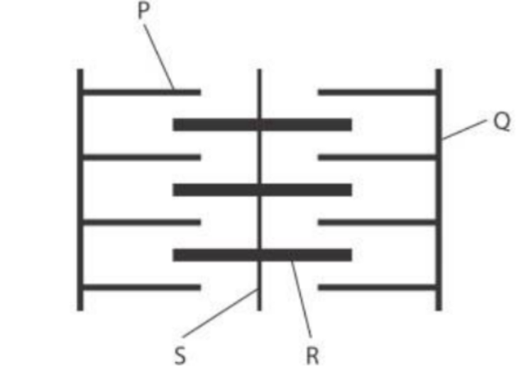
The diagram below shows a sarcomere of a muscle.
Label the following.
P = I band (where only thin actin present)
Q = Z- line
R = H band (where only thick myosin is present)
S = M line
Describe the role of calcium ions in the sliding filament theory of muscle contraction
(4 marks)
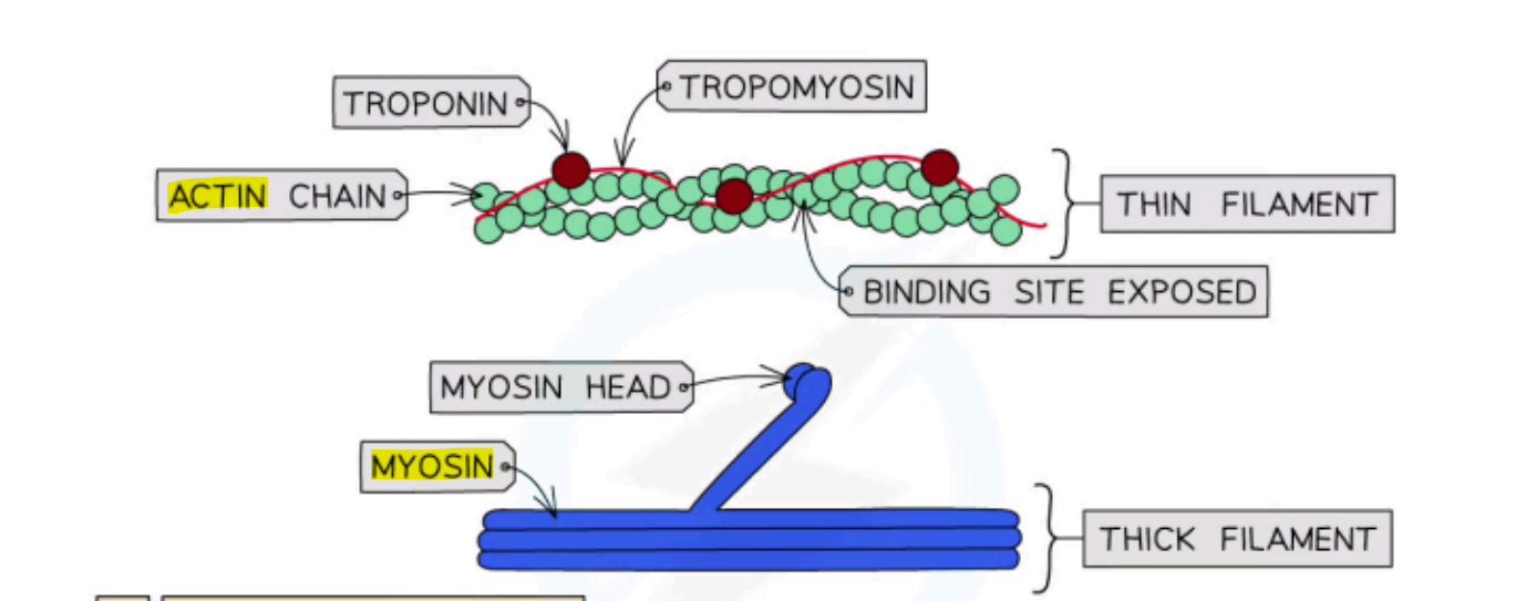
- calcium ions bind to troponin (ball thing)
- changing the shape of troponin
- this causes tropomyosin to move away from the myosin binding site
(tropomyosin is kinda like a thin string attached to troponin)
- myosin head then binds onto actin, forming an actin-myosin cross bridge
Describe the role of ATP in muscle contraction
(4 marks)
- ATP binds to the myosin head
- breaking the actin-myosin cross-bridge
(OR releasing the myosin head from the actin strand )
- ATP is hydrolysed by the myosin head
(ATP broken down into ADP)
- providing energy to move the myosin head back/upright
Compare and contrast the structure of fast-twitch and slow-twitch muscle fibres.
(5 marks)
- both contain actin & myosin
- both consist of sarcomeres
- slow twitch have more mitochondria than fast-twitch
(allows more respiration)
- slow-twitch has more myoglobin & capillaries than fast-twitch
(allows for greater store of O2)
- fast twitch are larger in diameter & longer than slow twitch
(slow-twitch muscle fibres contracts slowly and fast contracts rapidly)


- calcium ions bind to troponin, changing it’s shape
- this causes tropomyosin to move and allows myosin to bind to actin