VetPath exam 3 pictures (31-41)
 Call Kai
 Learn
Learn Practice Test
 Spaced Repetition
 Match
Match Flashcards
 Knowt Play
1/152

There's no tags or description
Looks like no tags are added yet.
Name | Mastery | Learn | Test | Matching | Spaced | Call with Kai | Chat |
|---|
No analytics yet
Send a link to your students to track their progress
153 Terms
acute
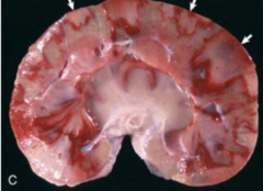
Which chronicity is this renal infarct considered?

subacute
Which chronicity is this renal infarct considered?
chronic
Which chronicity is this renal infarct considered?
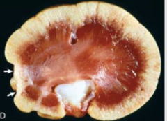
hemorrhage/thrombosis
What is going on in this image?

renal infarcts
Which histologic lesion is being shown?

Immune complex mediated glomerulonephritis
What disorder affecting the renal corpuscles is this gross image showing with lightly swollen, slightly pale kidneys and pinpoint foci on the cut surface?
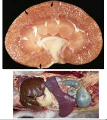
Canine Lyme nephritis
What disease is being shown where there is a unique triad of changes on histology including glomerulonephritis, tubular necrosis and regeneration, and lymphoplasmacytic intestinal nephritis?

porcine glomerulonephritis
What disease is being shown occurs in pigs which can be a sequela to chronic infection?
Glomerular amyloidosis
What disorder affecting the renal corpuscles is being shown in these images?
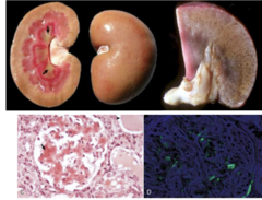
Acute suppurative glomerulonephritis
What disorder affecting the renal corpuscles Is being shown with gross lesions including multifocal, randomly distributed, pinpoint white to tan foci in the cortex?
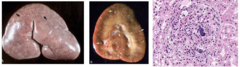
equine embolic glomerulonephritis
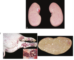
What disorder affecting the renal corpuscles is seen in these images in horses with gross lesions including numerous, random small to pinpoint raised tan foci that extend through the width of the cortex on cut section?

Diamond Skin disease
Which disease will have these renal lesions grossly in pig kidneys?
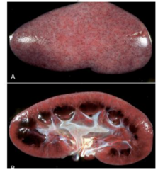
canine herpesvirus
Which disease will have these renal lesions seen in puppies younger than 3-5 weeks of age?

Chronic copper toxicity in sheep
Which disease affecting renal tubules is shown in this image and occurs in sheep?
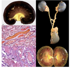
Bacillary hemoglobinuria
Which disease affecting the renal tubules is shown in the image and occurs in cattle?
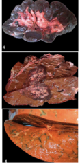
myoglobin
Which nephrotoxic pigment is being shown in this image?

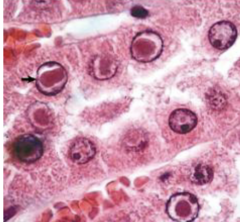
Intranuclear inclusions from lead toxicity
What is being shown in this histology image?
Ethylene glycol (antifreeze) toxicity
What is being shown in these gross images where there are pale yellow foci of calcium oxalate crystals that precipitate in renal tubular lumens, tubular epithelial cells, and the interstitum?

Ethylene glycol (antifreeze) toxicity
What is being shown on this histology where the crystals are birefringent under polarized light?

plant toxins
This image is showing the renal affects of what toxin on bovine kidneys?
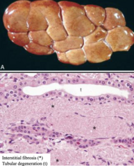
grape/rasin toxicosis
This image is showing the renal affects on histology of what toxin?

Pulpy kidney disease
What is being shown in the image?

Hydronephrosis
Which renal pelvis disorder is being shown on this gross image?

Pyelonephritis
Which renal pelvis disorder is being shown on this gross image?

Papillary necrosis (medullary crest necrosis)
Which renal pelvis disorder is being shown on this gross image?

Parasites of the renal pelvis
Which renal pelvis disorder is being shown on this gross image?

Lymphoplasmacytic interstitial nephritis
Which renal interstitium disorder is being shown on this gross image?

malignant catarrhal fever
Which renal interstitium disorder is being shown on this gross image?

Leptospirosis
Which renal interstitium disorder is being shown on this gross image?

Leptospirosis
Which renal interstitium disorder is being shown on this histology image?

Granulomatous nephritis
Which renal interstitium disorder is being shown on this histology image?

feline infectious peritonitis
Which renal interstitium disorder is being shown on this gross image?

Parasitis- toxocara canis
Which renal interstitium disorder is being shown on these images?

Hairy vetch (Vicia villosa)
Which toxicity is being shown on these images?

renal adenoma
Which renal neoplasia is being shown?

Renal carcinoma
Which renal neoplasia is being shown?

Nephroblastoma
Which renal neoplasia is being shown?

metastatic tumors
Which renal neoplasia is being shown?

renal lymphoma
Which renal neoplasia is being shown?
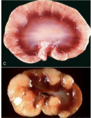
renal aplasia
Which renal developmental anomaly is being shown grossly?

Renal hypoplasia
Which renal developmental anomaly is being shown grossly?

renal dysplasia
Which renal developmental anomaly is being shown grossly?

renal cysts
Which renal developmental anomaly is being shown grossly?

Polycystic kidney disease
Which renal developmental anomaly is being shown grossly?

progressive juvenile nephropathy
Which renal developmental anomaly is being shown grossly?

ectopic and fused kidneys
Which renal developmental anomaly is being shown grossly?
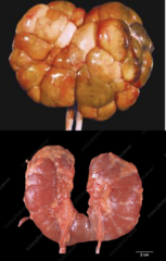
ureter
Which normal structure is being pointed out by the arrows?
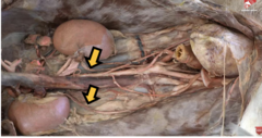
urinary bladder
Which normal structure is being pointed out by the arrow?

horse
This urine is considered normal in which species?

ectopic ureter
Which developmental anomaly is being shown on this image?

Urolithiasis
Which developmental anomaly is being shown on this image?

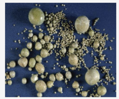
struvite calculi
Which type of canine calculi is seen in this image and is the historically most common crystal in dogs?
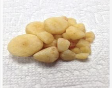
calcium oxalate
Which type of canine calculi is seen in this image that is more common in dogs and commonly seen with other diseases?
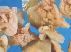
urate calculi
Which type of canine calculi is seen in this image that is common in Dalmatians?
urinary obstruction
What is being shown in this image that is more common in male species?
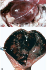
hydroureter
What is being shown in this gross image?

acute cystitis
What is being shown on this gross image?
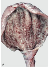
acute cystitis
What is being shown on these images?

diffuse
How would you classify this chronic cystitis pattern?
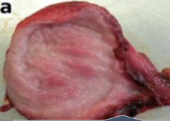
follicular
How would you classify this chronic cystitis pattern?

polypoid
How would you classify this chronic cystitis pattern?

cantharidin toxicosis
Which toxin caused these gross lesions in horses?

bracken fern cystitis
Which toxin caused these gross lesions?
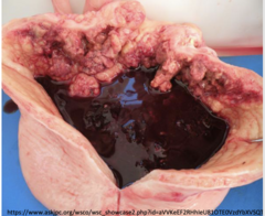
papilloma
Which type of epithelial tumor is shown in this image?
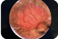
SCC, adenocarcinomas
Which type of epithelial tumor is shown in this image?
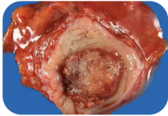
urothelial cell carcinoma
Which type of epithelial tumor is shown in this image?

renal fibrosis
What is being shown in this gross image?

uremia
What are these nonrenal lesions commonly caused from?

nephrocalcinosis
What is this gross and histology image showing where there is a gritty texture to the kidneys and calcification to the tubules and Bowmans capsule?

tubular necrosis, diffuse, severe, acute
What is the morphological diagnosis?

hemoglobinuric nephropathy or nephrosis = copper toxicity/toxicosis
What is the etiologic diagnosis?

cystitis, multifocal to coalescing, mild, acute, with intraluminal calculus
What is the morphological diagnosis?

calcium oxalate
If this was from a dog, which stone would this most likely be?
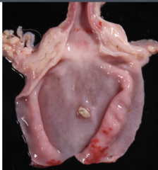
chronic
Which chronicity is accurate for this lesion?
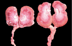
acute (left) and subacute (right)
Which chronicity is accurate for these lesions?

neutrophils
What are the cells surrounding the glomeruli?

glomerulonephritis, suppurative, multifocal to locally extensive, acute with bacterial emboli
What is the morphologic diagnosis of this kidney based on the histopathology?

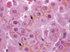
bile canaliculi
What are the arrows pointing to?
random hepatocellular degeneration and/or necrosis
What is being shown on this gross image?

zonal hepatocellular degeneration
What is being shown on this gross image?

centrilobular degeneration
What is being shown on this histology image?

panlobular necrosis
What is being shown on this histology image?

panlobular necrosis
What is being shown on this gross image?

icterus
Which clinical sign is shown here?

acute passive congestion
What is this gross image showing?

chronic passive congestion
What is this gross image showing?
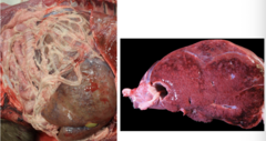
chronic passive congestion
What is this gross image showing?

anemia
What is this gross image showing?
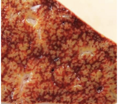
extrahepatic shunt
What is being shown on histology?

portal vein hypoplasia
What is being shown on histology?

telangiectasia
What is being shown grossly and histologically on this image?

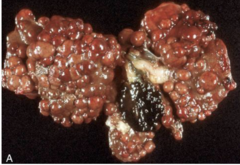
end-stage liver (cirrhosis)
How would you describe this liver?

cirrhosis
How would you describe this liver?
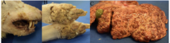
Hepatocutaneous syndrome
what are these images showing?
tension lipidosis (steatosis)
What is being shown on this gross image?
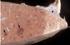
capsular fibrosis
What is being shown on this gross image?

acute hepatitis
How would you describe this liver grossly?

chronic hepatitis
How would you describe this liver grossly?

canine chronic hepatitis
What is going on in this histology slide?

lobular dissecting hepatitis
What is the gross and histology image showing?
