OPT 311 Midterm 1 Part 2 (Large + Tonic pupils)
1/56
There's no tags or description
Looks like no tags are added yet.
Name | Mastery | Learn | Test | Matching | Spaced |
|---|
No study sessions yet.
57 Terms
What are some etiologies with the muscle (non-neurological) causing a large pupil?
posterior synechiae
ocular trauma
angle closure glaucoma = also vomiting, cornea edema
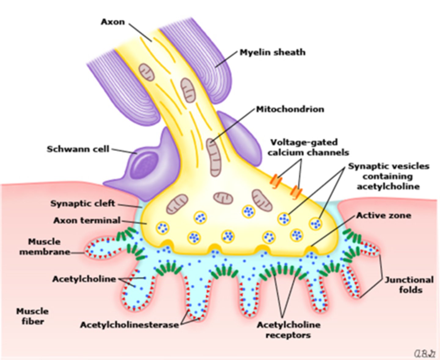
What are some etiologies with the NMJ causing a large pupil?
parasympatholytic drops like tropicamide = inhibits constriction = fixed to light
sympathomimetic drops like phenylephrine = promotes dilation = still some constriction
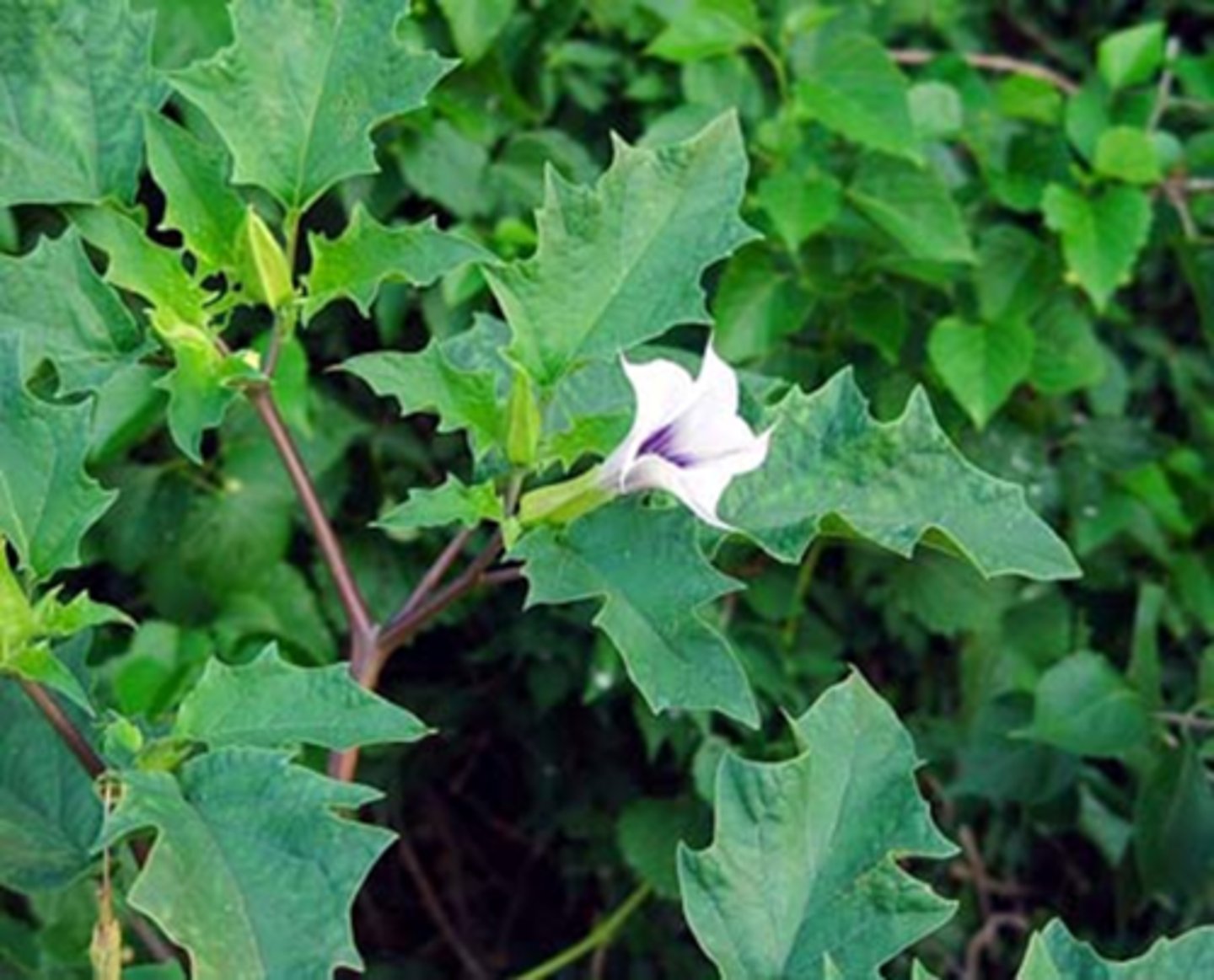
Jimson weed alkaloid ingested = causes atropine intoxication = "corn pickers pupil"
denervation supersensitivity
Recall that with a fixed, dilated pupil, we can differentiate whether it's an afferent or efferent problem using which test?
indirect APD test results being either...
(+) APD means afferent problem
(-) APD means efferent problem
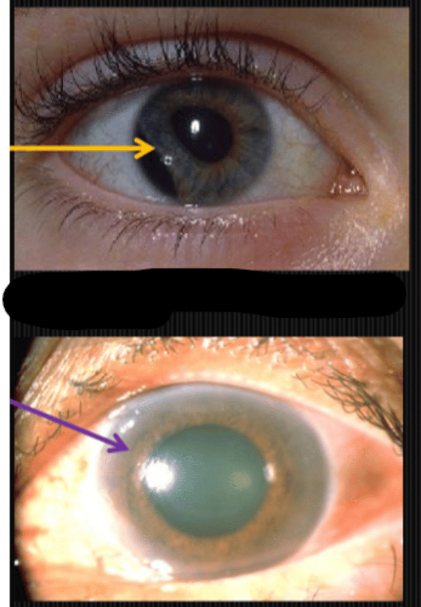
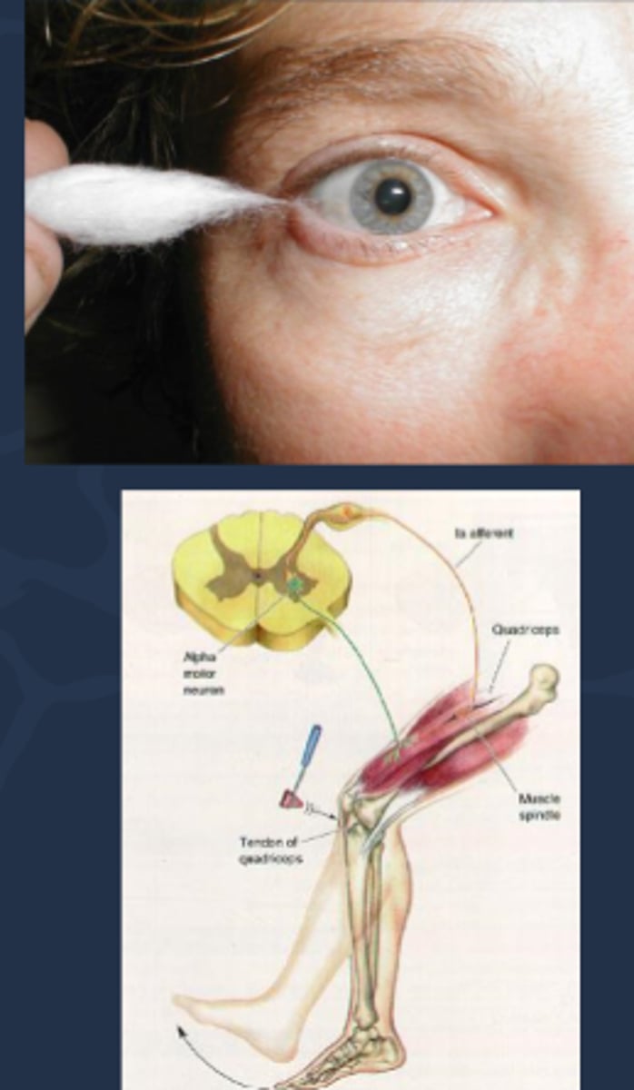
What is denervation supersensitivity causing a large pupil?
defect in parasymp fibers that travel with CN III = reduced ACh released at NMJ = sphincter mm does not receive ACh stimulation = sphincter mm becomes super-sensitive to low [ ] of any ACh-like neurotransmitter
![<p>defect in parasymp fibers that travel with CN III = reduced ACh released at NMJ = sphincter mm does not receive ACh stimulation = sphincter mm becomes super-sensitive to low [ ] of any ACh-like neurotransmitter</p>](https://knowt-user-attachments.s3.amazonaws.com/7772a7bd-b27a-4abf-96c4-489d718bd9a9.jpg)
Denervation supersensitivity of the iris sphincter mm is the basis for which test?
pilocarpine 0.125% test OU
How does a normal eye respond to the 0.125% pilocarpine test?
lots of ACh being released at NMJ = sphincter mm stimulated by a normal amount of ACh = sphincter mm will NOT respond to low [ ] of ACh
![<p>lots of ACh being released at NMJ = sphincter mm stimulated by a normal amount of ACh = sphincter mm will NOT respond to low [ ] of ACh</p>](https://knowt-user-attachments.s3.amazonaws.com/37da34a3-be23-4743-91ae-6132f51d6602.jpg)
How does a pharmacologically dilated eye respond to the 0.125% pilocarpine test?
ACh receptors blocked = adding ACh-like drops such as pilocarpine will NOT induce constriction of the sphincter mm

When is it typically okay to put pharmacological testing drops in the pt's eye when presenting with a large pupil?
as long as you have R/O a CN III palsy
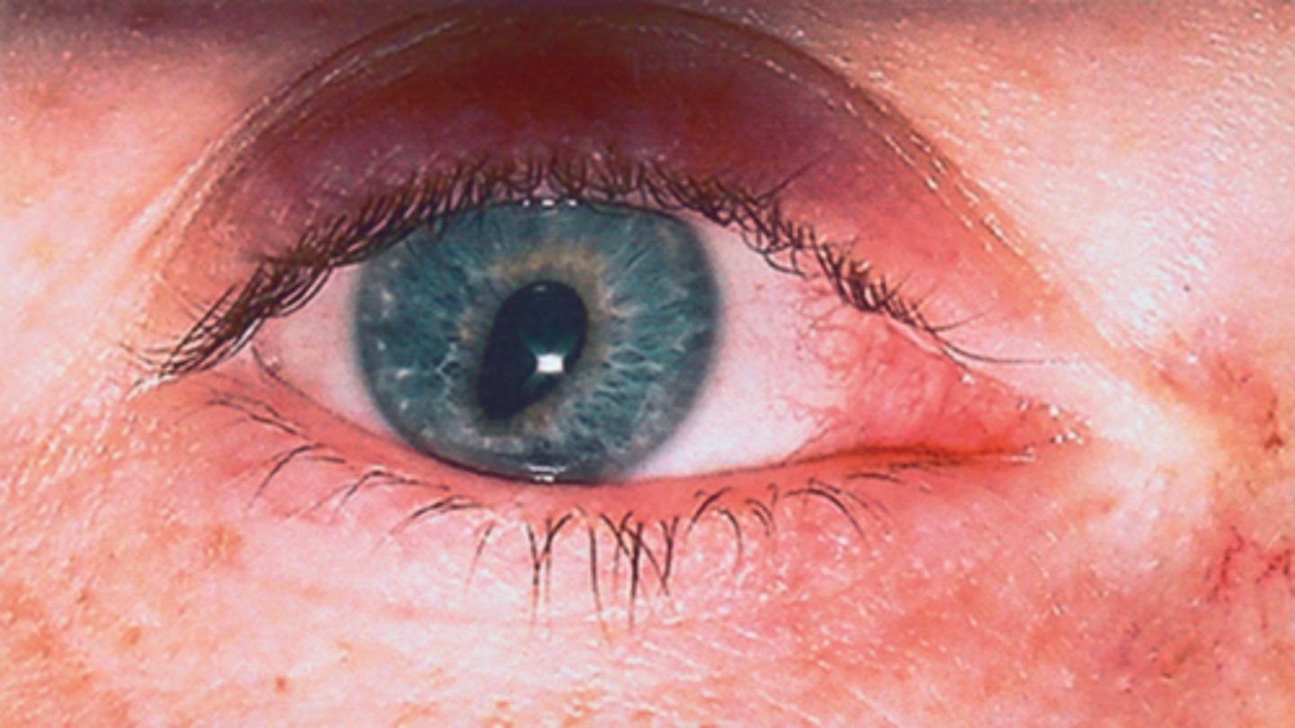
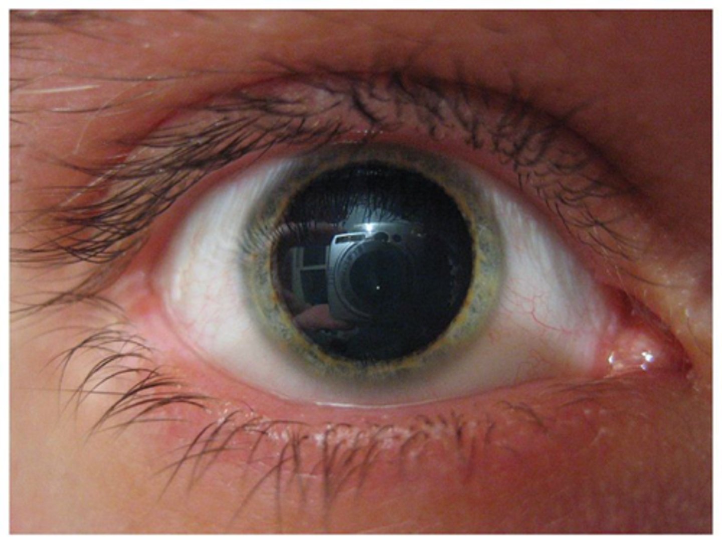
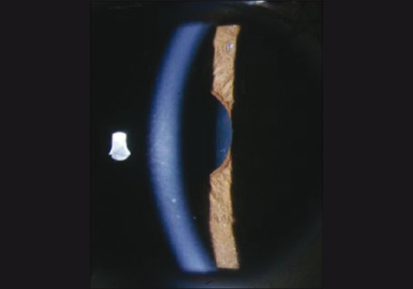
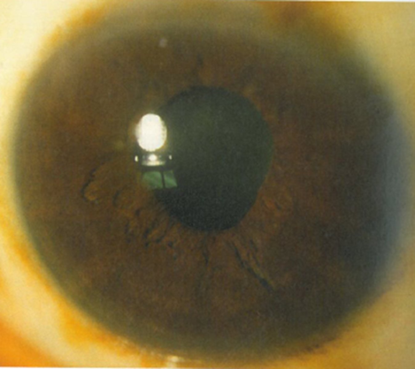
What 1/3 etiology with the nerve causing a large pupil is shown here?
tadpole pupil = sympathic irritation/spasm of iris dilator with unknown etiology = dilator pulls on sectoral area of pupil for 1-2 weeks
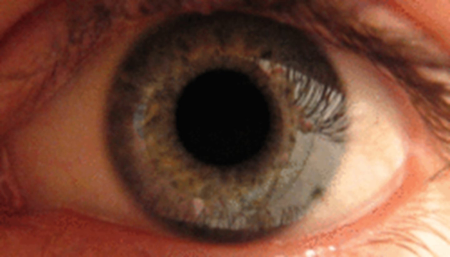
What 2/3 etiology with the nerve causing a large pupil is shown here?
tonic pupil

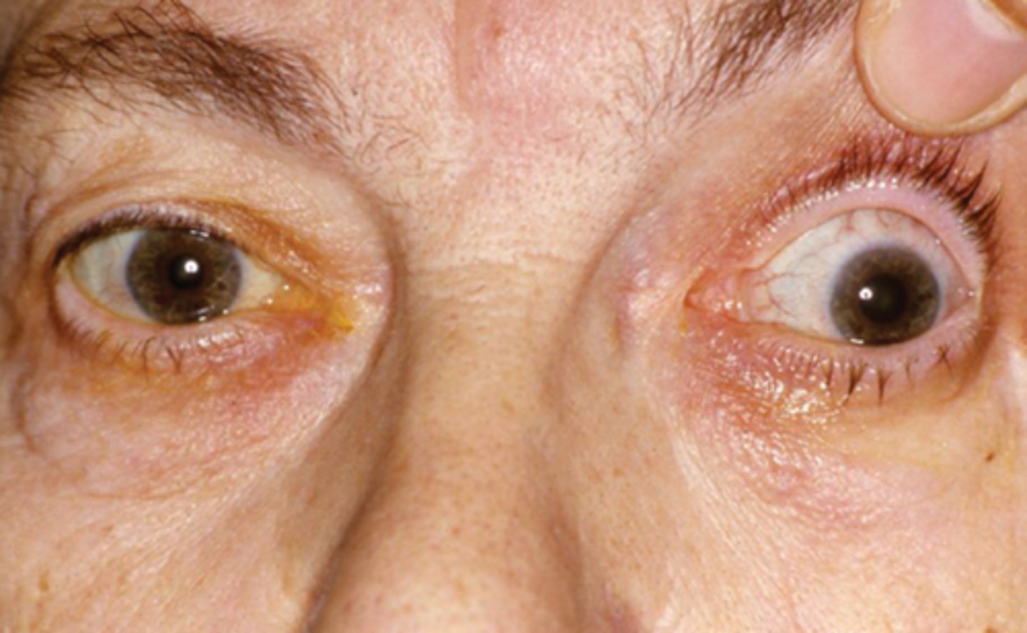
What 3/3 etiology with the nerve causing a large pupil is shown here?
CN III palsy = parasymp issue
What is meant by an isolated CN III palsy?
ONLY affecting CN III and not affecting any other CN (need to do a CN screening exam to R/O other CN issues)

What is meant by a complete CN III palsy? 2 factors.
complete CN III palsy = pupil involved, total ptosis
incomplete CN III palsy = pupil spared, partial ptosis
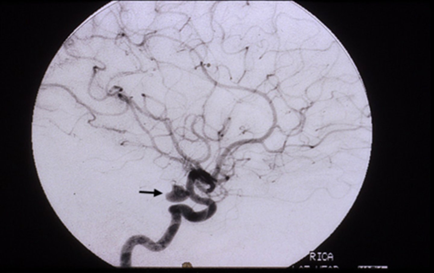
What is the difference between the cause of an isolated pupil-involved vs pupil-sparing CN III palsy?
fixed, dilated pupil (esp if painful like HA) = anuerysm at the junction of the ICA and posterior communicating artery
pupil-sparing (may or may not be painful like eye pain) = diabetes or ischemic vascular etiology
NOTE: can change from pupil-sparing to pupil-involved if it is an aneurysm
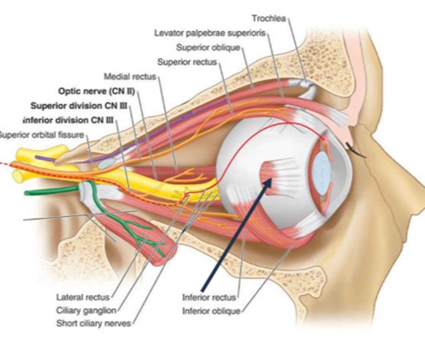
Explain the anatomy of the pupillomotor vs EOM fibers in the CN III.
pupillomotor fibers travel around the circumference of CN III
EOM fibers travel within the core of CN III
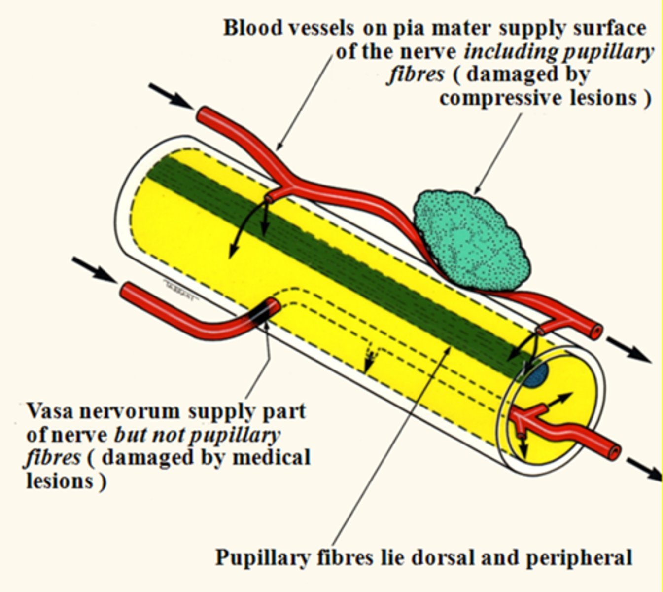
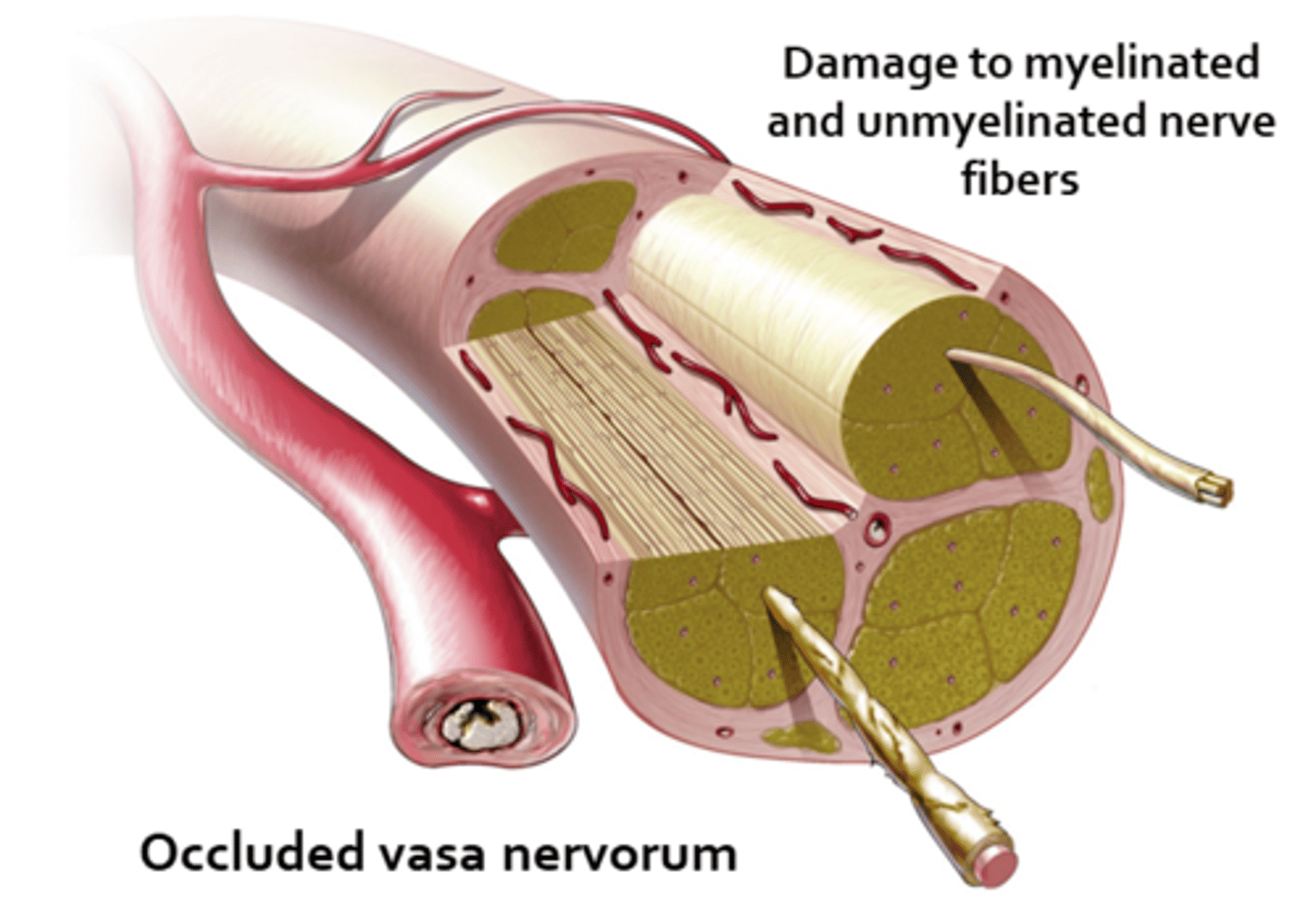
Why are most pupil-spared CN III palsies the result of DM or vascular etiologies?
vasa nervorum occlusion = infarcation of these small BV = focal areas of demyelination in the nerve core, while the outer pupillomotor fibers are spared
Where is the most likely location of the lesion in a CN III palsy (eye is down and out, ptosis) with a pupil that is larger in bright light, smaller in dim light?
cavernous sinus bc this is where the parasymp, symp, and CN III fibers are close together
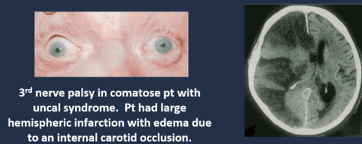
What is Uncal syndrome?
fast expanding mass, hemorrhage, tumor = increased ICP = brainstem compression and herniation down into foramen magnum (fatal!) = fixed, dilated pupil

What do we call the fixed, dilated pupil seen in Uncal syndrome?
Hutchinson pupil

Explain the progression of the Hutchinson pupil seen in Uncal syndrome.
1. miotic pupil due to initial cerbreal edema
2. ipsilateral dilated pupil due to expanding mass on that side
3. bilateral fixed and dilated pupils eventually
What are 3 other ocular signs of Uncal syndrome?
bilateral papilledema bc of increased ICP (bilateral even if only 1 pupil involved)
homonymous hemianopsia bc posterior cerebral arteries are occluded by mass = calcarine cortex ischemia
EOM paralysis = diplopia but mostly very late in the disease

What are some other S/S of Uncal syndrome?
HA
confusion
somnolence (sleepy)
coma
tenderness over clot region
Cheyne-Stokes respiration = forced breathing, deep and shallow, periods of no breath
overall, pt's will be very ill and presenting to the ER (or you should send them to ER immediately!)

What is the most common cause of Uncal syndrome?
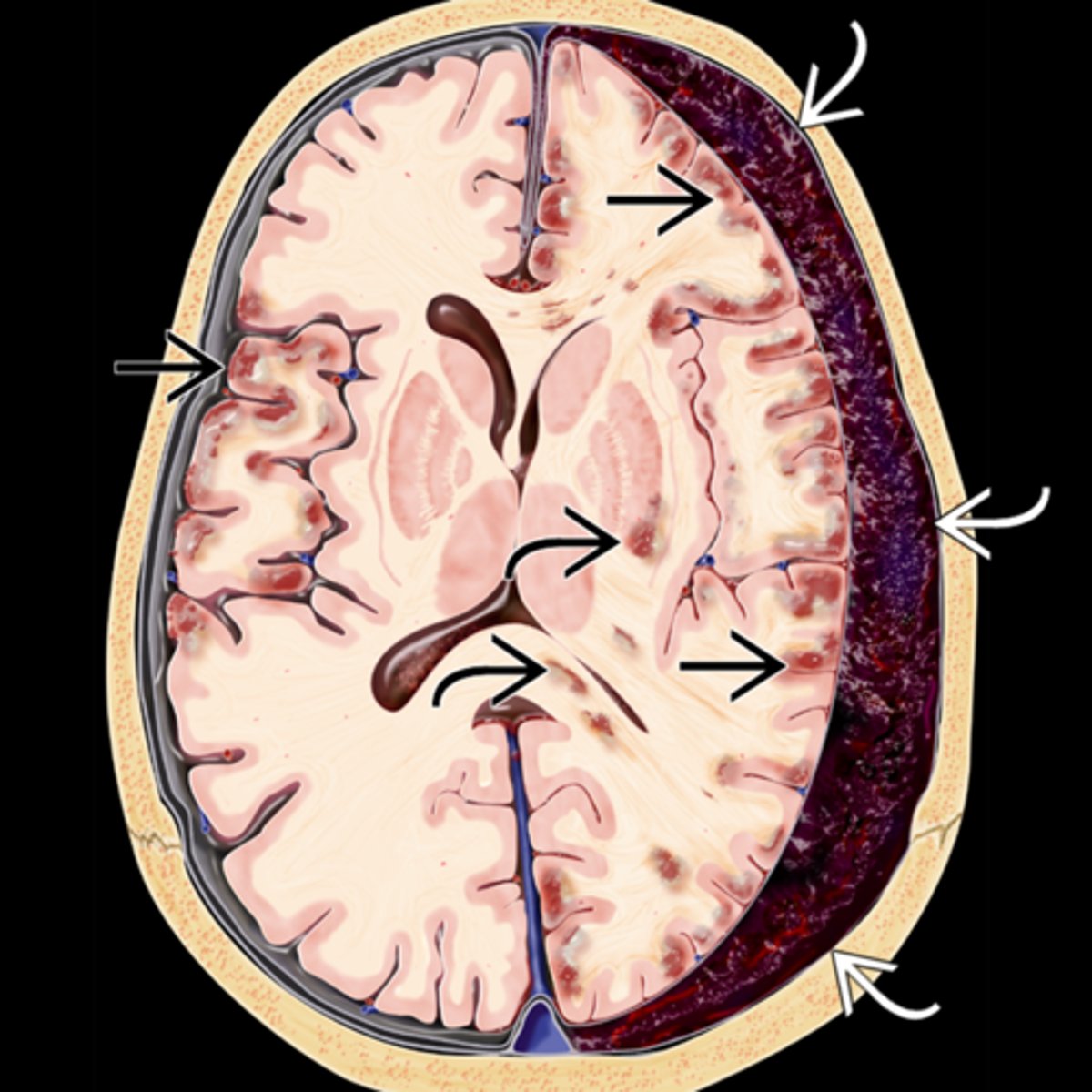
chronic subdural hematoma from head trauma esp in inebriated pt's

What are some less common causes of Uncal syndrome?
extradural hematoma from injury
subdural hematoma from drugs
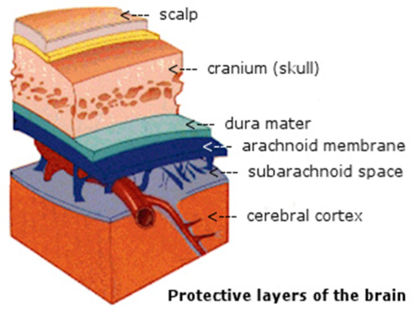
Explain the series of a events that causes a subdural hematoma, Uncal syndrome, and eventually death.
trauma = brain moves ant to post = cerebral veins in subdural space rip = blood fills subdural space = fibrous dura and arachnoid mesothelium beneath create a sac around the cot = sac breaks = blood enters brain = pt is sick and dies
Where is the Uncal region, affected in Uncal syndrome?
uncas of parahippocampal gyrus (inferior area of temporal lobe) = lateral to the brainstem = lateral to CN III in brainstem
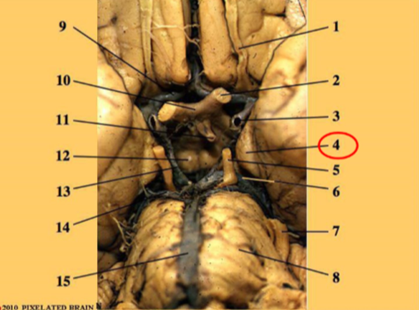
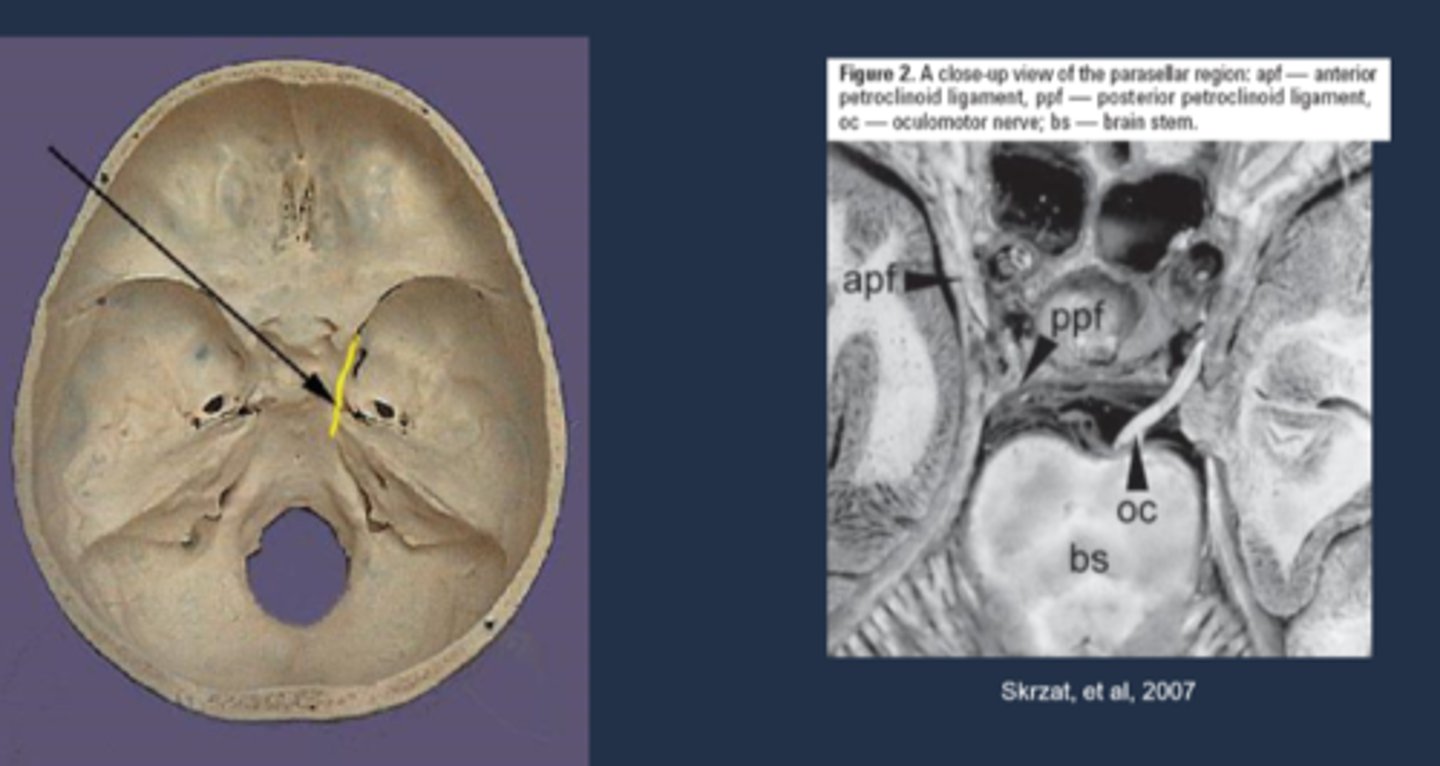
Explain how the Uncal region can affect CN III in Uncal syndrome.
CN III leaves brainstem = pupillomotor fibers move dorsomedially = uncus herniates = compresses CN III dorsomedially against petroclinoid ligament and dorsum sellae = pupil is blown
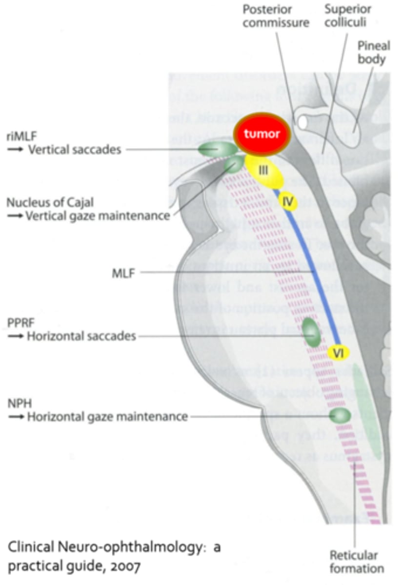
What is Parinaud's (dorsal midbrain) syndrome?
CNS lesion (tumor, MS, stroke, etc) causing compression on dorsal midbrain, affecting the...
superior colliculus = coordinating eye movement, convergence
MLF = vertical saccades
nucleus of Cajal = vertical gaze maintenance

What pupil finding is seen in Parinaud's (dorsal midbrain) syndrome?
light-near dissociated pupils
What are 3 other ocular findings seen in Parinaud's (dorsal midbrain) syndrome?
convergence retraction nystagmus = eyes turn in and retract (pulse in and out) when trying to look up
lid retraction = Collier's sign
inability to make saccades upwards (pursuit ability lost later as well)

What is a very common cause of Parinaud's (dorsal midbrain) syndrome?
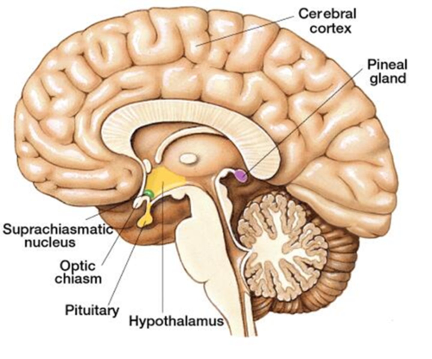
pineal gland tumor = would also show reduced melatonin and increased LH (precocious puberty)
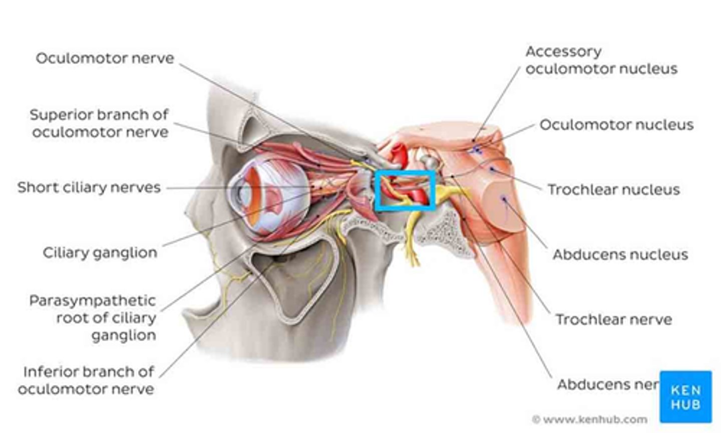
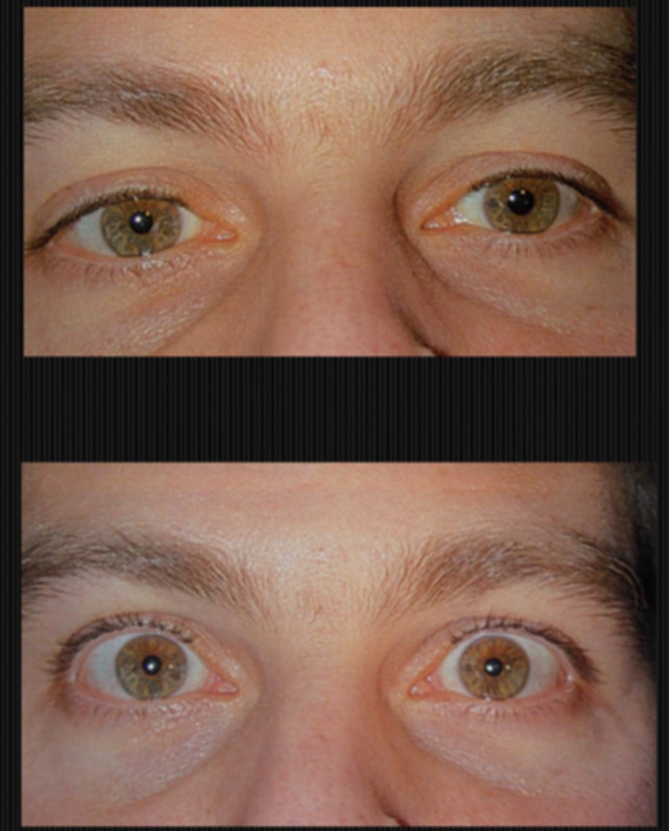
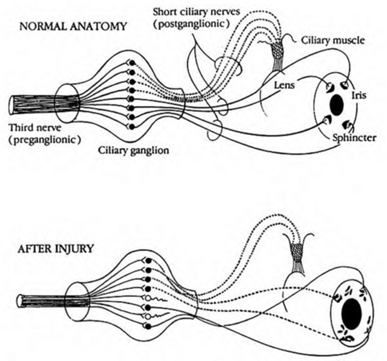
What is a tonic pupil?
ciliary ganglion damage due to trauma, infection, demyelinated, inflamed = parasymp fibers affected = pupil responds to near, but does not respond to light (light-near dissociation pupils) = large pupil

Ultimately, is a tonic pupil a large pupil caused by an issue with the muscle, NMJ, nerve, or brain?
nerve

What are the 4 S's that we expect to see when looking at the size and shape of a tonic pupil?
sector paralysis = abnormal shape due to sphincter/dilater paralysis
stromal spread = iris stroma spreads due to lack of innervation
pigment stream entropion = pupil border of iris is entropic adjacent to a part of the pupil border that is more flat
stromal streaming = stroma flows towards area that sphincter still works when light is turned on
What do we expect to see when looking at the direct response of a tonic pupil?
NO direct response (or may be extremely subtle)
What do we expect to see when looking at the near response of a tonic pupil?
slow, tonic constriction to near
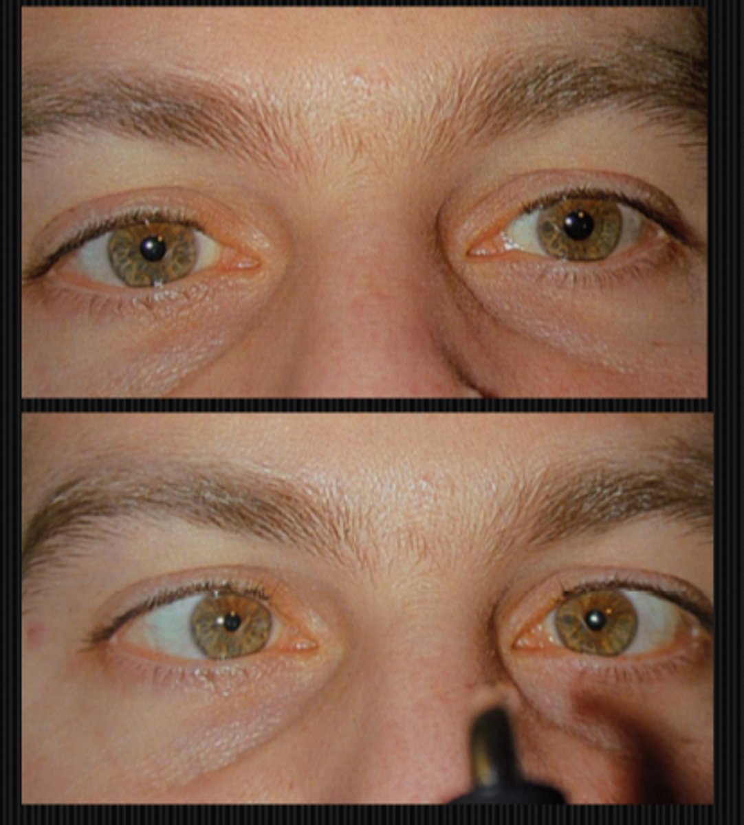
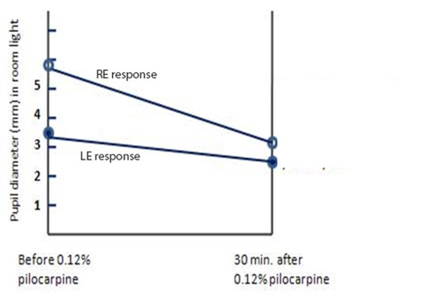
How will a tonic pupil present after the 0.125% pilocarpine denervation supersensitivity test?
tonic pupil will constrict more in response to dilute pilocarpine (bc the eye was starved)
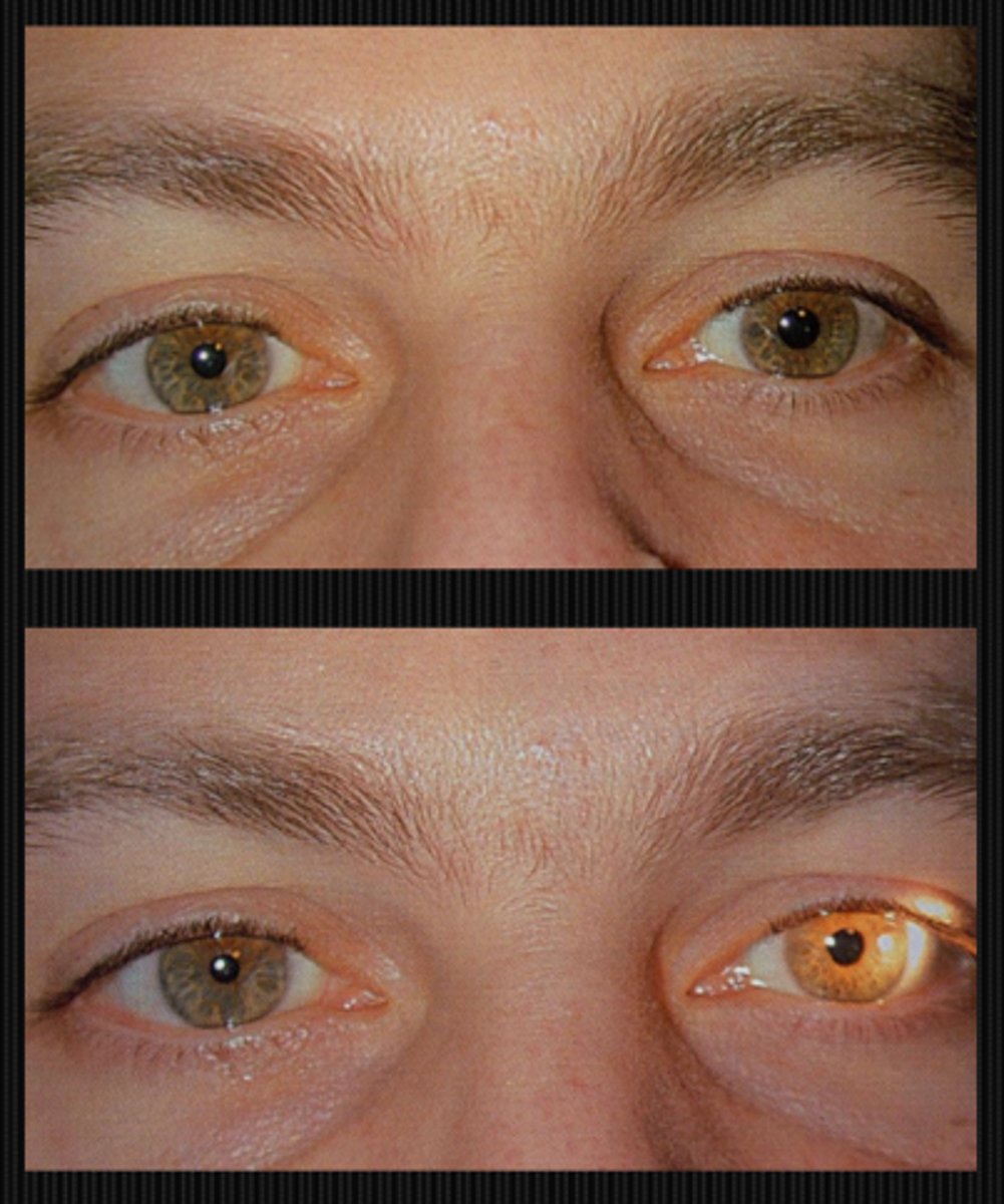
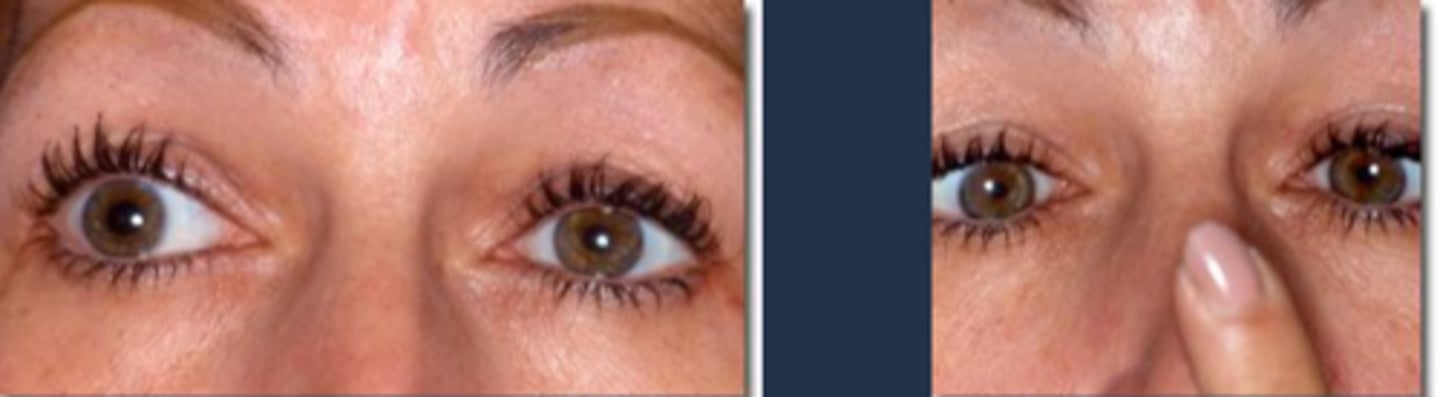
Ex) Based on the result of this 0.125% pilocarpine denervation supersensitivity test, which eye has the tonic pupil?
OD bc this is the diseased eye that is starved for ACh, so it will respond to dilute pilocarpine
What are the 3 types of tonic pupil?
local
neuropathic
Adies (idiopathic)

What are some causes of a local tonic pupil?
damage to ciliary ganglion via...
varicella
retrobulbar injection
orbital tumor
orbital surgery
traumatic iridoplegia = SPCN damage
post-ganglionic denervation
pre-ganglionic CN III palsy
abberrant regeneration of CN III
What are some causes of a neuropathic tonic pupil?
usually when pt's already have a known systemic disease such as...
DM
syphillis
Sarcoid
dysautonomia
Charcot-marie-Tooth syndrome
Ross' syndrome
NOTE: all are rare!
What are some causes of an Adie's tonic pupil?
idiopathic by definition
In which populations do we see Adie's tonic pupil?
females >>> males
age 20-40
THINK: Ladies get Aide's

What is the laterality of Adie's tonic pupil?
unilateral at first, then fellow eye can become involved
Why does the pupil react to near, but not to light in Adie's tonic pupil?
ciliary ganglion damage and recovery = nerve axoplasm that once went to accom now goes to pupil = pupillomotor fibers innervated by accom fibers = pupil still reacts to near
What are 3 ways that accom can be affected by Adie's tonic pupil?
normal
OR
accom paresis only in affected eye = unequal accom, HA
OR
CB spastic tonicity due to accom knocked out = denervation supersensitivity of ACh receptors = accom spasm, excess accom

Why is it that induced astig can show up in Adie's tonic pupil?
CB segmental paralysis = CB tugs on different parts of lens differently

What are some S/S of Adie's tonic pupil?
anisocoria
blurred vision
brow ache from accom issue
photophobia
dark adaptation issues
Pulfrich stereo phenomenon

What is the Pulfrich stereo phenomenon sometimes seen in Adie's tonic pupil?
anisocoria = one eye receives more light than the other = visual pigments more bleached in one eye = difference in signal transmission time from each eye = 2D objects appear to be 3D
Aside from the pupil not constricting to light, what are 2 other possible clinical findings seen in tonic pupil?
reduced corneal sensitivity bc sensory fibers from CN V1 travel through the damaged ciliary ganglion
decreased deep tendon reflexes for unknown reasons (called Holmes-Adie syndrome)
What is Holmes-Adie syndrome?
idiopathic tonic pupil
reduced deep tendon reflex
What is Ross's syndrome?
idiopathic tonic pupil
reduced deep tendon reflex
excess sweating
How do we perform supersensitivity testing of accom when accom is affected in a tonic pupil?
instil 0.25% pilocarpine OU = CB contracts = increased accom = re-refract pt = affected eye will have an increase in myopia with the pilocarpine (compared to the normal eye)
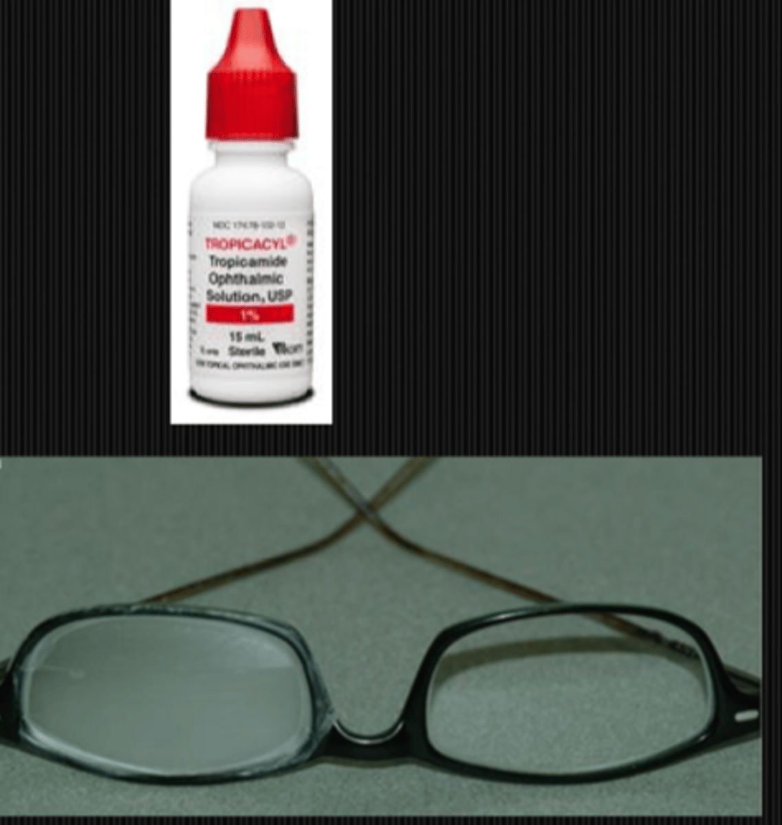
How do we treat the issue of CB spastic tonicity (tonic accom / accom spasm) sometimes seen in tonic pupil? 2 options
tropicamide or other atropine-like drug to relax the CB (but increases anisocoria)
OR
occlude/frost bottom part of glasses lens to prevent binocular vision at near (induce monovision)
How do we treat the issue of accom paresis (reduced accom) sometimes seen in tonic pupil? 3 options.
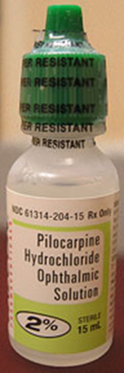
eserine or pilocarpine to stimulate the CB mm (but side effects of CB spasm, HA, myopia)
OR
MF CL in affected eye
OR
occlude/frost bottom part of glasses lens to prevent binocular vision at near (induce monovision)
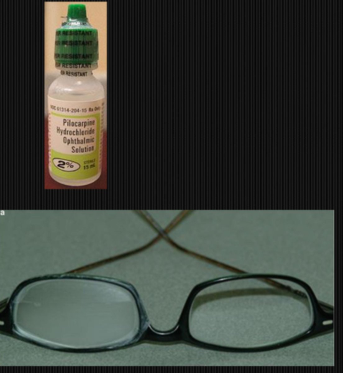
How do we treat the issue of anisocoria seen in tonic pupil? 3 options.
eye is super-sensitive to ACh and ACh-like molcules = prescribe 0.125% or 0.25% pilocarpine or Vuity = reduce pupil size (for cosmesis)
photochromic lenses for photophobia
cosmetic CL with a certain pupil size for cosmesis, photophobia
How long does it take for the pt to heal from a tonic pupil?
1-6 years, more on the longer end if accom is involved