Lecture 5 - Protein Structure and Function - part 1: Sequences Determines Structure
 Call Kai
 Learn
Learn Practice Test
 Spaced Repetition
 Match
Match Flashcards
 Knowt Play
1/22

There's no tags or description
Looks like no tags are added yet.
Name | Mastery | Learn | Test | Matching | Spaced | Call with Kai | Chat |
|---|
No analytics yet
Send a link to your students to track their progress
23 Terms
What drives protein folding
why do proteins want to fold - to produce what kind of structure
folding by covalent and non-covalent interactions
Proteins will fold to find the most stable structure
Three common themes in a 3D protein structure
flexible enough to function with other proteins
Stable enough that it will not change to another conformation
Exposed amino acids are compatible to the environment where proteins will function
Levels of protein structure
primary: linear sequence of AA Encoded by the DNA
Secondary: periodic, regular structures (folding of a -helix and B-beta strands)
Tertiary: Secondary structures folding into motifs and domains through non-covalent interactions
Quaternary: assembly of multiple polypeptides to form multiple subunit structures

Primary structure chains of amino acids
directionality
Which atom does the backbone of the peptide chain begin with?
Amino (N) terminal end is the beginning of the chain
Backbone consists of the peptide bonds and the a-carbons of each amino acidss
Peptide bond
characteristics
Polar, uncharged bonds
experiences resonance, and the characteristics of a double-bond
Rigid and planar - e.g. very little rotation

𝜙 and Ѱ angles
how does it rotate (which atoms)
How much can it twist?
rotation allowed around the bond linking of amide 𝜙 and carbony Ѱ from the
can twist range of -180 to +180, but depending on the structure, not all angles are premitted
Steric clashes need to be minimized to reduce hydrophobic strains, therefore should not twist with side chains are trans to one another

Levels of proteins structure: types of bonding
primary: peptide bonds
secondary: hydrogen bonds
tertiary and quaternary: disulfide bonds, electrostatic interactions hydrogen bonds, hydrophobic interactions
Secondary structure - H bonding
types of bonding
a-helix
B-strands and b-sheet: includes B-turns

a-helices: where in the strand does this folding occur in
intra-strand h-bonds forming between peptide backbone
Carboxyl bonds with an amino group 4 residues away
3.6 residues per 360 turn, 1.5 A high each

Proline: Helix breaker
what is special about proline
proline is an amino acid not typically present in a a-helix
This is because it is called a helix breaker
its side chains will covalently bind to the amino group in the peptide bond
Doesn’t allow for the proper 𝜙 and carbony Ѱ angle to form the coil we typically see in a helix

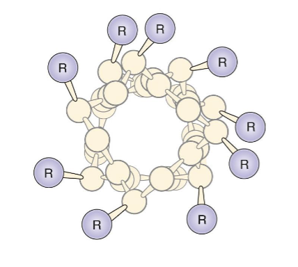
A-helices: projecting side-chains
shape of the helix + side chain facing what direction
right handed helix with side chains pointing out from the coil
B strands and B sheets
how does H-bonding work
H bond links to nearby or distant portions of the polypeptide to form b-sheets
R groups alternating to prevent steric strain
Strand either runs parallel, antiparallel, or mixed of both

B-pleated sheet
difference compared to a-helices
dimensions more extended when compared to the a-helices (spaced out)
Strands come together results in the sheet to get twisted

B-turns
a 4 residue segment allowing for turn of peptide 180 degrees
found on the surface of a globular proteins, connecting secondary structure
H bonds form between the carbonyl o2 and the amine hydrogen of the peptide backbone at position 1 and four
Proline amino acid common at position 2
Gly, Adn, and Ser is freq seen in turns \

Side chains and secondary structures
the allowable angles for the two bonds depend on the side chains

Tertiary structure
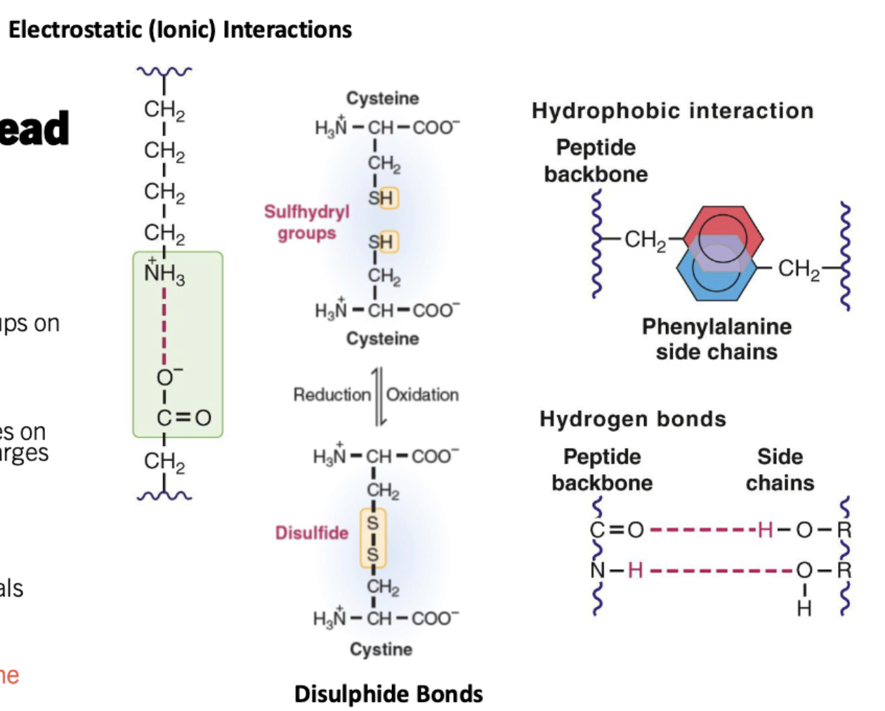
Electrostatic (ionic) Interactions
Hydrogen bonds
Hydrophobic Interactions
Disulphide bonds
Electrostatic (ionic) Interactions: interactions between charged groups on different amino acids
Hydrogen bonds: interactions between amino acids mediated by the partial positive charger on hydrogen and partial negative charges on other atoms
Hydrophobic interactions: grouping of non-polar amino acids (non-specific associations between closely packed atoms a.k.a Wan der Waals)
Disulphide bonds: a covalent bond only forms by two cyctseines to form a cystine
Tertiary structure
What are non-covalent bonds responsible for
What are ligands responsible for
What are post-translational modifications responsible for
non-covalent interactions and disulphide bonds helps form the 3D shape of the protein structure through brining distant regions of the polypeptide together
Ligands: Stabilizes or prevent specific interactions between the r groups
Post-translational modification: alter the functional group and therefore the structure of the protein
Protein Folding
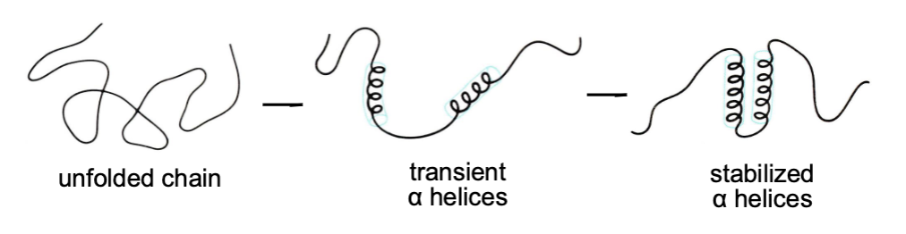
what interaction is the main driving force to protein folding
What do chaperones help with
Meaning of random coils
Why do proteins like being in their native structure
hydrophobic effect: main driving force in protein folding
Chaperones: prevent aggregation of the newly synthesizes and unfolded protein by binding to exposed hydrophobic regions
Random coils: no random - may be very stable
Native protein structure = best energetically stable conformation
Tertiary structure protein folding
what are motifs + functions
What are domains
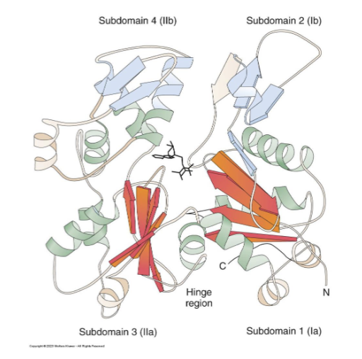
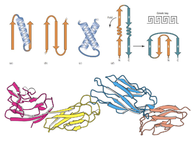
motifs: common combinations of secondary structures
Common motifs = common functions
Domains: independently folding regions within a polypeptide , connected by a short, flexible linker segment
Common domains = common functions - analysis of a protein sequence can help predict the functional domains
Post-translational modifications (PTMs)
what are ptm’s
What structure do they change
what opportunities do they provide
PTMs modify an amino acid within a protein
Altering interactions btw/ amino acids and change the teritary and quaternary structure
provides new sites for protein-protein interactions

Quaternary structure
the arrangwment of multiple subunits of polypeptide chains
may consist of non-identical or identical polypeptides
different subunits due to the multiple genes of the post-translational cleavage of precursors

Protein Denaturation
protein structure held together by numerous weak forces
Denaturation: the disruption of the weak forces by external stresses like heat, ph or chemical treatments
the result is the loss of structure and function

Human genome and the globin protein
the globin protein have 9 isoforms
Depending on the context, have similar functions
