Spinal Anatomy-Exam 1 General Characteristics Objectives
 Call Kai
 Learn
Learn Practice Test
 Spaced Repetition
 Match
Match Flashcards
 Knowt Play
1/52

There's no tags or description
Looks like no tags are added yet.
Name | Mastery | Learn | Test | Matching | Spaced | Call with Kai | Chat |
|---|
No analytics yet
Send a link to your students to track their progress
53 Terms
What is primary and secondary curvatures?
-primary= Kyphotic
-secondary= Lordotic
What regions of the spinal column belong to the primary curvatures?
Thoracic and Sacrococcygeal
What regions of the spinal column belong to the secondary curvatures?
Cervical and Lumbar
Is a Kyphotic curve more posterior or anterior? What about Lordotic?
-Kyphotic = curves posteriorly
-Lordotic= curves anteriorly
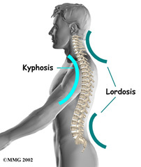
Kyphotic curve is formed due to greater ___ height than ___ height in the ____
posterior, anterior, vertebral body
Lordotic curve is formed due to greater ___ height than ___ height in the ___ of cervical and lumbar region
anterior, posterior, IVD
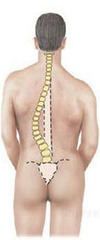
Where are lateral curvatures typically located? What are they thought to be a result of?
-They are typically in the Thoracic (upper portion) and Lumbar regions
-The deviations are a result from assymmetrical msucle use/tone
In a right handed lateral curve the convexity is open towards the ___
right
-thought to be associated with dominate hand
List some functions of the IVD
1. attach/separate vertebral bodies
2. helps shape the spine
3. acts as a powerful ligament
4. part of anterior border of vertebral canal and IVF
5. shock absorber
What two things form the IVD?
1. Annulus Fibrosus
2. Nucleus Pulposus
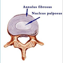
The annulus fibrosis attaches VB to each other, and is made of?
type 1-2 collagen
The nucleus pulposus separates VB from each other, and is made of ??
water, collagen, proteoglycans
What is the matrix of IVD made of?
proteoglycan gel and collagen fibers
What is a small rounded projection on a bone?
tubercle
What is a projection or outgrowth from a larger body on a bone?
Process
What is a small flat surface on a bone?
facet
term for "relating to a joint"
articular
term for "relating to, or situated to a rib"
costal
What forms the intervertebral foramina?
superior and inferior vertebral notches
The intervertebral Foramina located btwn T6 and T7 is formed from what?
Inferior vertebral notch of T6 and superior vertebral notch of T7
What gives rise to somites?
paraxial mesoderm
What do somites turn into?
sclerotomes, myotomes, and dermatomes
What do sclerotomes give rise to?
the vertebral column
what is the order of vertebral column formation?
membranous-->cartilaginous-->skeletal
Sclerotomes migrate to surround the notochord and form the ____
perchordal blastema
What is the fissure in the middle of a perichordal blastema?
intrasclerotomal fissure
The intrasclerotomal fissure gives rise to the ___ ___ which become the annulus fibrosus
perichordal discs
What are the centers of CHONDRIFICATION (not ossification) of a membranous vertebral blastema during week 6?
- one pair on the centrum
-one pair on the neural arch (left and right)
-one pair on the transverse process (left and right)
-total= 6!
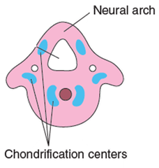
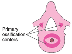
How many primary centres of OSSIFICATION are there?
-total= 3
-2 in the vertebral arch (L&R) and 1 in vertebral body
How many secondary centers of OSSIFICATION are there?
-total= 5
-2 in the epiphyseal rim (Sup & Inf.); 2 in the transverse process (L&R); 1 in the spinous process
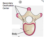
Where are there synchondroses centers?
-epiphyseal ring synchondrosis
-tip of TP synchondrosis
-tip of SP synchondrosis
Which somite within the paraxial mesoderm gives rise to the vertebral column?
Sclerotomes
The spinal canal is the union of ____ ___ into a singular canal
vertebral foramina
Where does the spinal cord end?
L2
List out the order of the spinal cord arteries starting with the aorta
aorta --> segmental branch A. --> spinal branch --> medullary feeder artery (post. & ant.) --> ant/post (2!) arteries & radicular arteries (spinal roots)
-Look at General Characteristics slide 31 for a visual image!
Which spinal canal artery directly feeds into the posterior spinal arteries?
medullary feeder artery
Where does the basivertebral venous foramina run?
through the vertebrae to allow venous return from canal
Where is the internal vertebral venous plexus?
epidural space
What are the other 2 spinal veins?
anterior/posterior spinal vein
List out the meninges from superficial to deep
-Dura Mater
-Arachnoid Mater
-Pia Mater
What meninge is tough and fibrous
Dura Mater
What meninge is web like?
Arachnoid Mater
List out the 3 spaces from Superficial to deep
-Epidural space
-Subdural space
-Subarachnoid space
The ___ space is external to the dura and is filled with fat
epidural
The ___ space is a POTENTIAL space between dura and arachnoid
subdural
The __ space is between arachnoid and pia and contains CSF
subarachnoid
Where is the anterior longitudinal ligament positioned?
On the anterior side of the vertebral bodies
Where is the posterior longitudinal ligament positioned?
On the posterior side of the vertebral bodies
What does the posterior longitudinal ligament prevent?
prevents hyperflexion and posterior spinal disc herniation
What ligament forms the ANTERIOR boundary of the spinal canal?
posterior longitudinal ligament
What ligament forms the POSTERIOR boundary of the spinal canal?
Ligamentum Flavum
The____ is an extension of pia mater, and anchors the spinal cord to the Sacrum and Coccyx. It prevents_____ movement.
Filum Terminale; vertical movement
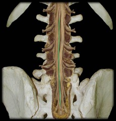
The___ is an extension of pia mater, and anchors the spinal cord to the dura mater. It prevents ____ movement.
Denticulate ligaments; side movement