PSYC 110: Nervous System; Cell Structure/Function; Neurodevelopment
1/95
Earn XP
Description and Tags
PSYC110: The Brain & Behavior (SP 2025). Covers Lectures 1, 2, and 3.
Name | Mastery | Learn | Test | Matching | Spaced | Call with Kai |
|---|
No analytics yet
Send a link to your students to track their progress
96 Terms
Neurodevelopment
Development of the central nervous system; how axons find their end targets.
Neural Tube
A early hollow, elongated form/structure that the CNS begins embryonic life as.
Starts as a single cell (fertilized egg).
Pluripotent Cells
Early-stage cells that can become any cell type in the body.
Referred to as embryonic stem cells.
Stem Cells
Cells that can divide or develop into adult cells.
Neurulation
The creation of the neural tube and neural crest; happens ~24 days after conception.
Differentiation
The process of structures becoming more complex and functionally specialized.
Neuronal Proliferation
The creation of neurons.
3 Primary Vesicles (4 Weeks) of the Neural Tube
Prosencephalon, Mesencephalon, Rhombencephalon
5 Secondary Brain Vesicles (5 Weeks) of the Neural Tube
Telencephalon, Diencephalon, Mesencephalon, Metencephalon, Myelencephalon.
Symmetrical Cell Division
Progenitor cells in the ventricular zone split to produce 2 new progenitor cells.
Creates the ventricular (VZ) & subventricular zones (SVZ).
Asymmetrical Cell Division
After 7 weeks, cells divide into 1 progenitor + 1 brain cell.
First brain cells produced are radial glia.
Radial Migration
Radial glia extends fibers from the ventricular zone (VZ) to the pia mater.
Glia fibers grow longer as new layers are added to the cortex
Neurons migrate along the radial glia to their place.
Tangential Migration
New cells push out previously formed cells from the proliferation zone.
Proliferation Zone
Where neurons were generated, in the ventricular or subventricular zones.
Spatiotemporal Gradient
Structures that are layered/constructed from the inside out.
Usually present for inner structures: thalamus, hypothalamus, cortex, etc.
Synaptogenesis
Neurons at their final location begin to form connections with other neurons by growing axons & dendrites and creating synapses.
Apoptosis
Death of neurons.
[e.g.]: Radial glial cells go through apoptosis and are transformed into astrocytes after the layers are created.
Synaptic Pruning
Death of synapses by apoptosis.
Continues after birth into the late-20s.
Myelination
Forming of the myelin sheath on axons.
Postnatal Brain Development
The human brain continues to grow after birth for ~25 years, growing up to 5x its size:
Growth of synaptic connections between neurons.
Myelination of axons.
Neurogenesis
Production of new neurons in the adult brain, which still contains some stem cells.
Neuroplasticity
The ability of the nervous system to change its activity to intrinsic or extrinsic stimuli by reorganizing its structure, functions, and connections.
Learning strengthens brain connections, which decay when neglected.
Biopsychology in Ancient Cultures
Ancient Chinese, Egyptian, and Indian cultures believed that the heart was the seat of thoughts and emotions; behavior was dependent on our body’s inner physiology.
Reticular Theory
Camillo Golgi’s hypothesis: Neurites of different neurons are fused together to form a continuous reticulum.
Early 1950s microscopes showed that neurites are not continuous with other neurons.
Neuron Doctrine
Santiago Ramon y Cajal’s hypothesis: Neurons are separate cells that communicate by contact.
Shared a Nobel Prize (Physiology or Medicine) with Golgi in 1908.
Visualizing Cells: Golgi Stain
Makes only a few neurons stand out so they can be seen separately.
Developed by Golgi and applied by Cajal to prove that brain cells are separate.
Immunofluorescent Staining
Uses protein-specific antibodies chemically labeled with fluorescent dyes.
Allows visualization of those proteins using light microscopy.
Brainbow
Genetic-cell labeling method that allows visualization of individual neurons in a brain slice using hundreds of hues of fluorescent proteins.
Dr. Suzanne Herculano-Houzel
Used a “brain soup” method to count the number of human brain cells:
Liquefied the brain & took a sample.
Stained the neurons’ nuclei.
Counted them under a microscope.
Number of Neurons in a Human Brain
~86 billion neurons that receive an avg. of ~5000 synaptic contacts each.
DNA Transportation Out the Nucleus
DNA cannot leave the nucleus on it’s own. It must go through:
Transcription: DNA ⇒ mRNA
Translation: mRNA ⇒ Proteins
Transportation: Proteins ⇒ Out
Nucleus
Located in the neuron’s cell body (soma) and contains genetic material.
Semipermeable Membrane
Outer surface of the neuron; allows smaller molecules without electric charges to pass through it. Stops larger or highly charged molecules.
Soma
Cell body of the cell; makes up the gray matter of the brain.
Dendrites
Antennae that protrude from a neuron’s soma to receive synapses.
Covered in dendritic spikes that change over time with specialized receptors.
Classifies neurons.
Axon Hillock
A part of the soma that integrates all messages in the neuron & initiates action potentials.
Axon
A wirelike structure that protrudes from the soma to axon terminals.
Insulated by a myelin sheath.
Myelin Sheath
A fatty membrane that insulates the axon and helps speed neural impulses.
Nodes of Ranvier
Gaps in the myelin sheath.
Axon Terminals
Branches at the end of an axon (on the opposite side of the dendrites & soma) that forms junctions with other cells.
Contains the terminal buttons & synaptic vesicles.
Terminal Buttons
The region in the axon terminal where signals traveling down the axons end at.
Synaptic Vesicles
Parts of the terminal buttons (in the axon terminal) that house neurotransmitters.
Glial Cells
Provide physical & metabolic support to neurons.
Notable Neuroglia: Oligodendrocytes (CNS), Schwann Cells (PNS), Microglia, Astrocytes
Astrocytes
Clears neurotransmitters from the synaptic cleft.
Secretes proteins that help neurons from connections.
Most numerous glial cell in the brain.
Astrocytic Process
The process of astrocytes wrapping around synapses to isolate them ®ulate neurotransmitter levels.
Microglia (Phagocytes)
Clean up cellular debris, pathogens, foreign material, etc. produced by damage
Oligodendrocytes
Central nervous system (CNS) cells that produce the myelin sheath.
Forms multiple (>30) sheaths around different axons.
Schwann Cells
Peripheral nervous system (PNS) cells that form the myelin sheath, regenerate nerves, and provide nutrients to axons.
Forms a single sheath around one axon.
Central Nervous System (CNS)
Part of the nervous system composed of the brain + spinal cord.
Peripheral Nervous System (PNS)
Part of the nervous system composed of the autonomic + somatic nervous system.
Gray Matter
The part of the brain that contains the cell bodies (somas) in the CNS.
White Matter
The part of the brain that contains myelinated axons running to and from neurons.
Somatic Nervous System
Part of the PNS; relays sensory & motor information to/from the CNS.
Connects the CNS to the organs, limbs, and skin.
Autonomic Nervous System
Part of the PNS; controls our internal organs & glands.
Can be divided into the sympathetic + parasympathetic nervous systems.
Sympathetic Nervous System
“Fight or Flight”: Physiological changes that direct blood flow and oxygen to skeletal muscles.
Enables the organism to move quickly if threatened.
Parasympathetic Nervous System
“Rest & Digest”: Physiological changes that signify resting and gathering energy.
Meninges
3 protective layers around the brain:
Dura Mater “Tough Mother”
Arachnoid Mater
Pia Mater “Tender Mother”
Dura Mater “Tough Mother”
Tough, outermost, flexible layer of meninge.
Arachnoid Mater
Middle meninge layer, resembles spiderwebs.
Subarachnoid Space
Hollow-ish space between the arachnoid and pia mater meninges that is filled with cerebrospinal fluid (CSF).
Pia Mater “Tender Mother”
Delicate, innermost layer of meninge.
Cerebrospinal Fluid (CSF)
A filtrate of blood plasma that provides nourishment, removes waste, and protects the brain.
Fills the ventricular system and subarachnoid space.
Ventricular System
Consists of ventricles: interconnected cavities in the brain.
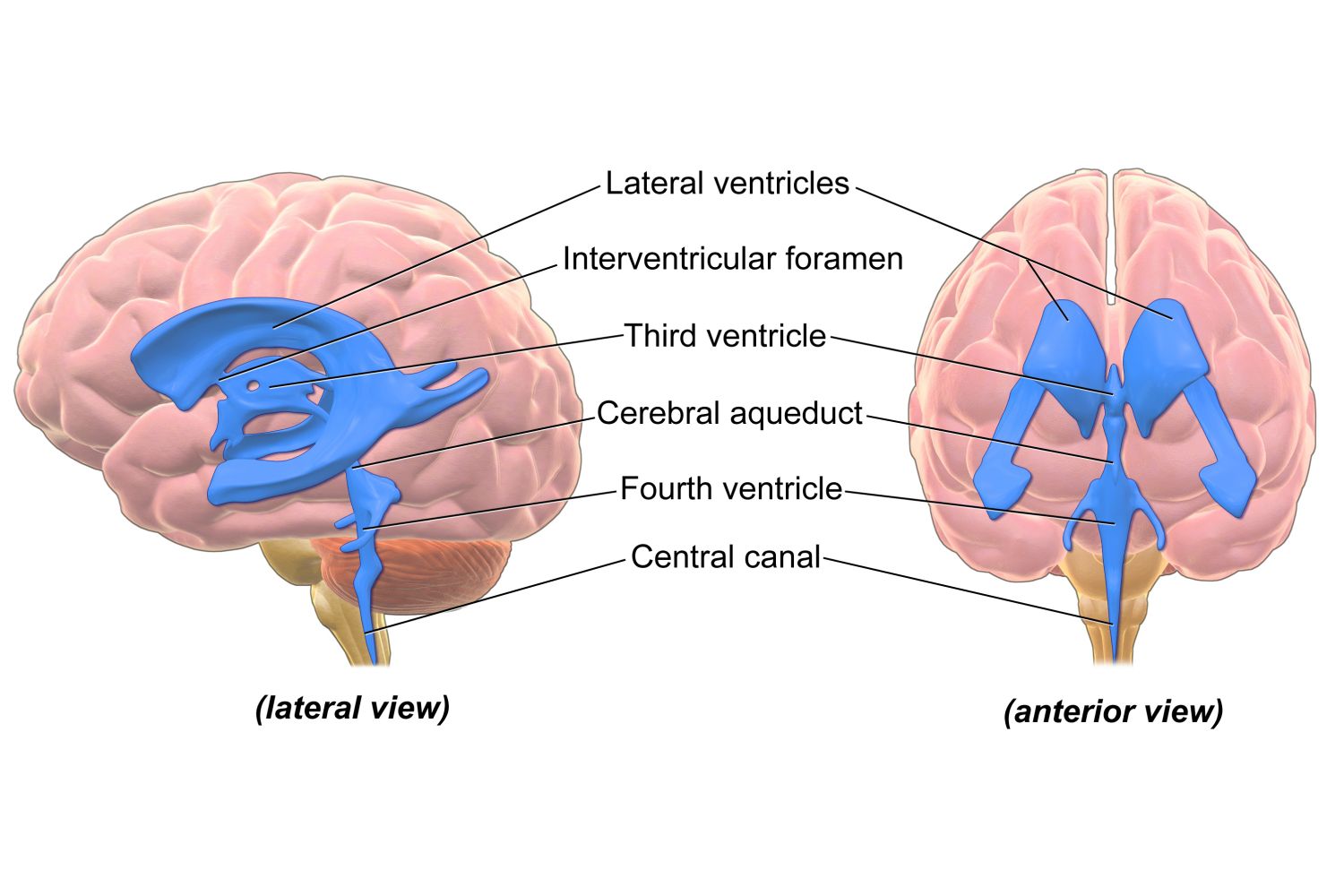
Cerebral Aqueduct
The ventricle that connects the 3rd ⇔ 4th ventricles.
Interventricular Foramen
The ventricle that connects the lateral ⇔ third ventricles
Choroid Plexus
A network of blood vessels in each ventricle of the brain; derived from the pia mater
Secretes cerebrospinal fluid.
Provides a barrier between the blood and the brain.
Spinal Cord
Part of the CNS; acts as a relay system connecting the brain to the world.
Starts at the brain stem and ends just below the ribs.
Surrounded by spinal meninges.
Dorsal Horn
“In the door…”: Sensory/Afferent information IN → from the sensory pathways (gray matter) of the PNS
Ventral Horn
“Out the vent…”: Motor/Efferent information OUT ← from the sensory pathways (gray matter) of the PNS
Neuron Circuit
Groups of neurons connected by synapses that process specific types of information.
Gyri
The bumps on the cerebrum’s (brain’s) surface.
Sulci
The grooves in the cerebrum’s (brain’s) surface.
Fissures
Especially deep sulci (grooves) in the brain’s surface.
[e.g.]: Medial longitudinal, lateral, central
Medial Longitudinal Fissure
The fissure that splits the brain from front to back into its two hemispheres.
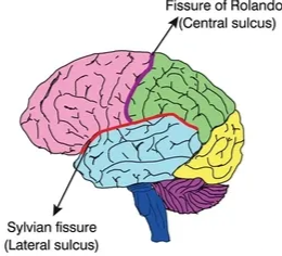
Lateral (Sylvian) Fissure
The fissure in between the frontal/parietal and temporal lobes

Central Fissure
The fissure in between the frontal and parietal lobes.
Resting Membrane Potential
The tendency of the cell’s inside to be more negative (-65mV) compared to the outside when at rest.
Membrane Potential
The difference in charge between the intracellular and extracellular space of a neuron.
Potential
The separation of electrical charge across the membrane; has the potential to be excitable.
Sodium (NA+)
A positively-charged ion that is more concentrated outside the cell in extracellular fluid.
Tendency to move inside the cell.
Potassium (K+)
A positively-charged ion that is more concentrated inside the cell near the membrane in intracellular fluid.
Tendency to move outside the cell.
Chloride (CL-)
A negatively-charged ion that is evenly distributed on both sides of the membrane.
All-or-None Phenomenon
An incoming signal from another neuron will either be sufficient or insufficient to reach the threshold of excitation.
No in-between; no stopping it when it has started.
The action potential is propagated at its full strength at every gap of the axon.
Stronger stimulus = more frequent signal.
Action Potential
Ions are lined up on either side of the membrane
NA+ outside; K+ inside
A neuron receives a signal/stimulus from the dendrites and abruptly changes its state
Depolarization
Threshold of excitement is triggered for an action potential
The peak of the NA+ ion surge is reached
Repolarization
Hyperpolarization
Resumes resting potential
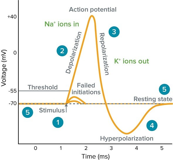
Depolarization
Sodium (NA+) channels open and NA+ ions flow into the cell.
Threshold of Excitement
When enough NA+ is inside the cell (making it more positively-charged), a threshold (-50 to -55 mV) is triggered for an action potential.
An action potential only happens when the threshold is reached by the signal/stimulus.
Repolarization
Potassium (K+) channels open and K+ ions flow out the cell.
Hyperpolarization
The state after repolarization; the cell overcorrects the positive charge and becomes too negative.
Saltatory Conduction
The process by which NA+ ions skip from one Node of Ranvier to the next.
NA+ ions enter the axon via depolarization
NA+ ions repel each other down the axon (positive charges push against one another)
The next area in the axon becomes overly positive (polarized)
New NA+ channels open
More NA+ surges into the axon
The previous area hyperpolarizes (becomes overly negative)
NA+ channels cannot open there
An action potential only goes in one direction
No new sodium channels where myelin covers the axon ⇒ NA+ “skips” ahead, speeding up the process
2 Types of Neuronal Communication
Neurons send both chemical & electrical signals:
Chemical: Neurotransmitters transmit intercommunication.
Electrical: Transfer of ions
Chemical Communication
Neurotransmitter (NT) synthesis
NTs are loaded into synaptic vesicles at the terminal buttons (end of axons)
Waiting for an action potential to reach the presynaptic terminal to be released
The action potential reaches the presynaptic cleft
NTs release
Vesicles (loaded with NTs) fuse with the presynaptic terminal
NTs bind to postsynaptic receptors
Biochemical/electrical changes in postsynaptic cells occur
Excess NTs are cleared:
Reuptake
Get broken down
Drift away naturally/get taken away by astrocytes
Neurotransmitter Reuptake
Neurotransmitters are reabsorbed back into the axon terminal they were released from.
Electrical Communication
Electrical signals are passed through the connexon between two neurons.
Faster at conducting nerve impulses, but can only produce simple behavior.
Connexon
A protein structure that physically bridges two neurons at their gap junctions and allows for electrical communication.
Allows both electrical signals and small molecules to pass between neurons in both directions.
Excitatory Synapse
When the synapse releases positively-charged ions; triggers depolarization and a reactionary potential in the next synapse.
Inhibitory Synapse
When the synapse releases negatively-charged ions; triggers hyperpolarization and stops the neuron action chain.