Testable SOT term 2

0.0(0)
learnLearn
examPractice Test
spaced repetitionSpaced Repetition
heart puzzleMatch
flashcardsFlashcards
Card Sorting

1/86

encourage image

There's no tags or description

Looks like no tags are added yet.

Study Analytics
Name
Mastery
Learn
Test
Matching
Spaced

No study sessions yet.

87 Terms

1
New cards

Upper Limb Tests

2
New cards

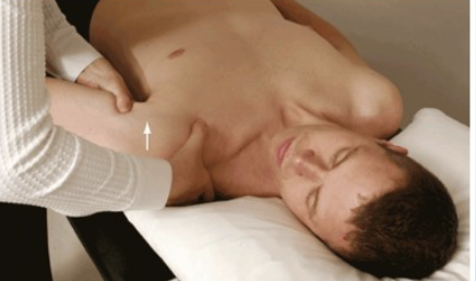
Anterior Drawer Test

Purpose: tests for anterior GH instability

Positive signs: Hypermobility, clicking, apprehension

Method:

  • Patient lies supine

  • Abduct shoulder to 80-120 degrees, flexed forward to 20 degrees, laterally rotated up to 30 degrees

  • Therapist stabilizes scapula

  • Draws humerus forward

<p><strong>Purpose:</strong> tests for anterior GH instability</p><p><strong>Positive signs:</strong> Hypermobility, clicking, apprehension</p><p><strong>Method:</strong></p><ul><li><p>Patient lies supine</p></li></ul><ul><li><p>Abduct shoulder to 80-120 degrees, flexed forward to 20 degrees, laterally rotated up to 30 degrees</p></li><li><p>Therapist stabilizes scapula</p></li><li><p>Draws humerus forward</p></li></ul><p></p>
3
New cards

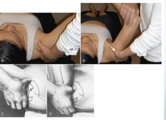
Posterior Drawer

Purpose: Tests for posterior instability

Positive Signs: Hypermobility, pain, apprehension

Method:

  • Start same position as anterior drawer- supine

  • Medially rotate arm and flex forward to 60-80°

  • Apply posterior force

<p><strong>Purpose:</strong> Tests for posterior instability</p><p><strong>Positive Signs:</strong> Hypermobility, pain, apprehension</p><p><strong>Method:</strong></p><ul><li><p>Start same position as anterior drawer- supine</p></li><li><p>Medially rotate arm and flex forward to 60-80°</p></li><li><p>Apply posterior force</p></li></ul><p></p>
4
New cards

Apprehension/Crank Test

Purpose: Tests for gross instability of GH joint

Positive Signs: Apprehension, muscle spasm

Method:

  • Patient supine- at edge of table so arm can go off edge

  • Abduct to 90 degrees and fully externally rotate

  • Apply overpressure if no pain (push down on their arm)
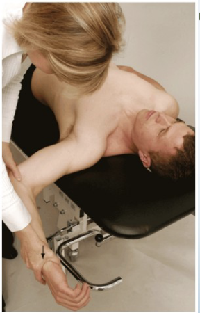
<p><strong>Purpose:</strong> Tests for gross instability of GH joint</p><p><strong>Positive Signs:</strong> Apprehension, muscle spasm</p><p><strong>Method:</strong></p><ul><li><p>Patient supine- at edge of table so arm can go off edge</p></li><li><p>Abduct to 90 degrees and fully externally rotate</p></li><li><p>Apply overpressure if no pain (push down on their arm)</p></li></ul><p></p>
5
New cards

Push Pull Test

Purpose: Tests for posterior instability

Positive Signs: Hypermobility, pain, apprehension

Method:

  • Patient supine

  • Abduct arm to 90 degrees, flex to 30 degrees

  • Downward push at shoulder and upward pull at wrist
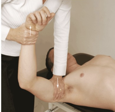
<p><strong>Purpose:</strong> Tests for posterior instability</p><p><strong>Positive Signs:</strong> Hypermobility, pain, apprehension</p><p><strong>Method:</strong></p><ul><li><p>Patient supine</p></li><li><p>Abduct arm to 90 degrees, flex to 30 degrees</p></li><li><p>Downward push at shoulder and upward pull at wrist</p></li></ul><p></p>
6
New cards

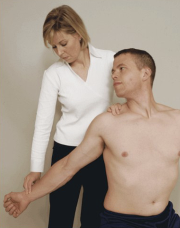
Sulcus Sign

  • Purpose: Tests for inferior G/H instability

  • Positive Signs: Hypermobility, sulcus sign, with pain/apprehension

  • Method:

  • Arm relaxed at side

  • Distal pull of humerus

<ul><li><p><strong>Purpose:</strong> Tests for inferior G/H instability</p></li><li><p><strong>Positive Signs:</strong> Hypermobility, sulcus sign, with pain/apprehension</p></li><li><p><strong>Method:</strong></p></li><li><p>Arm relaxed at side</p></li><li><p>Distal pull of humerus</p></li></ul><p></p>
7
New cards

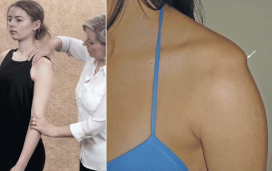
Hawkins-Kennedy Test

Purpose: Tests for supraspinatus tendinopathy/impingement

Positive Signs: Pain, apprehension

Method:

  • Seated or standing

  • Forward flex arm to 90 degrees, medially rotate
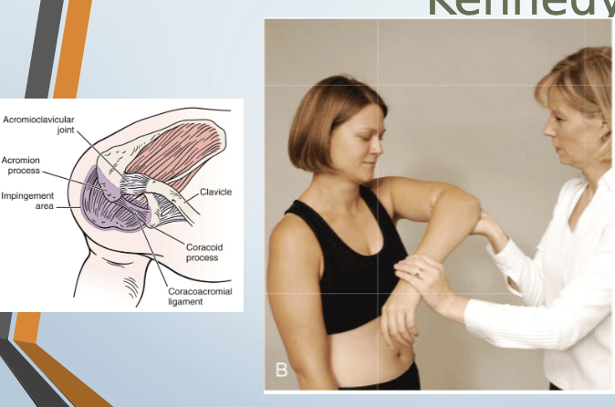
<p><strong>Purpose:</strong> Tests for supraspinatus tendinopathy/impingement</p><p><strong>Positive Signs:</strong> Pain, apprehension</p><p><strong>Method:</strong></p><ul><li><p>Seated or standing</p></li><li><p>Forward flex arm to 90 degrees, medially rotate</p></li></ul><p></p>
8
New cards

Neer Impingement Test

Purpose: Tests for supraspinatus overuse injury

Positive Signs: Pain, apprehension

Method:

  • Seated or standing

  • Abduct arm in scaption, medially rotated
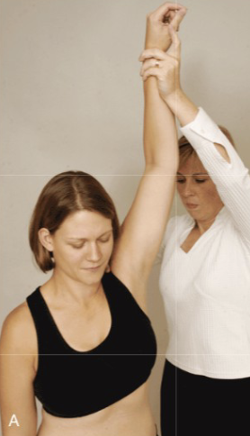
<p><strong>Purpose:</strong> Tests for supraspinatus overuse injury</p><p><strong>Positive Signs:</strong> Pain, apprehension</p><p><strong>Method:</strong></p><ul><li><p>Seated or standing</p></li><li><p>Abduct arm in scaption, medially rotated</p></li></ul><p></p>
9
New cards

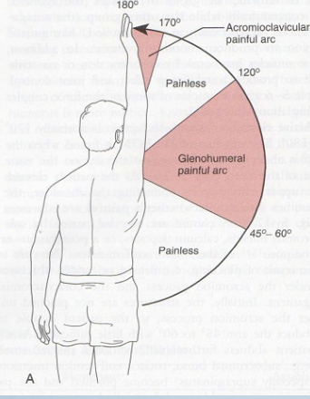
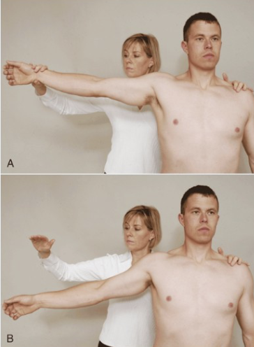
Painful Arc

Purpose: Tests for compression of subacromial structures

Positive Signs: Pain during mid-arc, pain at 170-180° indicates AC injury

Method:

  • AFROM abduction

<p><strong>Purpose:</strong> Tests for compression of subacromial structures</p><p><strong>Positive Signs:</strong> Pain during mid-arc, pain at 170-180° indicates AC injury</p><p><strong>Method:</strong></p><ul><li><p>AFROM abduction</p></li></ul><p></p>
10
New cards

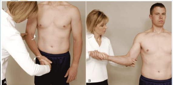
Apley’s Scratch Test

Purpose: Tests combined shoulder movements- internal and external rotation

Positive Signs: Normal fingertip touching range

Method:

  • Demonstrate to client to mimic

  • one arm over and behind shoulder

  • One arm reaching under and up to touch other hand
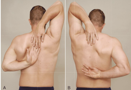
<p><strong>Purpose:</strong> Tests combined shoulder movements- internal and external rotation</p><p><strong>Positive Signs:</strong> Normal fingertip touching range</p><p><strong>Method:</strong></p><ul><li><p>Demonstrate to client to mimic</p></li><li><p>one arm over and behind shoulder</p></li><li><p>One arm reaching under and up to touch other hand</p></li></ul><p></p>
11
New cards

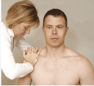
AC Shear Test

Purpose: Tests acromioclavicular joint pathology

Positive Signs: Pain, hypermobility

Method:

  • Seated

  • Hands on clavicle and spine of scapula

  • Interlock fingers one hand on each side of shoulder

  • Squeeze together

<p><strong>Purpose:</strong> Tests acromioclavicular joint pathology</p><p><strong>Positive Signs:</strong> Pain, hypermobility</p><p><strong>Method:</strong></p><ul><li><p>Seated</p></li><li><p>Hands on clavicle and spine of scapula</p></li><li><p>Interlock fingers one hand on each side of shoulder</p></li><li><p>Squeeze together</p></li></ul><p></p>
12
New cards

Drop Arm

Purpose: Tests for rotator cuff tear

Positive Signs: Inability to slowly lower arm, pain

Method:

  • Abduct shoulder to 90 degrees

  • Client lowers arm slowly to side

<p><strong>Purpose:</strong> Tests for rotator cuff tear</p><p><strong>Positive Signs:</strong> Inability to slowly lower arm, pain</p><p><strong>Method:</strong></p><ul><li><p>Abduct shoulder to 90 degrees</p></li><li><p>Client lowers arm slowly to side</p></li></ul><p></p>
13
New cards

Speed’s Test

Purpose: Tests for bicipital tendinopathy

Positive Signs: Pain in bicipital groove

Method:

  • Seated

  • Resists shoulder forward flexion in supination then pronation
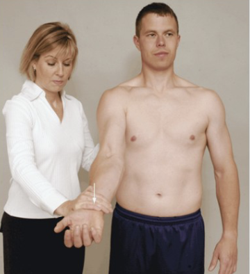
<p><strong>Purpose:</strong> Tests for bicipital tendinopathy</p><p><strong>Positive Signs:</strong> Pain in bicipital groove</p><p><strong>Method:</strong></p><ul><li><p>Seated</p></li><li><p>Resists shoulder forward flexion in supination then pronation</p></li></ul><p></p>
14
New cards

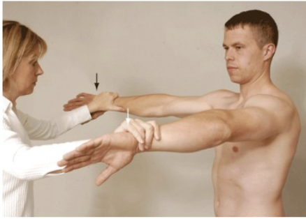
Empty Can/ Jobes

Purpose: Tests for supraspinatus muscle/tendon tear

Positive Signs: Muscle weakness, pain

Method:

  • Abducted arm to 90 degrees in neutral, resist

  • Rotate medially into scaption and resist again

  • Resist at wrists pushing downward

<p><strong>Purpose:</strong> Tests for supraspinatus muscle/tendon tear</p><p><strong>Positive Signs:</strong> Muscle weakness, pain</p><p><strong>Method:</strong></p><ul><li><p>Abducted arm to 90 degrees in neutral, resist</p></li><li><p>Rotate medially into scaption and resist again</p></li><li><p>Resist at wrists pushing downward</p></li></ul><p></p>
15
New cards

Yergason’s Test

Purpose: Tests for transverse humeral ligament integrity

Positive Signs: Biceps tendon pops out, pain in biccipital groove

Method:

  • Seated

  • Palpate biccipital groove

  • Elbow flexed medially and pronated then

  • Resist supination and lateral rotation with elbow flexed

  • Like a hitchhike

<p><strong>Purpose:</strong> Tests for transverse humeral ligament integrity</p><p><strong>Positive Signs:</strong> Biceps tendon pops out, pain in biccipital groove</p><p><strong>Method:</strong></p><ul><li><p>Seated</p></li><li><p>Palpate biccipital groove</p></li><li><p>Elbow flexed medially and pronated then</p></li><li><p>Resist supination and lateral rotation with elbow flexed</p></li><li><p>Like a hitchhike</p></li></ul><p></p>
16
New cards

Adson’s Test

Purpose: Tests for anterior scalene-related TOS

Positive Signs: Pain into arm, loss of pulse

Method:

  • Seated

  • Palpate radial pulse, rotate head towards test shoulder

  • Extend head

  • Therapist externally rotates and extends shoulder

  • Patient takes deep breath and holds

<p><strong>Purpose:</strong> Tests for anterior scalene-related TOS</p><p><strong>Positive Signs:</strong> Pain into arm, loss of pulse</p><p><strong>Method:</strong></p><ul><li><p>Seated</p></li><li><p>Palpate radial pulse, rotate head towards test shoulder</p></li><li><p>Extend head</p></li><li><p>Therapist externally rotates and extends shoulder</p></li><li><p>Patient takes deep breath and holds</p></li></ul><p></p>
17
New cards

Travell’s Test

Purpose: To assess for compression of the brachial plexus or subclavian artery ( Thoracic Outlet syndrome

Positive Signs: Decrease in radial pulse, Pain into arm

Method:

  • Client Sits

  • monitor radial pulse

  • Rotate and extend head away from test shoulder

  • Unlike Adsons which is toward affected side

18
New cards

Wright’s Hyperabduction Test

Purpose: Tests for pec minor-related TOS

Positive Signs: Increased symptoms, decreased radial pulse

Method:

  • Seated

  • Passively fully abduct arm to 180 degrees with slight extension

  • Monitor the radial pulse

<p><strong>Purpose:</strong> Tests for pec minor-related TOS</p><p><strong>Positive Signs:</strong> Increased symptoms, decreased radial pulse</p><p><strong>Method:</strong></p><ul><li><p>Seated</p></li><li><p>Passively fully abduct arm to 180 degrees with slight extension</p></li></ul><ul><li><p>Monitor the radial pulse</p></li></ul><p></p>
19
New cards

Halstead’s Test

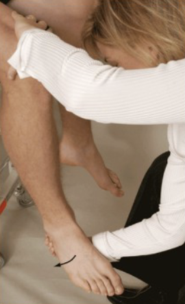
Purpose: Tests for scalene-related TOS

Positive Signs: Pain into arm, neurological symptoms

Method:

  • Seated

  • Rotate head AWAY from test shoulder then extend head

  • Therapist externally rotates and extends shoulder with downward traction
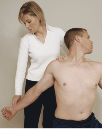
<p><strong>Purpose:</strong> Tests for scalene-related TOS</p><p><strong>Positive Signs:</strong> Pain into arm, neurological symptoms</p><p><strong>Method:</strong></p><ul><li><p>Seated</p></li><li><p>Rotate head AWAY from test shoulder then extend head</p></li><li><p>Therapist externally rotates and extends shoulder with downward traction</p></li></ul><p></p>
20
New cards

Eden’s Test (Military Brace)

Purpose: Tests for costoclavicular-related TOS

Positive Signs: Increased neurological symptoms, decreased radial pulse

Method:

  • Monitor radial pulse,

  • Therapist depress and retract affected arm
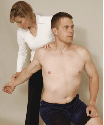
<p><strong>Purpose:</strong> Tests for costoclavicular-related TOS</p><p><strong>Positive Signs:</strong> Increased neurological symptoms, decreased radial pulse</p><p><strong>Method:</strong></p><ul><li><p>Monitor radial pulse,</p></li><li><p>Therapist depress and retract affected arm</p></li></ul><p></p>
21
New cards

Shoulder Depression Test

Tests for:
• Brachial plexus
compression/irritation
• Multiple cervical nerve root
irritation
• Foraminal encroachment on
compressed side (osteophytes)
• Hypomobile joint capsule on
elongated side
•Positive sign:
• Increased pain and neurological symptoms
•Method:
• Patient seated
• Therapist applies downward
pressure to shoulder while side
flexing head to opposite side
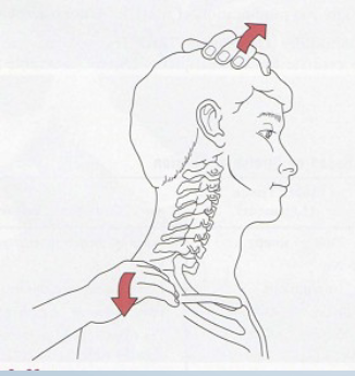
<p><span><strong>Tests for:</strong><br>• Brachial plexus<br>compression/irritation<br>• Multiple cervical nerve root<br>irritation<br>• Foraminal encroachment on<br>compressed side (osteophytes)<br>• Hypomobile joint capsule on<br>elongated side<br><strong>•Positive sign:</strong><br>• Increased pain and neurological symptoms<br><strong>•Method:</strong><br>• Patient seated<br>• Therapist applies downward<br>pressure to shoulder while side<br>flexing head to opposite side</span></p>
22
New cards

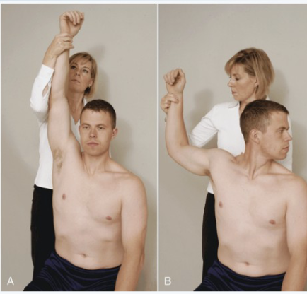
Brakody’s Test/ Shoulder abduction

•Tests for:
• C4, C5, C6 nerve root
compression
• Herniated disc
•Positive sign:
• Decreased pain and
neurological symptoms (also
known as Brakody’s sign)
•Method:
• Patient seated or lying down
• Therapist passively (or
patient actively) elevates
arm through abduction so
that the hand or forearm
rests on top of the head
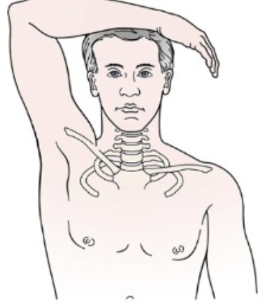
<p><span><strong>•Tests for:</strong><br>• C4, C5, C6 nerve root<br>compression<br>• Herniated disc<br><strong>•Positive sign:</strong><br>• Decreased pain and<br>neurological symptoms (also<br>known as Brakody’s sign)<br><strong>•Method:</strong><br>• Patient seated or lying down<br>• Therapist passively (or<br>patient actively) elevates<br>arm through abduction so<br>that the hand or forearm<br>rests on top of the head</span></p>
23
New cards

Elbow Tests

24
New cards

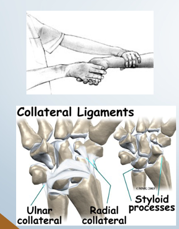
Varus/ Valgus

•Tests for:
• Medial (ulnar) and lateral (radial) collateral ligament instability
• Varus stress also can stress the anular ligament of the radius
•Positive sign:
• Hypermobility/Pain
•Method:
• Patients elbow in slight flexion
• A varus (adduction) force is applied by the therapist to the distal forearm
to test the lateral collateral and a valgus (abduction) stress to test the
medial collateral
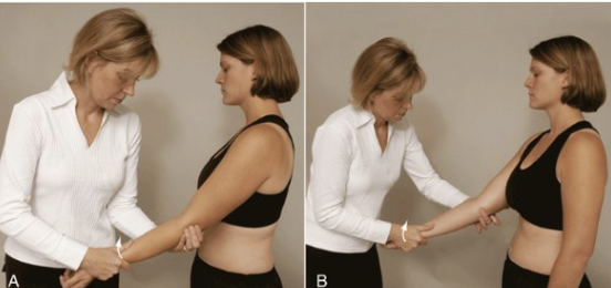
<p><span><strong>•Tests for:</strong><br>• Medial (ulnar) and lateral (radial) collateral ligament instability<br>• Varus stress also can stress the anular ligament of the radius<br><strong>•Positive sign:</strong><br>• Hypermobility/Pain<br><strong>•Method:</strong><br>• Patients elbow in slight flexion<br>• A varus (adduction) force is applied by the therapist to the distal forearm<br>to test the lateral collateral and a valgus (abduction) stress to test the<br>medial collateral</span></p>
25
New cards

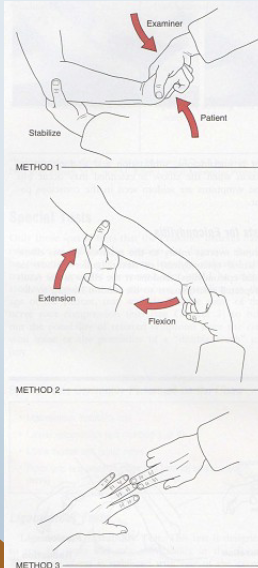
Mill’s

Tests for:
• Inflammation at the lateral epicondyle
• Commonly called tennis elbow
•Positive sign:
• Severe pain @ lateral epicondyle

•Method 2: (AKA- Mill’s)
• Therapist palpates the lateral
epicondyle , then passively
pronates, flexes wrist, and extends

<p><span><strong>Tests for:</strong><br>• Inflammation at the lateral epicondyle<br>• Commonly called tennis elbow<br><strong>•Positive sign:</strong><br>• Severe pain @ lateral epicondyle</span></p><p><span><strong>•Method 2: (AKA- Mill’s)</strong><br>• Therapist palpates the lateral<br>epicondyle , then passively<br>pronates, flexes wrist, and extends</span></p>
26
New cards

Reverse Mill’s

Tests For:

epicondyle of the humerus
• Commonly called Golfer’s elbow
•Positive sign:
• Severe pain medial epicondyle

• Reverse Mill’s
• Therapist palpates the medial
epicondyle
• Patients forearm is passively
supinated while the elbow and wrist

<p><strong>Tests For:</strong></p><p><span>epicondyle of the humerus<br>• Commonly called Golfer’s elbow<br><strong>•Positive sign:</strong><br>• Severe pain medial epicondyle</span></p><p><span><strong>• Reverse Mill’s<br></strong>• Therapist palpates the medial<br>epicondyle<br>• Patients forearm is passively<br>supinated while the elbow and wrist</span></p>
27
New cards

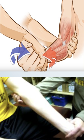
Cozen

Tests for:
• Inflammation at the lateral epicondyle
• Commonly called tennis elbow
•Positive sign:
• Severe pain @ lateral epicondyle
•Method 1: (AKA- Cozen’s)
• Patients elbow stabilized by
therapists thumb resting on lateral
epicondyle
• Patient makes fist, pronates,
radially deviates, and extends the
wrist with the therapist resisting
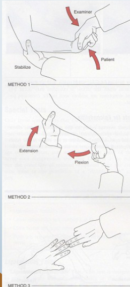
<p><span><strong>Tests for:</strong><br>• Inflammation at the lateral epicondyle<br>• Commonly called tennis elbow<br><strong>•Positive sign:</strong><br>• Severe pain @ lateral epicondyle<br><strong>•Method 1: (AKA- Cozen’s)</strong><br>• Patients elbow stabilized by<br>therapists thumb resting on lateral<br>epicondyle<br>• Patient makes fist, pronates,<br>radially deviates, and extends the<br>wrist with the therapist resisting</span></p>
28
New cards

Reverse Cozen’s

• Inflammation of the medial
Tests For:

epicondyle of the humerus
• Commonly called Golfer’s elbow
•Positive sign:
• Severe pain medial epicondyle
•Method:
• Reverse Cozen’s

• Patients elbow stabilized by
therapists thumb resting on medial
epicondyle
• Patient makes fist, supinates, ulnar
deviates, and flexes wrist with the
therapist resisting
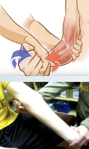
<p><span style="color: transparent">• Inflammation of the medial</span><br><strong>Tests For:</strong></p><p><span>epicondyle of the humerus<br>• Commonly called Golfer’s elbow<br><strong>•Positive sign:</strong><br>• Severe pain medial epicondyle<br><strong>•Method:<br>• Reverse Cozen’s</strong><br>• Patients elbow stabilized by<br>therapists thumb resting on medial<br>epicondyle<br>• Patient makes fist, supinates, ulnar<br>deviates, and flexes wrist with the<br>therapist resisting</span></p>
29
New cards

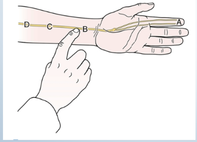
Tinels: Elbow

•Tests for:
• Ulnar nerve compression/
regeneration status
•Positive sign: • Tingling sensation in

ulnar distribution (the distribution of these
symptoms informs you how far the nerve has
regenerated/or the level of damage)
•Method:
• Tap the groove between
the olecranon and medial
epicondyle
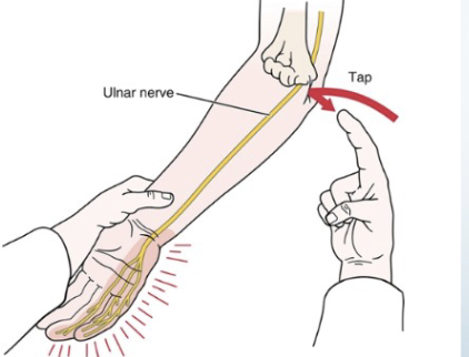
<p><span><strong>•Tests for:</strong><br>• Ulnar nerve compression/<br>regeneration status<br><strong>•Positive sign: </strong>• Tingling sensation in </span><br><span>ulnar distribution (the distribution of these<br>symptoms informs you how far the nerve has<br>regenerated/or the level of damage)<br><strong>•Method:</strong><br>• Tap the groove between<br>the olecranon and medial<br>epicondyle</span></p>
30
New cards

Wrist Tests

31
New cards

Radial Ligamentous

• Tests for: Ulnar collateral ligament
• Positive sign: • Pain and hypermobility
• Method:
• Place client in supination one
hand stabilizes proximal to
wrist
• Passively move hand into
radial deviation with

<p><span><strong>• Tests for:</strong> Ulnar collateral ligament<br><strong>• Positive sign: </strong>• Pain and hypermobility<br><strong>• Method:</strong><br>• Place client in supination one<br>hand stabilizes proximal to<br>wrist<br>• Passively move hand into<br>radial deviation with</span></p>
32
New cards

Ulnar Ligamentous Stress Test

• Tests for:• Radial collateral ligament
• Positive sign: • Pain and hypermobility
• Method:
• Place client in supination one
hand stabilizes proximal to
wrist
• Passively move hand into ulnar
deviation with overpressure
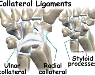
<p><span><strong>• Tests for:</strong>• Radial collateral ligament<br><strong>• Positive sign: </strong>• Pain and hypermobility<br><strong>• Method:</strong><br>• Place client in supination one<br>hand stabilizes proximal to<br>wrist<br>• Passively move hand into ulnar<br>deviation with overpressure</span></p>
33
New cards

Finklestein’s Test

•Tests for:
• DeQuervain’s
disease/tenosynovitis/paratenonitis
(APL/EPB)
•Positive sign:
• Pain over the abductor pollicis longus and
extensor pollicis brevis tendon at the
wrist
• Most people have some degree of
discomfort with this test, comparing
bilaterally confirms
•Method:
• Patient makes a fist with the thumb
inside the fingers
• Examiner stabilizes forearm and deviates
wrist toward ulnar side
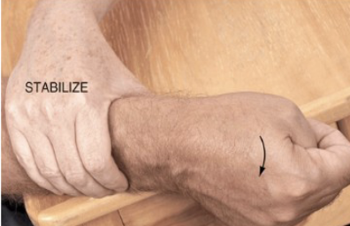
<p><span><strong>•Tests for:</strong><br>• DeQuervain’s<br>disease/tenosynovitis/paratenonitis<br>(APL/EPB)<br><strong>•Positive sign:</strong><br>• Pain over the abductor pollicis longus and<br>extensor pollicis brevis tendon at the<br>wrist<br>• Most people have some degree of<br>discomfort with this test, comparing<br>bilaterally confirms<br><strong>•Method:</strong><br>• Patient makes a fist with the thumb<br>inside the fingers<br>• Examiner stabilizes forearm and deviates<br>wrist toward ulnar side</span></p>
34
New cards

Phalen’s Test

•Tests for: Carpal tunnel syndrome
•Positive sign:
• Tingling sensation in the
thumb index finger and
middle and lateral half
of ring finger (median
nerve distribution)
•Method:
• Examiner flexes patients
wrists maximally and

holds this position for 1 min pushing the wrists
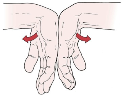
<p><span><strong>•Tests for:</strong> Carpal tunnel syndrome<br><strong>•Positive sign:</strong><br>• Tingling sensation in the<br>thumb index finger and<br>middle and lateral half<br>of ring finger (median<br>nerve distribution)<br><strong>•Method:</strong><br>• Examiner flexes patients<br>wrists maximally and</span><br><span>holds this position for 1 min pushing the wrists</span></p>
35
New cards

Reverse Phalen’s Test

•Tests for: Median nerve
pathology
•Positive sign: Same as Phalen’s- Tingling sensation…

Tingling sensation in the
thumb index finger and middle and lateral half
of ring finger (median nerve distribution)
•Method:
• Therapist places patients wrists in full extension and then
draws downward
• Apply overpressure forst
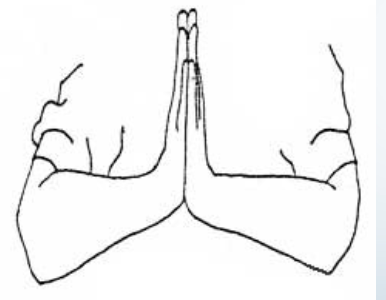
<p><span><strong>•Tests for:</strong> Median nerve<br>pathology<br><strong>•Positive sign: </strong>Same as Phalen’s- Tingling sensation…</span></p><p><span><strong>•</strong>Tingling sensation in the<br>thumb index finger and middle and lateral half<br>of ring finger (median nerve distribution)<br><strong>•Method:</strong><br>• Therapist places patients wrists in full extension and then<br>draws downward<br>• Apply overpressure forst</span></p>
36
New cards

Leg Tests

37
New cards

Craig’s Test

  • Tests for: Femoral head anteversion or retroversion

  • Positive sign: Excessive anteversion (>15°) leading to squinting patella or retroversion (<5°)

  • Method:

    1. Patient lies prone with knee flexed to 90°

    2. Therapist palpates posterior aspect of greater trochanter

    3. Passively rotate the hip medially and laterally until parallel with the table

    4. Estimate the degree of anteversion based on lower leg angle
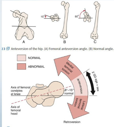
<ul><li><p><strong>Tests for</strong>: Femoral head anteversion or retroversion</p></li><li><p><strong>Positive sign</strong>: Excessive anteversion (&gt;15°) leading to squinting patella or retroversion (&lt;5°)</p></li><li><p><strong>Method</strong>:</p><ol><li><p>Patient lies prone with knee flexed to 90°</p></li><li><p>Therapist palpates posterior aspect of greater trochanter</p></li><li><p>Passively rotate the hip medially and laterally until parallel with the table</p></li><li><p>Estimate the degree of anteversion based on lower leg angle</p></li></ol></li></ul><p></p>
38
New cards

Gaenslen’s Test

  • Tests for: Ipsilateral SI joint lesion, hip pathology, L4 nerve root lesion

  • Positive sign: Pain

  • Method:

    1. Patient lies sidelying on the unaffected side

    2. Flex the hip and knee closest to the table

    3. Therapist passively extends the hip while stabilizing the pelvis
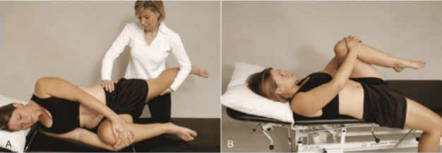
<ul><li><p><strong>Tests for</strong>: Ipsilateral SI joint lesion, hip pathology, L4 nerve root lesion</p></li><li><p><strong>Positive sign</strong>: Pain</p></li><li><p><strong>Method</strong>:</p><ol><li><p>Patient lies sidelying on the unaffected side</p></li><li><p>Flex the hip and knee closest to the table</p></li><li><p>Therapist passively extends the hip while stabilizing the pelvis</p></li></ol></li></ul><p></p>
39
New cards

SI Gapping

•Tests for:
• SI joint mobility/posterior sacroiliac ligaments
•Positive sign: Difference in
quality/quantity of movement from side to side
•Method:
• Patient lies prone examiner stabilizes the pelvis on the opposite side
• Patients knee is flexed to 90+ degrees
• Therapist medially rotates hip as far as possible while palpating the SI joint on the same side

<p><span><strong>•Tests for:</strong><br>• SI joint mobility/posterior sacroiliac ligaments<br><strong>•Positive sign: </strong>Difference in<br>quality/quantity of movement from side to side<br><strong>•Method:</strong><br>• Patient lies prone examiner stabilizes the pelvis on the opposite side<br>• Patients knee is flexed to 90+ degrees<br>• Therapist <strong>medially rotates hip</strong> as far as possible while palpating the SI joint on the same side</span></p>
40
New cards

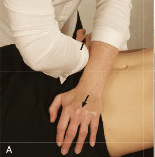
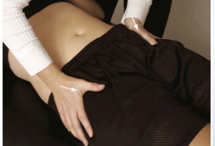
Squish Test

  • Tests for: Sprain in posterior sacroiliac joints

  • Positive sign: SI pain

  • Method: Patient supine while examiner applies medial and inferior pressure to ASIS

    • can be done in sideline

<ul><li><p><strong>Tests for</strong>: Sprain in posterior sacroiliac joints</p></li><li><p><strong>Positive sign</strong>: SI pain</p></li><li><p><strong>Method</strong>: Patient supine while examiner applies medial and inferior pressure to ASIS</p><ul><li><p>can be done in sideline</p></li></ul></li></ul><p></p>
41
New cards

Faber’s/ Patricks Test

•Tests for:
• Hip joint pathology
• Iliopsoas pathology
•Positive sign: Test side knee remains
above opposite knee, pain
•Method:
• Client is supine
• Therapist places patients leg so that the foot of the test leg is on top of the knee of
the opposite leg
• Allow knee to drop laterally
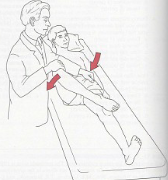
<p><span><strong>•Tests for:</strong><br>• Hip joint pathology<br>• Iliopsoas pathology<br><strong>•Positive sign: </strong>Test side knee remains<br>above opposite knee, pain<br><strong>•Method</strong>:<br>• Client is supine<br>• Therapist places patients leg so that the foot of the test leg is on top of the knee of<br>the opposite leg<br>• Allow knee to drop laterally</span></p>
42
New cards

Apparent/ True Leg Length Test

How the test is performed
• Client lies supine
• A measurement between ASIS and one of the maleoli (usually medial)
• Measure both legs
• Positive sign: A difference of more than 1.5 cm
• Tests for
• Slight difference but within the normal range: leg length usually stems from the position of the innominate
• If beyond normal indicates unequal bone length
NOTE: Apparent leg length is umbilicus to medial malleoli and is often a positional
issue
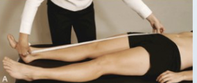
<p><span><strong>How the test is performed</strong><br>• Client lies supine<br>• A measurement between ASIS and one of the maleoli (usually medial)<br>• Measure both legs<br><strong>• Positive sign: </strong>A difference of more than 1.5 cm<br><strong>• Tests for</strong><br>• Slight difference but within the normal range: leg length usually stems from the position of the innominate<br>• If beyond normal indicates unequal bone length<br><strong>NOTE: Apparent leg length is umbilicus to medial malleoli and is often a positional<br>issue</strong></span></p>
43
New cards

Ely’s Test

  • Tests for: Contracture of rectus femoris

  • Positive sign: Pelvis raises off the table on same side, Pain in anterior thigh

  • Method: Patient lies prone, therapist flexes knee, observe movement of pelvis

    NOTE: This is sometimes called the
    Prone Knee Bending or Nachlas test
    • Additional +ves:
    • Pain in lumbar= L3 nerve root, lumbar
    joint compression
    • Pain at SI= hypomobile SI joint
    Muscle length: Ely’s Test
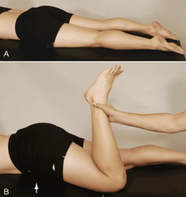
<ul><li><p><strong>Tests for</strong>: Contracture of rectus femoris</p></li><li><p><strong>Positive sign</strong>: Pelvis raises off the table on same side, Pain in anterior thigh</p></li><li><p><strong>Method</strong>: Patient lies prone, therapist flexes knee, observe movement of pelvis</p><p><span><em>NOTE: This is sometimes called the<br>Prone Knee Bending or Nachlas test<br>• Additional +ves:<br>• Pain in lumbar= L3 nerve root, lumbar<br>joint compression<br>• Pain at SI= hypomobile SI joint<br>Muscle length: Ely’s Test</em></span></p></li></ul><p></p>
44
New cards

90/90 Test

  • Tests for: Contractured hamstrings

  • Positive sign: Extended knee within 20° of full extension

  • Method: Patient is supine with knees flexed, actively extend one knee at a time

45
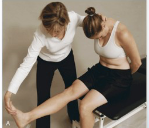
New cards

Straight Leg Raise Test

  • Tests for:

  • Disc herniation

  • Dura matter impingement/irritation

  • Sciatic root impingement

  • Lumbar facet irritation

  • SI pathology

  • Hamstring length

  • Positive sign:

  • With neck and dorsiflexion most likely dura matter irritation or a lesion within the spinal cord (disc herniation, tumor etc.)

  • Back/hip pain with hip flexion below 70 degrees indicates SI pathology

  • Pain with hip flexion over 70 degrees indicates lumbar vertebral joint (facet) involvement

  • Back and leg pain with hip flexion up to 70 degrees indicates sciatic nerve root irritation (L5, S1, 52)

  • Method:

  • Patient in supine position with hip medially rotated and adducted, knee extended

  • Therapist flexes hip until pain occurs

  • Therapist reduces hip flexion until comfortable for patient and then asks patient to flex neck while therapist dorsiflexes foot (This step is sometimes called Lasegue's
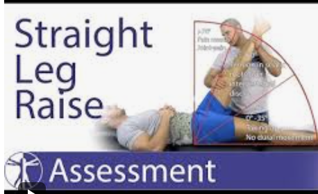
<ul><li><p>Tests for:</p></li><li><p>Disc herniation</p></li><li><p>Dura matter impingement/irritation</p></li><li><p>Sciatic root impingement</p></li><li><p>Lumbar facet irritation</p></li><li><p>SI pathology</p></li><li><p>Hamstring length</p></li><li><p>Positive sign:</p></li><li><p>With neck and dorsiflexion most likely dura matter irritation or a lesion within the spinal cord (disc herniation, tumor etc.)</p></li><li><p>Back/hip pain with hip flexion below 70 degrees indicates SI pathology</p></li><li><p>Pain with hip flexion over 70 degrees indicates lumbar vertebral joint (facet) involvement</p></li><li><p>Back and leg pain with hip flexion up to 70 degrees indicates sciatic nerve root irritation (L5, S1, 52)</p></li><li><p>Method:</p></li><li><p>Patient in supine position with hip medially rotated and adducted, knee extended</p></li><li><p>Therapist flexes hip until pain occurs</p></li><li><p>Therapist reduces hip flexion until comfortable for patient and then asks patient to flex neck while therapist dorsiflexes foot (This step is sometimes called Lasegue's</p></li></ul><p></p>
46
New cards

Ober’s Test

•Tests for: TFL/ITB contracture
•Positive sign:
• Leg does not lower to table
• Leg flexed likely TFL
• Leg extended likely ITB
• Pain can indicate trochanteric bursitis
•Method:
• Patient lays sidelying with lower leg flexed at hip and knee for stability
• Therapist abducts and extends the patients upper leg with knee straight or flexed to 90 degrees
• Therapist then lowers the limb
• Therapist should stabilize clients pelvis so they don’t feel like they are falling backward
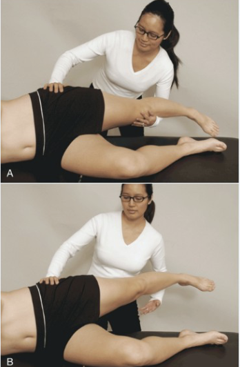
<p><span><strong>•Tests for: </strong>TFL/ITB contracture<br><strong>•Positive sign:</strong><br>• Leg does not lower to table<br>• Leg flexed likely TFL<br>• Leg extended likely ITB<br>• Pain can indicate trochanteric bursitis<br><strong>•Method:</strong><br>• Patient lays sidelying with lower leg flexed at hip and knee for stability<br>• Therapist abducts and extends the patients upper leg with knee straight or flexed to 90 degrees<br>• Therapist then lowers the limb<br>• Therapist should stabilize clients pelvis so they don’t feel like they are falling backward</span></p>
47
New cards

Piriformis Length Test

  • Tests for: Piriformis contracture, Piriformis Syndrome

  • Positive sign:

    • Pain in the piriformis muscle (stretch) or

    • neurological pain in a sciatic distribution

  • Method:

    • Client in sidelying (test leg on top) , knee and hip flexed (60º),

    • Stabilize pelvis and apply downward pressure to test leg

    • Kind of feel like they are going to all off of the table
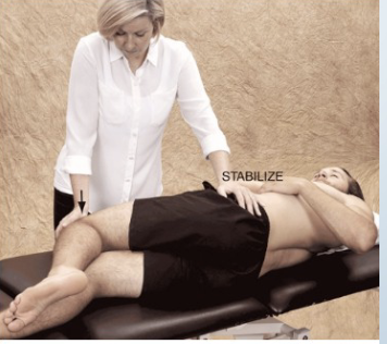
<ul><li><p><strong>Tests for</strong>: Piriformis contracture, Piriformis Syndrome</p></li><li><p><strong>Positive sign</strong>:</p><ul><li><p>Pain in the piriformis muscle (stretch) or</p></li><li><p>neurological pain in a sciatic distribution</p></li></ul></li><li><p><strong>Method</strong>:</p><ul><li><p>Client in sidelying (test leg on top) , knee and hip flexed (60<span>º)</span>,</p></li><li><p>Stabilize pelvis and apply downward pressure to test leg</p></li><li><p>Kind of feel like they are going to all off of the table</p></li></ul></li></ul><p></p>
48
New cards

Trendelenburg’s Test

Test’s For:
• Weak gluteus medius
Muscle Strength: Trendelenburg’s

Positive sign:
• Client is observed for the pelvis dropping on the
non-weight bearing side

Method:
• Client is standing
• Therapist asks the client to stand on one leg
• Can increase difficulty by adding a squat motionS
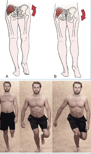
<p><strong> Test’s For:</strong><br>• Weak gluteus medius<br>Muscle Strength: Trendelenburg’s</p><p><strong>Positive sign:</strong><br>• Client is observed for the pelvis dropping on the<br>non-weight bearing side</p><p><strong>Method:</strong><br>• Client is standing<br>• Therapist asks the client to stand on one leg<br>• Can increase difficulty by adding a squat motion<span style="color: transparent">S</span></p>
49
New cards

Thomas Test

  • Tests for: Hip flexor contracture

  • Positive sign: Opposing leg remains slightly flexed, excessive lordosis

    • Cannot fully extend leg, gap under knee

  • Method: Patient supine, one leg slowly extends while other remains flexed to chest
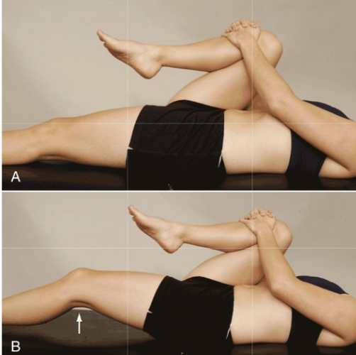
<ul><li><p><strong>Tests for</strong>: Hip flexor contracture</p></li><li><p><strong>Positive sign</strong>: Opposing leg remains slightly flexed, excessive lordosis</p><ul><li><p>Cannot fully extend leg, gap under knee</p></li></ul></li><li><p><strong>Method</strong>: Patient supine, one leg slowly extends while other remains flexed to chest</p></li></ul><p></p>
50
New cards

Knee SOTs

51
New cards

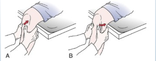
Varus Stress Test

•Tests for: Lateral instability/ lateral collateral ligament
•Positive sign:
Hypermobility
•Method:
• Patient is supine
• Therapist places a varus stress on the knee
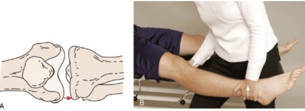
<p><span><strong>•Tests for:</strong> Lateral instability/ lateral collateral ligament<strong><br>•Positive sign: </strong>Hypermobility<br><strong>•Method:</strong><br>• Patient is supine<br>• Therapist places a varus stress on the knee</span></p>
52
New cards

Valgus Stress Test

•Tests for: Medial instability/medial collateral ligament
•Positive sign: Hypermobility
•Method:
• Patient is supine
• Therapist places a valgus stress on the knee
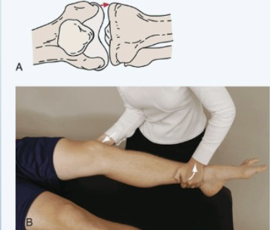
<p><span><strong>•Tests for: </strong>Medial instability/medial collateral ligament<br><strong>•Positive sign: </strong>Hypermobility<br><strong>•Method:</strong><br>• Patient is supine<br>• Therapist places a valgus stress on the knee</span></p>
53
New cards

Anterior Drawer of the Knee

  • Tests for: Anterior cruciate ligament instability

  • Positive sign: More than 6mm anterior translation

  • Method: Patient knee flexed to 90°, therapist stabilizes foot (Sit on it with towel underneath) and draws tibia forward on the femur
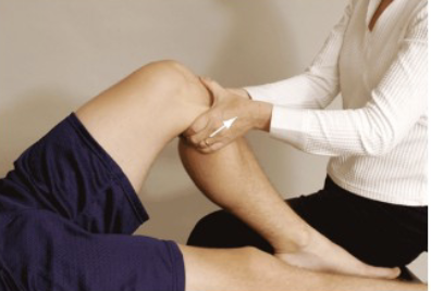
<ul><li><p><strong>Tests for</strong>: Anterior cruciate ligament instability</p></li><li><p><strong>Positive sign</strong>: More than 6mm anterior translation</p></li><li><p><strong>Method</strong>: Patient knee flexed to 90°, therapist stabilizes foot (Sit on it with towel underneath) and draws tibia forward on the femur</p></li></ul><p></p>
54
New cards

Posterior Drawer of the Knee

  • Tests for: PCL instability (Posterior Cruciate ligament)

  • Positive sign: Hypermobility and pain

  • Method: Patient knee flexed to 90° and hip flexed to 45°

    • • First observe for “Sag sign” a
      sulcus inferior to the femur
      • If not present therapist stabilizes
      clients foot with body weight (sit on it with towel underneath)
      • Therapist then pushes the tibia backward on the femur
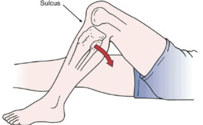
<ul><li><p><strong>Tests for</strong>: PCL instability (Posterior Cruciate ligament)</p></li><li><p><strong>Positive sign</strong>: Hypermobility and pain</p></li><li><p><strong>Method</strong>: Patient knee flexed to 90° and hip flexed to 45°</p><ul><li><p><span>• First observe for “Sag sign” a<br>sulcus inferior to the femur<br>• If not present therapist stabilizes<br>clients foot with body weight (sit on it with towel underneath)<br>• Therapist then pushes the tibia backward on the femur</span></p></li></ul></li></ul><p></p>
55
New cards

Lachman’s Test

  • Tests for: ACL injury (anterior cruciate Ligament)

  • Positive sign: Soft/mushy end feel/ hypermobility

  • Method: Patient supine with knee at 30° Flexion, therapist pulls tibia anteriorly and pushes femur posteriorly
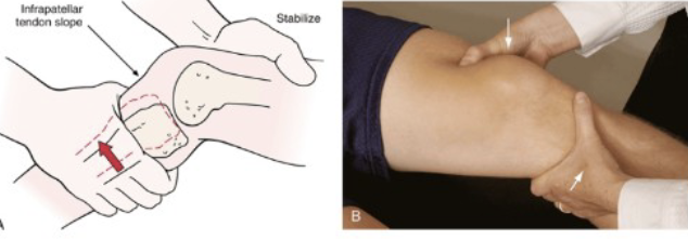
<ul><li><p><strong>Tests for</strong>: ACL injury (anterior cruciate Ligament)</p></li><li><p><strong>Positive sign</strong>: Soft/mushy end feel/ hypermobility</p></li><li><p><strong>Method</strong>: Patient supine with knee at 30° Flexion, therapist pulls tibia anteriorly and pushes femur posteriorly</p></li></ul><p></p>
56
New cards

Noble’s Compression Test

Tests For: ITB friction syndrome = inflammation of the ITB as it rubs

Positive sign: Pain at approximately 30º of flexion
Method:

• Client lies supine with hip and knee
flexed
• Therapist places pressure on the lateral
femoral epicondyle over the ITB
• Client extends
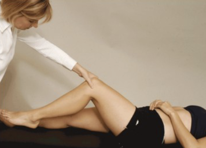
<p><span><strong>Tests For: </strong>ITB friction syndrome = inflammation of the ITB as it rubs</span></p><p><span><strong>Positive sign: </strong>Pain at approximately 30º of flexion<strong><br>Method:</strong><br>• Client lies supine with hip and knee<br>flexed<br>• Therapist places pressure on the lateral<br>femoral epicondyle over the ITB<br>• Client extends</span></p>
57
New cards

Slocum’s Test Part 1

• Tests for:
• Anterolateral rotary instability
• Some degree of injury to the following:
• ACL/PCL/LCL/ITB
• Posterolateral capsule
• Arcuate-popliteus complex
• Positive sign: Pain in the lateral knee, Excessive movement
• Method:
• This is anterior drawer with a twist
• Client supine
• Knee flexed to 90º, hip flexed to 45º, knee
medially rotated to 30º
• Therapist sits on client’s foot for stability
• Tibia is drawn forward
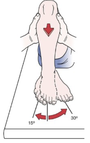
<p><strong>• Tests for:</strong><br><span>• Anterolateral rotary instability</span><br><span>• Some degree of injury to the following:</span><br><span>• ACL/PCL/LCL/ITB</span><br><span>• Posterolateral capsule</span><br><span>• Arcuate-popliteus complex</span><br><strong>• Positive sign: </strong><span>Pain in the lateral knee, Excessive movement</span><br><strong>• Method:<br></strong><span>• This is anterior drawer with a twist</span><br><span>• Client supine</span><br><span>• Knee flexed to 90º, hip flexed to 45º, knee</span><br><span>medially rotated to 30º</span><br><span>• Therapist sits on client’s foot for stability</span><br><span>• Tibia is drawn forward</span></p>
58
New cards

Slocum’s Part 2

Tests For: Anteromedial instability
Method:
• Externally rotate to 15 degrees and pull anteriorly
again
Tests for:
• MCL/ACL, posteromedial capsule, posterior oblique

59
New cards

Hughston’s Test

Tests for
• Posterolateral/posteromedial rotary instability
• Some degree of injury to the following:
• Postlat=PCL/MCL
• Postmed=PCL/LCL
Positive sign: Excessive movement of tibia

Method:
• This is posterior drawer with a twist
• Client supine/seated knee flexed to 90º
• Medial/lateral rotation
n of

<p><span><strong>Tests for</strong><br>• Posterolateral/posteromedial rotary instability<br>• Some degree of injury to the following:<br>• Postlat=PCL/MCL<br>• Postmed=PCL/LCL<br><strong>Positive sign: </strong>Excessive movement of tibia</span></p><p><span><strong>Method:</strong><br>• This is posterior drawer with a twist<br>• Client supine/seated knee flexed to 90º<br>• Medial/lateral rotation</span><span style="color: transparent">n of</span></p>
60
New cards

Lateral Pivot Test

•Tests for: Anterolateral rotary instability, ACL rupture, ITB injury

•Positive sign: Client reports a “giving away”

•Method:

•Client in supine

•Hip flexed and abducted 30º slight medial rotation 20º

•Therapist holds foot with one hand, knee with other (hand placed posterior to the head of the fibula)

•Knee is extended
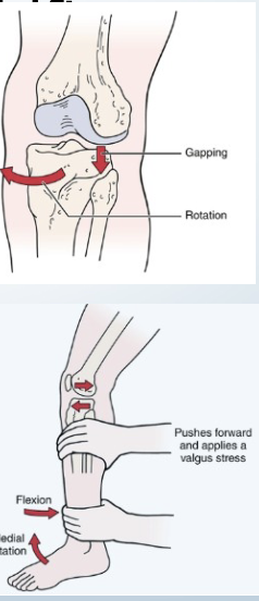
<p><span><strong>•Tests for: </strong>Anterolateral rotary instability, ACL rupture, ITB injury</span></p><p><span><strong>•Positive sign: </strong>Client reports a “giving away”</span></p><p><span><strong>•Method:</strong></span></p><p><span>•Client in supine</span></p><p><span>•Hip flexed and abducted 30º slight medial rotation 20º</span></p><p><span>•Therapist holds foot with one hand, knee with other (hand placed posterior to the head of the fibula)</span></p><p><span>•Knee is extended</span></p>
61
New cards

Apley’s Compression and Distraction

Tests for: Meniscus injury/ligament injury
•Positive sign:
• If rotation with distraction is more painful or
hypermobile indicates a ligamentous injury
• If rotation with compression is more painful then
meniscus
•Method:
• Client prone with knee flexed to 90 degrees
• Therapist stabilizes clients thigh with their knee
• Therapist then medially and laterally rotates the tibia combined
with distraction
• Repeat process with compression
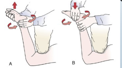
<p><span><strong>Tests for:</strong> Meniscus injury/ligament injury<br><strong>•Positive sign:</strong><br>• If rotation with distraction is more painful or<br>hypermobile indicates a ligamentous injury<br>• If rotation with compression is more painful then<br>meniscus<br><strong>•Method:</strong><br>• Client prone with knee flexed to 90 degrees<br>• Therapist stabilizes clients thigh with their knee<br>• Therapist then medially and laterally rotates the tibia combined<br>with distraction<br>• Repeat process with compression</span></p>
62
New cards

Bounce Home Test

Tests for: Meniscus injury/foreign body
•Positive sign: If the knee doesn’t reach full extension/ rubbery end feel/pain
•Method:
• Client lies in supine with heel cupped in therapists hand
• Clients knee is fully flexed and then allowed to passively come
into extension
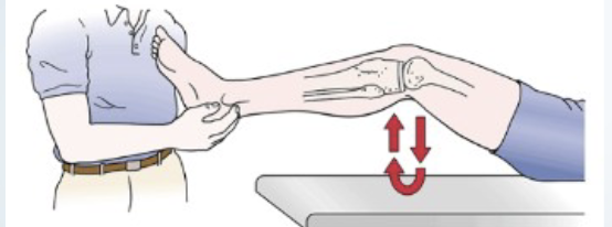
<p><span><strong>Tests for: </strong>Meniscus injury/foreign body<br><strong>•Positive sign: </strong>If the knee doesn’t reach full extension/ rubbery end feel/pain<br><strong>•Method:</strong><br>• Client lies in supine with heel cupped in therapists hand<br>• Clients knee is fully flexed and then allowed to passively come<br>into extension</span></p>
63
New cards

McMurray’s Test

•Tests for: Meniscal injury
•Positive sign: Clicking/popping/snapping
• With medial rotation=lateral meniscus
• With lateral rotation= medial meniscus
•Method:
• Patient lies supine with the knee completely flexed
• Therapist then medially rotates the tibia and extends the knee
• Repeat with knee laterally rotated

  • This one you be kind of rough with the knee
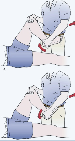
<p><span><strong>•Tests for: </strong>Meniscal injury<br><strong>•Positive sign: </strong>Clicking/popping/snapping<br>• With medial rotation=lateral meniscus<br>• With lateral rotation= medial meniscus<br><strong>•Method:</strong><br>• Patient lies supine with the knee completely flexed<br>• Therapist then medially rotates the tibia and extends the knee<br>• Repeat with knee laterally rotated</span></p><ul><li><p>This one you be kind of rough with the knee</p></li></ul><p></p>
64
New cards

Helfet’s Test

• Tests for:
• Meniscus tear, impaired
quadriceps function leading to
lack of “screw home” mechanism
• Positive sign:
• Tib tub not aligning with midline
of patella @ 90 degrees flexion
• Tib tub not aligning with lateral
border of patella in extension

Method:

  • client sits with legs of edge of table

  • feel tibial tuberosity when leg is bent

  • The feel its position when leg is straight
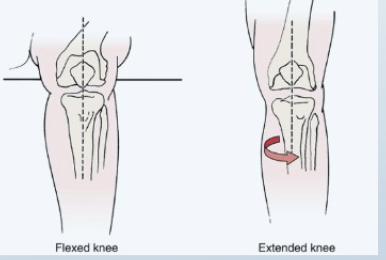
<p><span><strong>• Tests for:</strong><br>• Meniscus tear, impaired<br>quadriceps function leading to<br>lack of “screw home” mechanism<br><strong>• Positive sign:</strong><br>• Tib tub not aligning with midline<br>of patella @ 90 degrees flexion<br>• Tib tub not aligning with lateral<br>border of patella in extension</span></p><p><span>Method:</span></p><ul><li><p>client sits with legs of edge of table</p></li><li><p>feel tibial tuberosity when leg is bent</p></li><li><p>The feel its position when leg is straight</p></li></ul><p></p>
65
New cards

Brush Effusion

•Tests for: Minimal effusion
•Positive sign: Wave of fluid passes to the medial side of the joint and bulges
just below medial distal portion of the patella (pes anserine)
•Method:
• Client is supine
• Therapist commences just below joint line on medial side of the patella
and strokes proximal toward patients hip up to the suprapatellar
pouch, two or three time
• With opposite hand therapist strokes down lateral side of patella

Very light stroking movements
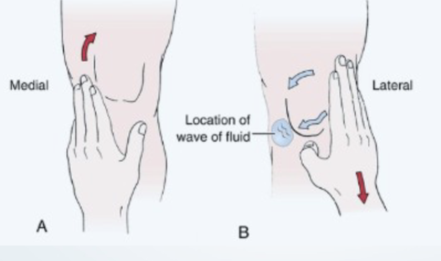
<p><span><strong>•Tests for:</strong> Minimal effusion<br><strong>•Positive sign</strong>: Wave of fluid passes to the medial side of the joint and bulges<br>just below medial distal portion of the patella (pes anserine)<br><strong>•Method:</strong><br>• Client is supine<br>• Therapist commences just below joint line on medial side of the patella<br>and strokes proximal toward patients hip up to the suprapatellar<br>pouch, two or three time<br>• With opposite hand therapist strokes down lateral side of patella</span></p><p><span><strong>Very light stroking movements</strong></span></p>
66
New cards

Clarks Patellofemoral- patella grind test

•Tests for: Patellofemoral dysfunction
•Positive sign: Pain
•Method:
Client is supine
• Therapist presses down with the
web space of their hand on the
superior patella while asking the
client to contract their quadriceps
• Can be repeated at 30, 60 and 90
degrees of flexion to test the
different contact surfaces of the
patella
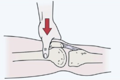
<p><span><strong>•Tests for:</strong> Patellofemoral dysfunction<br><strong>•Positive sign: </strong>Pain<br><strong>•Method:</strong><br>Client is supine<br>• Therapist presses down with the<br>web space of their hand on the<br>superior patella while asking the<br>client to contract their quadriceps<br>• Can be repeated at 30, 60 and 90<br>degrees of flexion to test the<br>different contact surfaces of the<br>patella</span></p>
67
New cards

McConnell’s Test

•Tests for: Patellofemoral dysfunction/chondromalacia

•Positive sign: Pain disappears with medial patella accommodation

•Method:

•Client is seated with femur laterally rotated

•Patient performs isometric quadriceps contractions at 120, 90, 60, 30, 0 and holds each for 10 seconds

•If pain is produced at any level the leg is returned to full extension and the therapist then pushes the patella medially

•The client then tries the contraction from the previously painful range again
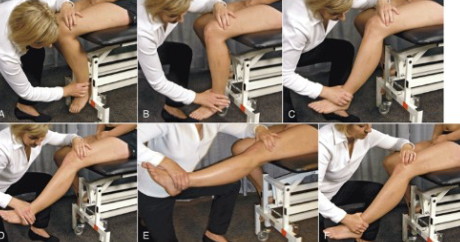
<p><span><strong>•Tests for:</strong> Patellofemoral dysfunction/chondromalacia</span></p><p><span><strong>•Positive sign: </strong>Pain disappears with medial patella accommodation</span></p><p><span><strong>•Method:</strong></span></p><p><span>•Client is seated with femur laterally rotated</span></p><p><span>•Patient performs isometric quadriceps contractions at 120, 90, 60, 30, 0 and holds each for 10 seconds</span></p><p><span>•If pain is produced at any level the leg is returned to full extension and the therapist then pushes the patella medially</span></p><p><span>•The client then tries the contraction from the previously painful range again</span></p>
68
New cards

Waldron’s Test

Tests for:
• Patellofemoral dysfunction
Positive sign:
• Pain, crepitus, tracking dysfunction
Method:
• Client performs AFROM or squat with therapist palpating patella

69
New cards

Ankle SOTs

70
New cards

Anterior Drawer of the Ankle

•Tests for:
• Anterior talofibular ligament
instability
•Positive sign:
• Excessive anterior movement
• Depression of the skin on both sides
of the achilles
•Method:
• Patient lies prone, or supine with feet
extending over end of table
• Therapist pushes the heel forward, or
pulls foot forward while pushing tibia
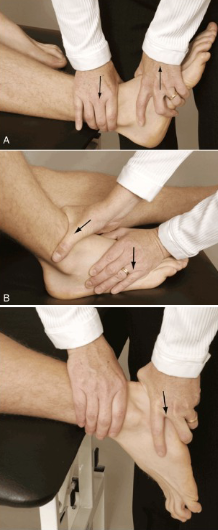
<p><span><strong>•Tests for:</strong><br>• Anterior talofibular ligament<br>instability<br><strong>•Positive sign:</strong><br>• Excessive anterior movement<br>• Depression of the skin on both sides<br>of the achilles<br><strong>•Method:</strong><br>• Patient lies prone, or supine with feet<br>extending over end of table<br>• Therapist pushes the heel forward, or<br>pulls foot forward while pushing tibia</span></p>
71
New cards

External Rotation Test/ Kleiger’s

  • Tests for: Deltoid ligament sprain or syndesmosis tear

  • Positive sign: Pain medial and lateral, potential talus displacement from medial malleolus

  • Method: Client seated, Knee flexed foot relaxed

    • Therapist rotates foot laterally

<ul><li><p><strong>Tests for</strong>: Deltoid ligament sprain or syndesmosis tear</p></li><li><p><strong>Positive sign</strong>: Pain medial and lateral, potential talus displacement from medial malleolus</p></li><li><p><strong>Method</strong>: Client seated, Knee flexed foot relaxed</p><ul><li><p>Therapist rotates foot laterally</p></li></ul></li></ul><p></p>
72
New cards

Talar Tilt

•Tests for: Calcaneofibular ligament tear, deltoid ligament
•Positive sign:
• Excessive movement with adduction= calcaneofibular ligament
(some anterior talofibular ligament)
• Excessive movement with abduction= deltoid ligament
(tibionavicular, tibiocalcaneal, posterior tibiotalar ligaments)
•Method:
• Client is prone, supine or sidelying with knee slightly flexed to
relax the gastrocnemius
• Therapist holds foot in 90 degree position
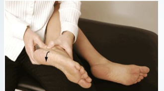
<p><span><strong>•Tests for: </strong>Calcaneofibular ligament tear, deltoid ligament<br><strong>•Positive sign:</strong><br>• Excessive movement with adduction= calcaneofibular ligament<br>(some anterior talofibular ligament)<br>• Excessive movement with abduction= deltoid ligament<br>(tibionavicular, tibiocalcaneal, posterior tibiotalar ligaments)<br><strong>•Method:</strong><br>• Client is prone, supine or sidelying with knee slightly flexed to<br>relax the gastrocnemius<br>• Therapist holds foot in 90 degree position</span></p>
73
New cards

Homan’s Test

•Tests for: DVT (deep vein thrombosis)
•Positive sign: Pain in calf
• Can be accompanied by pallor,
swelling, loss of dorsalis pedis pulse
•Method:
• Therapist passively dorsiflexes patients foot
with the knee extended
• Pain also on palpationc
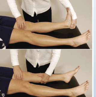
<p><span><strong>•Tests for: </strong>DVT (deep vein thrombosis)<br><strong>•Positive sign: </strong>Pain in calf<br>• Can be accompanied by pallor,<br>swelling, loss of dorsalis pedis pulse<br><strong>•Method:</strong><br>• Therapist passively dorsiflexes patients foot<br>with the knee extended<br>• Pain also on palpationc</span></p>
74
New cards

Morton’s Test

•Tests for: Morton’s neuroma, stress fracture
•Positive sign: Pain
•Method:
• Patient lies supine
• Therapist grasps foot at metatarsal heads and squeezes them together
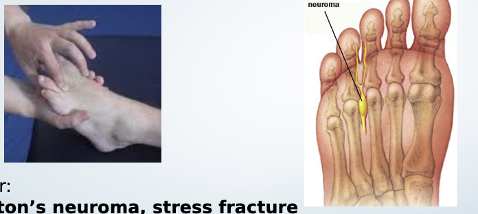
<p><span><strong>•Tests for: </strong>Morton’s neuroma, stress fracture<br><strong>•Positive sign: </strong>Pain<br><strong>•Method:</strong><br>• Patient lies supine<br>• Therapist grasps foot at metatarsal heads and squeezes them together</span></p>
75
New cards

Spine SOTs

76
New cards

Spurlings Test

  • Tests For: Identifies nerve root compression.

  • Positive Sign: Pain radiating into the arm on the affected side, increased neurological symptoms.

    • Identify the affected dermatome/ nerve root involved through pain pattern

  • Method:

    • Patient sidebends cervical spine towards the unaffected side first.

    • Therapist applies straight downward pressure on the head.
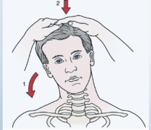
<ul><li><p><strong>Tests For:</strong> Identifies nerve root compression.</p></li><li><p><strong>Positive Sign:</strong> Pain radiating into the arm on the affected side, increased neurological symptoms.</p><ul><li><p>Identify the affected dermatome/ nerve root involved through pain pattern</p></li></ul></li><li><p><strong>Method:</strong></p><ul><li><p>Patient sidebends cervical spine towards the unaffected side first.</p></li><li><p>Therapist applies straight downward pressure on the head.</p></li></ul></li></ul><p></p>
77
New cards

Cervical Distraction Test

  • Purpose: Tests for nerve root compression.

  • Positive Sign: Decreased pain when traction is applied.

  • Method:

    • Therapist places one hand under the patient's chin and the other cradling the occiput.

    • Slowly lift the patient's head to traction the cervical spine.
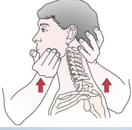
<ul><li><p><strong>Purpose:</strong> Tests for nerve root compression.</p></li><li><p><strong>Positive Sign:</strong> Decreased pain when traction is applied.</p></li><li><p><strong>Method:</strong></p><ul><li><p>Therapist places one hand under the patient's chin and the other cradling the occiput.</p></li><li><p>Slowly lift the patient's head to traction the cervical spine.</p></li></ul></li></ul><p></p>
78
New cards

Scalene Cramp Test

  • Tests For: Identifies scalene trigger points.

  • Positive Sign: Increased pain due to scalene trigger points of the side rotated toward.

  • Method:

    • Patient rotates head towards the affected side and pulls chin towards the clavicle.
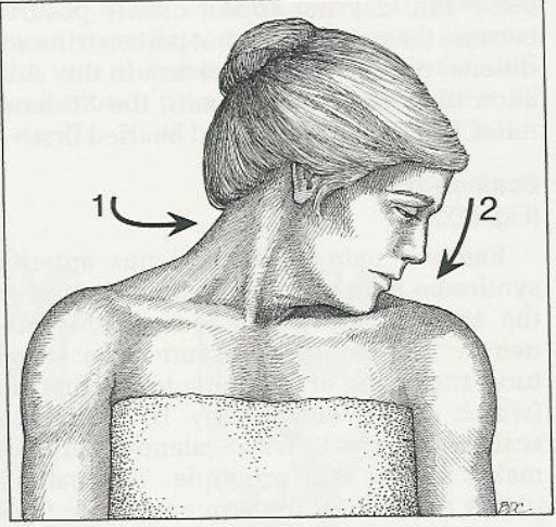
<ul><li><p><strong>Tests For:</strong> Identifies scalene trigger points.</p></li><li><p><strong>Positive Sign:</strong> Increased pain due to scalene trigger points of the side rotated toward.</p></li><li><p><strong>Method:</strong></p><ul><li><p>Patient rotates head towards the affected side and pulls chin towards the clavicle.</p></li></ul></li></ul><p></p>
79
New cards

Vertebral Artery Test

Tests For:
The Vertebral Artery Test assesses the integrity of the vertebral arteries and checks for potential vascular insufficiency related to cervical spine movement.

Method:
The patient is positioned supine. The examiner rotates the patient's head to one side while observing for signs of vascular insufficiency.


Positive Sign:
Dizziness, tinnitus, visual disturbances, or other neurological symptoms indicate a positive test, suggesting potential vertebrobasilar insufficiency, eyes wavering

80
New cards

Hautant Test

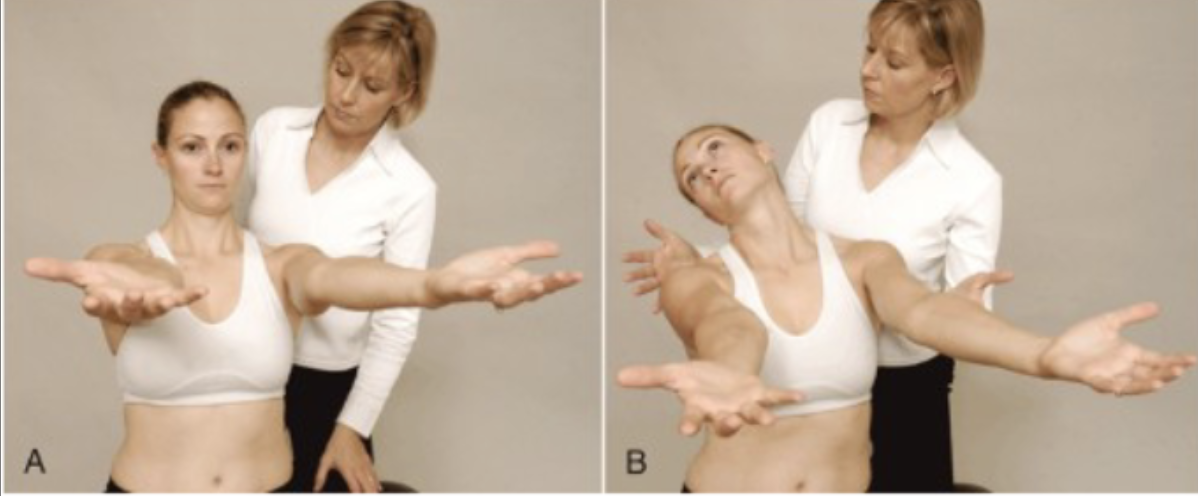
  • Tests for : Differentiates between dizziness caused by articular problems and vascular problems.

  • Positive Sign:

    • step 1: arms move= nonvascular dizziness

    • step 2: Arms move = vascular impairment

  • Method:

    • Step 1: Patient flexes arms to 90 degrees, closes eyes for 10-30 seconds.

    • Watch for Loss of arm position = potential vascular issue.

    • Step 2: Patient rotates and extends neck (arms out eyes closed) while observing for loss of arm position.

    • hold 30 seconds

<p></p><ul><li><p><strong>Tests for :</strong> Differentiates between dizziness caused by articular problems and vascular problems.</p></li><li><p><strong>Positive Sign:</strong></p><ul><li><p>step 1: arms move= nonvascular dizziness</p></li><li><p>step 2: Arms move = vascular impairment</p></li></ul></li><li><p><strong>Method:</strong></p><ul><li><p>Step 1: Patient flexes arms to 90 degrees, closes eyes for 10-30 seconds.</p></li><li><p>Watch for Loss of arm position = potential vascular issue.</p></li><li><p>Step 2: Patient rotates and extends neck (arms out eyes closed) while observing for loss of arm position.</p></li><li><p>hold 30 seconds</p></li></ul></li></ul><p></p>
81
New cards

Adam’s Forward Bend

Tests for: scoliosis

Positive sign: Assymetry (one side is higher than the other)

Method:

  • patient needs to bend forward, starting at the waist until the back comes in the horizontal plane, with feet together, arms hanging, amd the knees in extension. The palms are held together
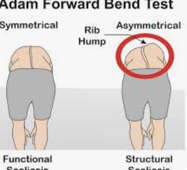
<p><strong>Tests for:</strong> scoliosis</p><p><strong>Positive sign:</strong> Assymetry (one side is higher than the other)</p><p><strong>Method:</strong></p><ul><li><p>patient needs to bend forward, starting at the waist until the back comes in the horizontal plane, with feet together, arms hanging, amd the knees in extension. The palms are held together</p></li></ul><p></p>
82
New cards

Valsalva

  • Tests for: Tests for space-occupying lesions (herniated disc, tumors).

  • Positive Sign: Increased pain indicating pressure on the spinal cord.

  • Method: Patient takes deep breath, holds it, then pushes down as if blowing up a balloon.
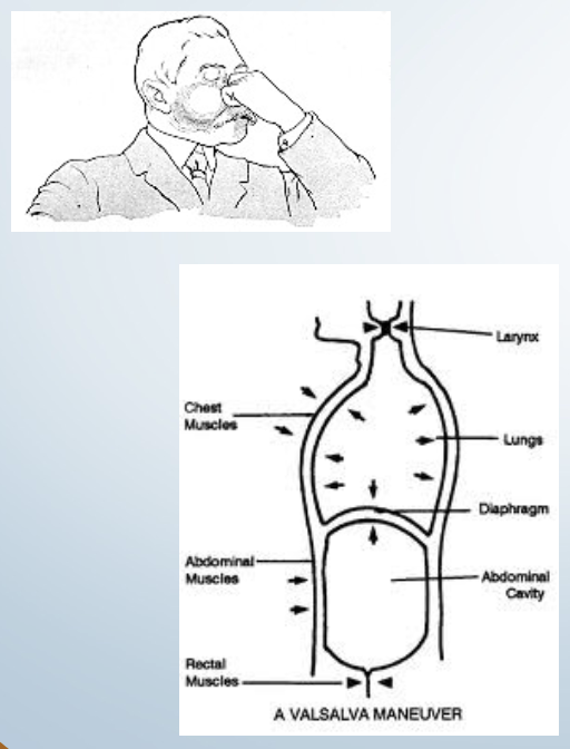
<ul><li><p><strong>Tests for:</strong> Tests for space-occupying lesions (herniated disc, tumors).</p></li><li><p><strong>Positive Sign:</strong> Increased pain indicating pressure on the spinal cord.</p></li><li><p><strong>Method:</strong> Patient takes deep breath, holds it, then pushes down as if blowing up a balloon.</p></li></ul><p></p>
83
New cards

Kemp’s Quadrant

  • Purpose: Evaluates facet joint and nerve root compression.

  • Positive Sign: Local pain on facet side, recreation of neuro symptoms indicate nerve root involvement.

  • Method: Client stands, puts hand on posterolateral thigh

  • therapist directs them to extend, sidebend, and rotate (slide hand down towards knee) while therapist applies overpressure.
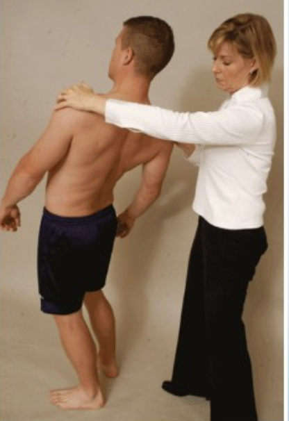
<ul><li><p><strong>Purpose:</strong> Evaluates facet joint and nerve root compression.</p></li><li><p><strong>Positive Sign:</strong> Local pain on facet side, recreation of neuro symptoms indicate nerve root involvement.</p></li><li><p><strong>Method:</strong> Client stands, puts hand on posterolateral thigh</p></li><li><p>therapist directs them to extend, sidebend, and rotate (slide hand down towards knee) while therapist applies overpressure.</p></li></ul><p></p>
84
New cards

Kernig’s Test

Tests for: meningitis, Kernig's test is a physical examination maneuver used to assess for meningeal irritation, often indicative of meningitis.

Positive sign: Pain in the neck,
Resistance or pain when attempting to straighten the knee indicates a positive Kernig's test, suggesting meningeal irritation.

Method: client lies supine,
The patient lies on their back, and the examiner flexes one knee and hip to 90 degrees. The knee is then straightened.
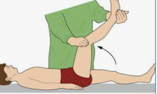


<p><strong>Tests for: </strong>meningitis, Kernig's test is a physical examination maneuver used to assess for meningeal irritation, often indicative of meningitis.</p><p><strong>Positive sign:</strong> Pain in the neck, <br> Resistance or pain when attempting to straighten the knee indicates a positive Kernig's test, suggesting meningeal irritation.</p><p><strong>Method: </strong>client lies supine,<br> The patient lies on their back, and the examiner flexes one knee and hip to 90 degrees. The knee is then straightened.</p><p></p><p><br></p><p><br></p>
85
New cards

Slump Test

  • Purpose: Tests for impingement of dura, spinal cord, or nerve roots.

  • Positive Sign: Neurological symptoms such as sciatica or electric nerve pain anywhere.

  • Method: Ask client slumps forward (leave head elevated), chin to chest, therapist applies overpressure.

  • If symptoms do not occur, Extend one knee and dorsiflex foot to provoke symptoms.

  • (SOTO- HALL- same thing but supine)

<ul><li><p><strong>Purpose:</strong> Tests for impingement of dura, spinal cord, or nerve roots.</p></li><li><p><strong>Positive Sign:</strong> Neurological symptoms such as sciatica or electric nerve pain anywhere.</p></li><li><p><strong>Method:</strong> Ask client slumps forward (leave head elevated), chin to chest, therapist applies overpressure.</p></li><li><p>If symptoms do not occur, Extend one knee and dorsiflex foot to provoke symptoms.</p></li><li><p>(SOTO- HALL- same thing but supine)</p></li></ul><p></p>
86
New cards

Rebound Tenderness Test

Tests for: appendix

Positive: Pain continues when pressure released

Method: Client supine, push right above iliac crest with some pressure then releasee

87
New cards

SLR- straight leg raise

  • Tests for:

  • Disc herniation

  • Dura matter impingement/irritation

  • Sciatic root impingement

  • Lumbar facet irritation

  • SI pathology

  • Hamstring length

  • Positive sign:

  • With neck and dorsiflexion most likely dura matter irritation or a lesion within the spinal cord (disc herniation, tumor etc.)

  • Back/hip pain with hip flexion below 70 degrees indicates SI pathology

  • Pain with hip flexion over 70 degrees indicates lumbar vertebral joint (facet) involvement

  • Back and leg pain with hip flexion up to 70 degrees indicates sciatic nerve root irritation (L5, S1, 52)

  • Method:

  • Patient in supine position with hip medially rotated and adducted, knee extended

  • Therapist flexes hip until pain occurs

  • Therapist reduces hip flexion until comfortable for patient and then asks patient to flex neck while therapist dorsiflexes foot (This step is sometimes called Lasegue's