Bio D170 Lab Practical 2
 Call Kai
 Learn
Learn Practice Test
 Spaced Repetition
 Match
Match Flashcards
 Knowt Play
1/180

There's no tags or description
Looks like no tags are added yet.
Name | Mastery | Learn | Test | Matching | Spaced | Call with Kai | Chat |
|---|
No analytics yet
Send a link to your students to track their progress
181 Terms
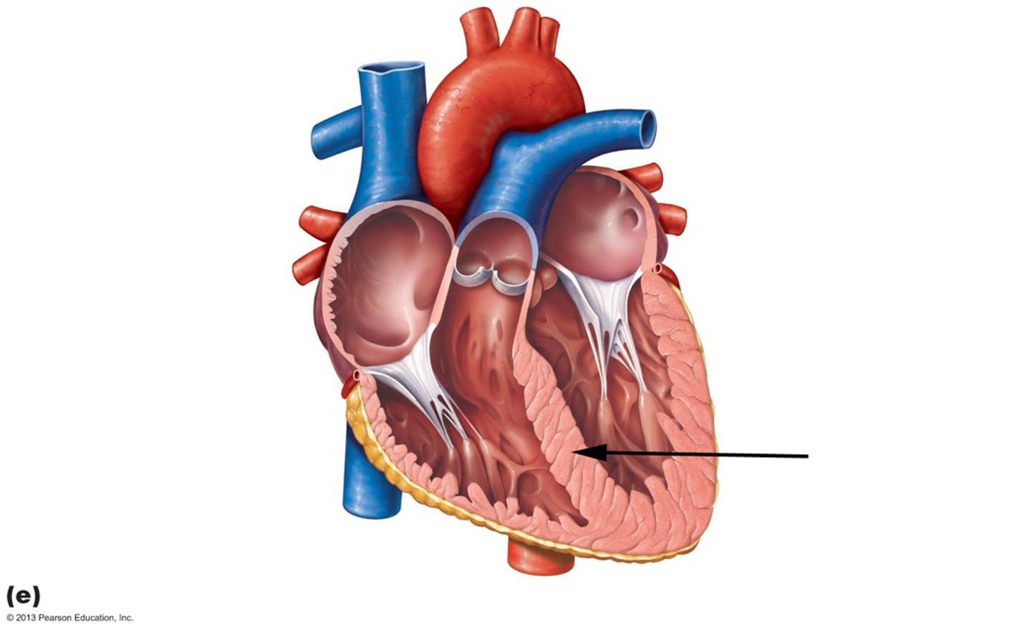
Interventricular Septum
separates ventricles
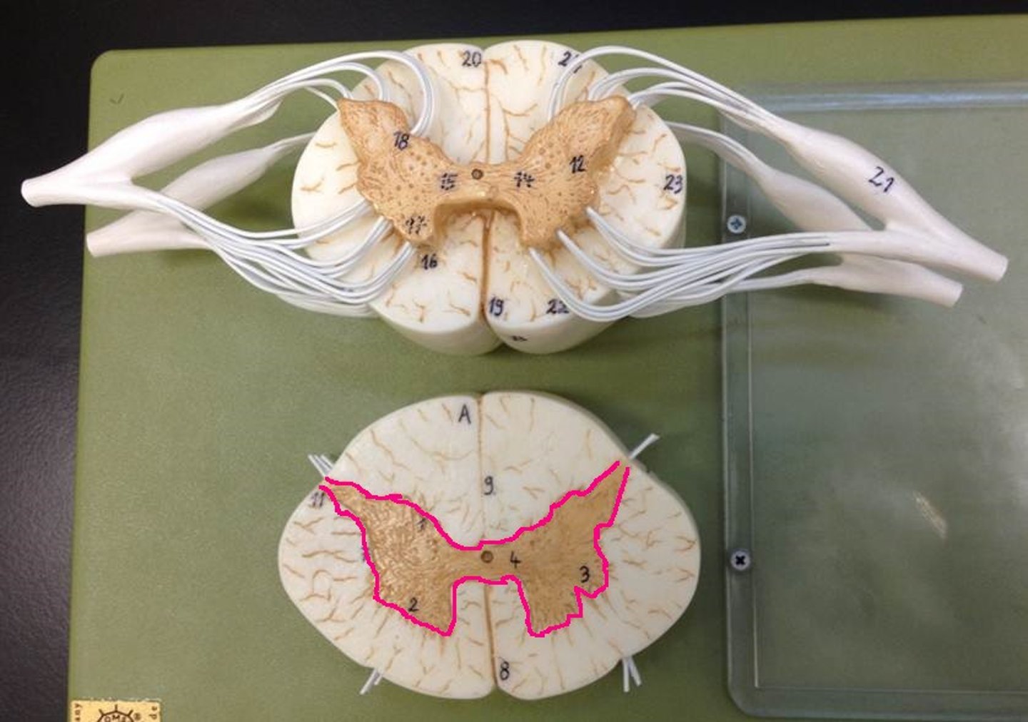
Gray Matter
Dendrites, short unmyelinated, neuroglia
White Matter
-surrounds gray matter
-myelinated axons + neuroglia; NO cell bodies
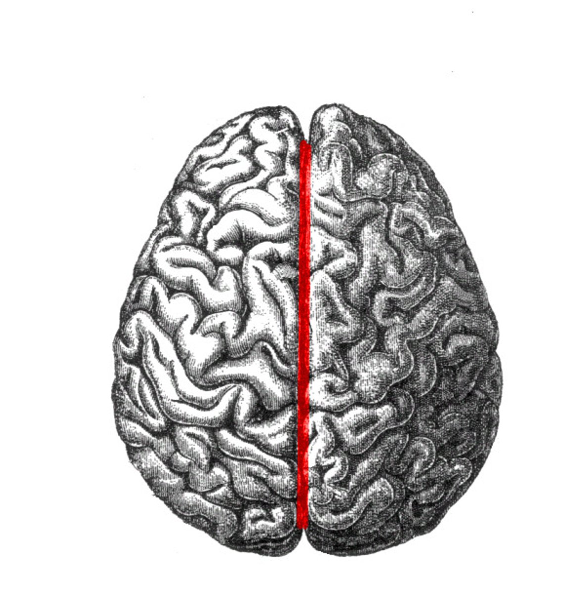
Longitudinal Fissure
separates left and right hemispheres
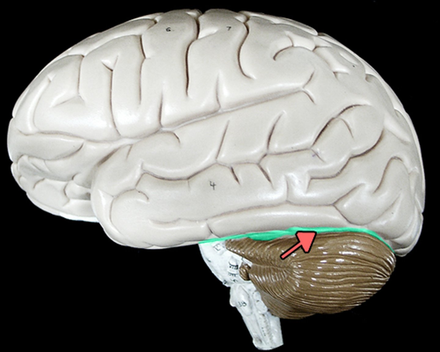
Transverse Fissure
-separates cerebrum from cerebellum
-caudal/inferior to occipital lobe
Parieto-Occipital Sulcus
separates parietal and occipital lobes
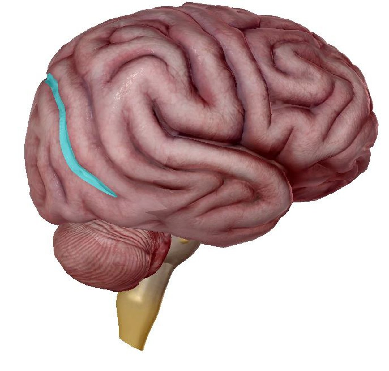
Lateral Sulcus
separates the parietal and temporal lobes
Central Sulcus
separates frontal and parietal lobes
Precentral Gyrus
-in front of central sulcus
-lies on frontal lobe
Postcentral Gyrus
-behind central sulcus
-part of parietal lobe
Olfactory Bulb (I)
-above the nasal cavity
-beneath the frontal lobes
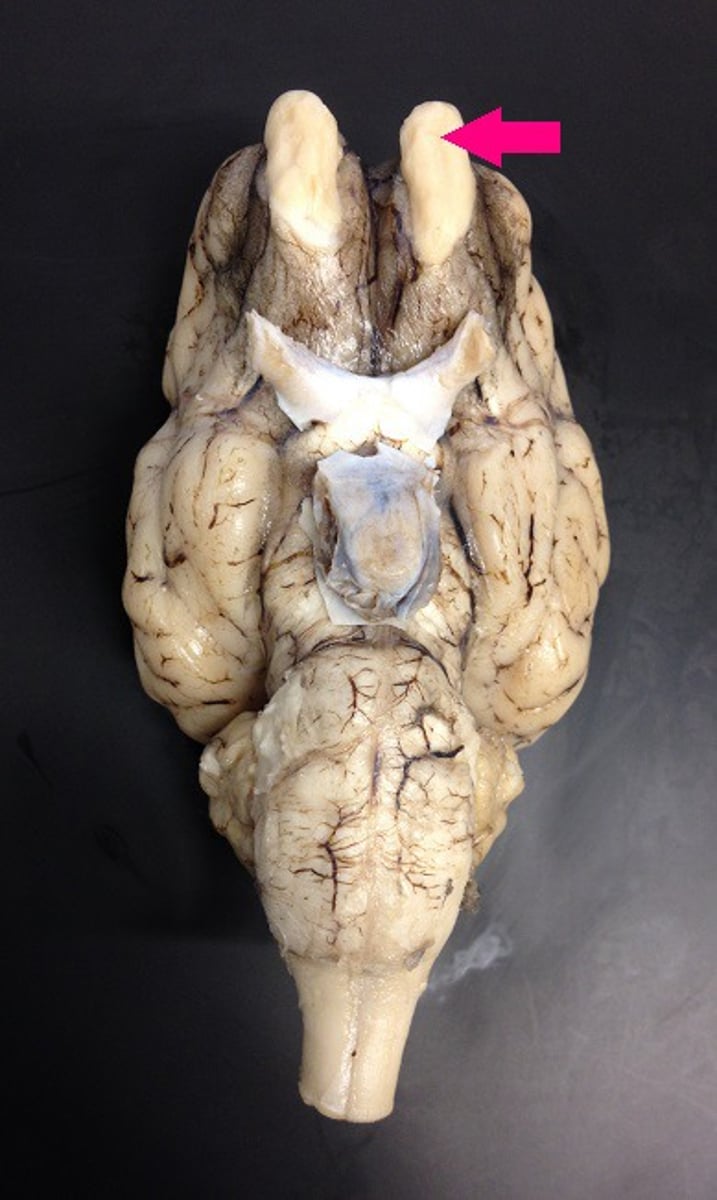
Optic Nerve (brain, II)
-carries neural impulses from the eye to the brain
-inferior to olfactory bulb
-originates from forebrain
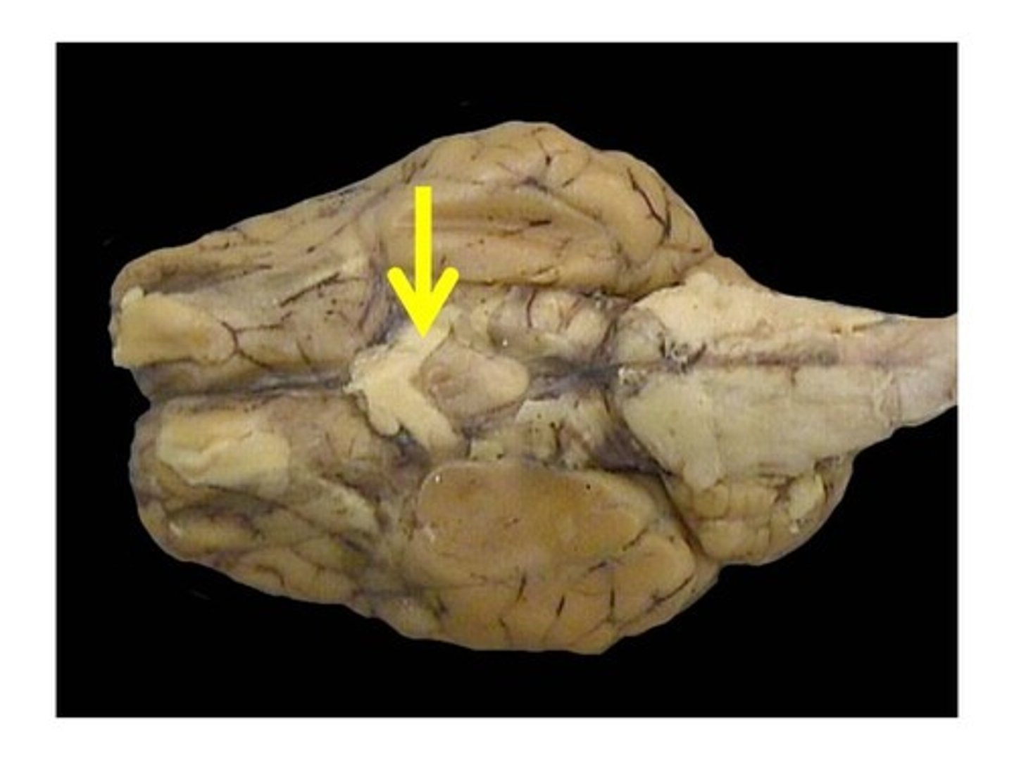
Optic Chiasm
point at which optic nerve fibers cross in the brain
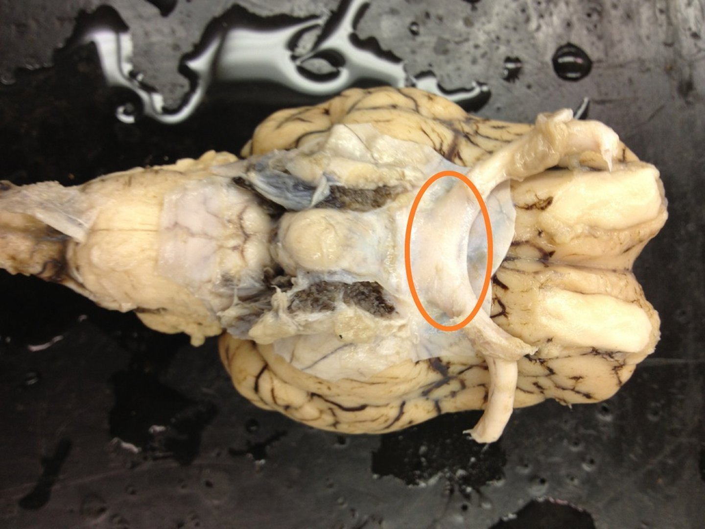
Cerebellum
between brainstem and cerebrum
Brainstem
-midbrain, pons, medulla oblongata
-caudal to cerebellum
-rostral to spinal cord
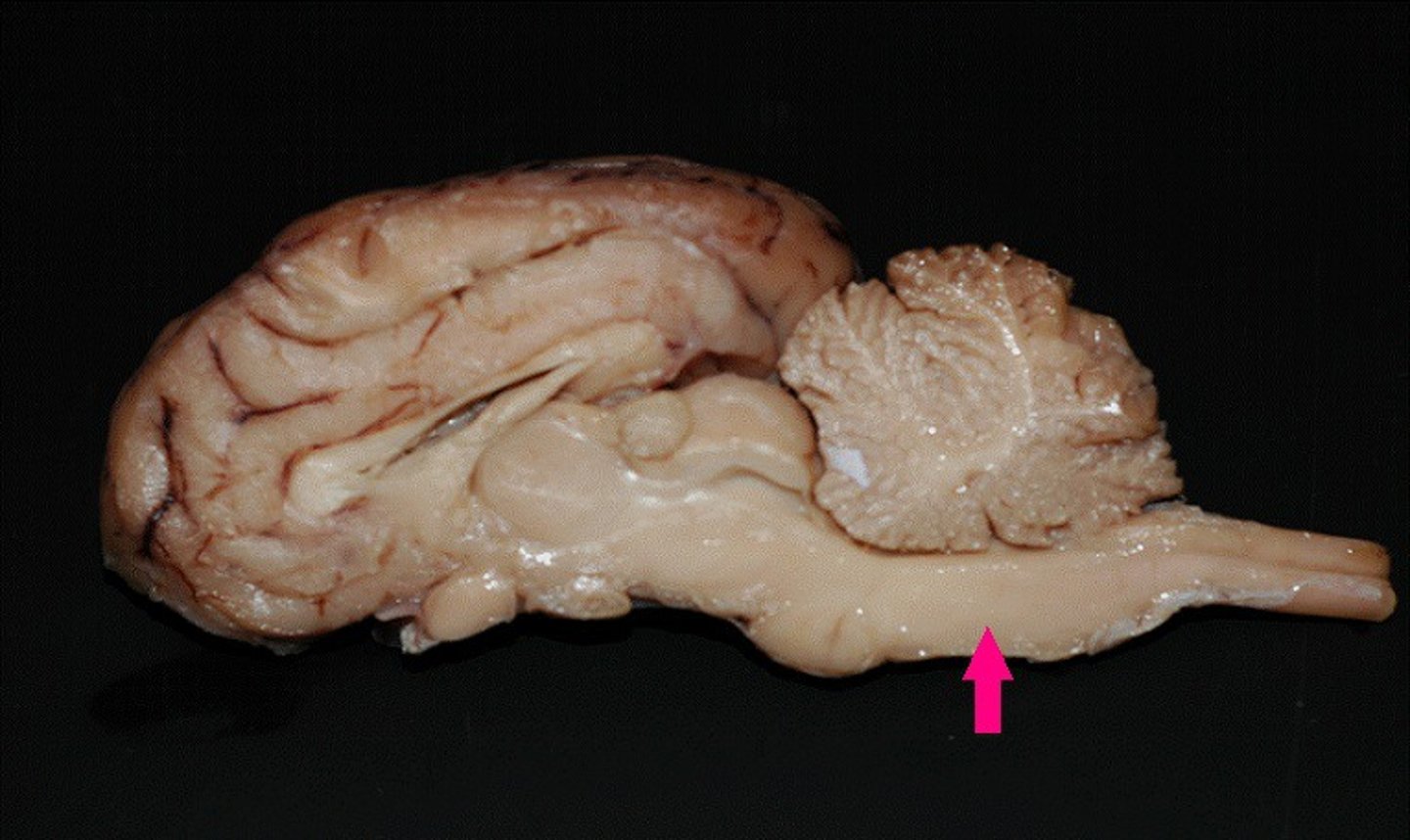
Corpora Quadrigemina (sheep)
dorsal midbrain
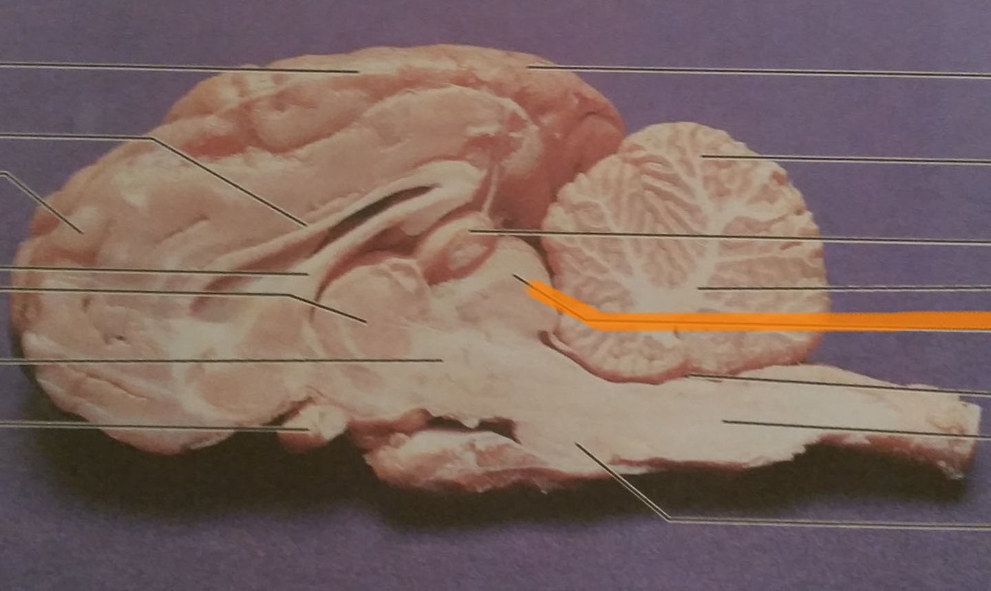
Middle Cerebellar Peduncle
connects cerebellum to pons
Midbrain
-topmost brainstem
-connects cerebrum with spinal cord
-rostral to pons and thalamus
Hypothalamus
below the thalamus
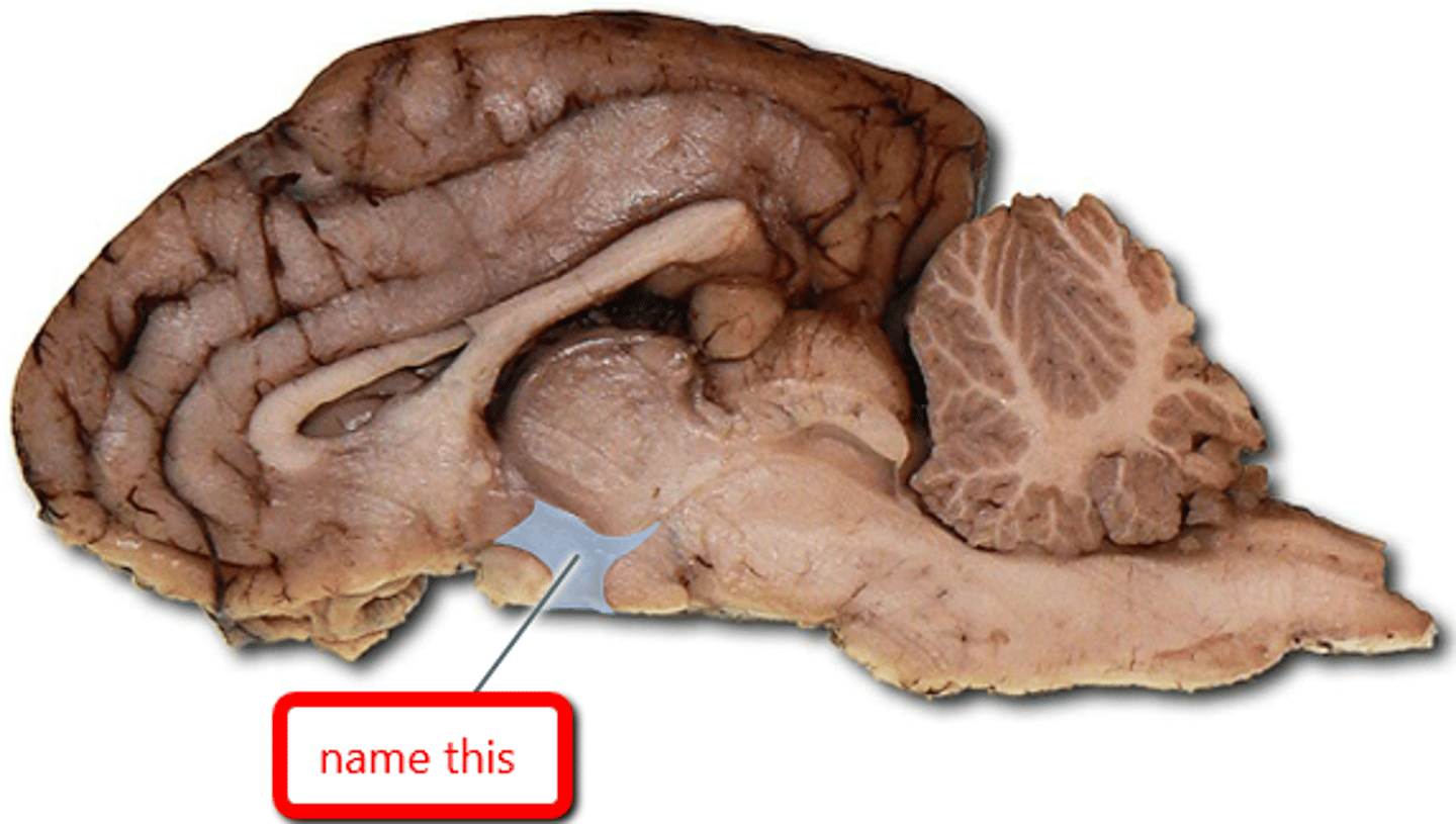
Thalamus
-below third ventricle
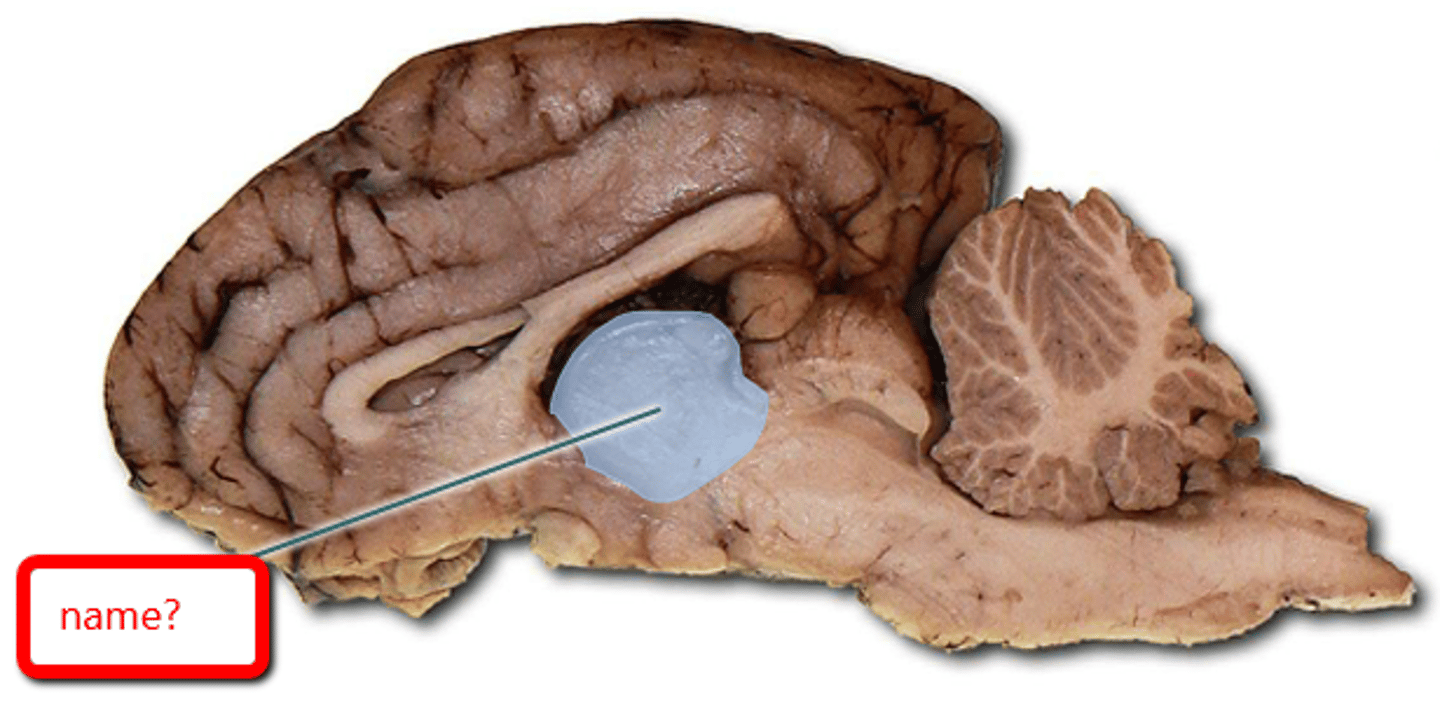
Corpus Callosum
-joins both hemispheres
Third Ventricle
-diencephalon
-inferior/dorsal/caudal to corpus callosum
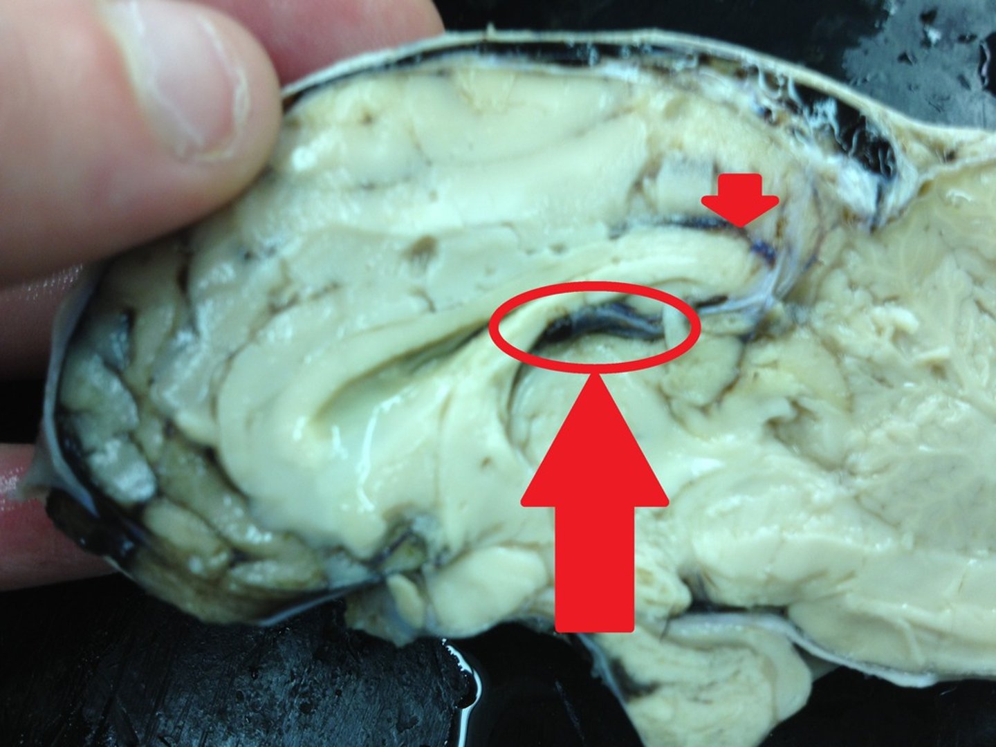
Arbor Vitae
white matter of the cerebellum
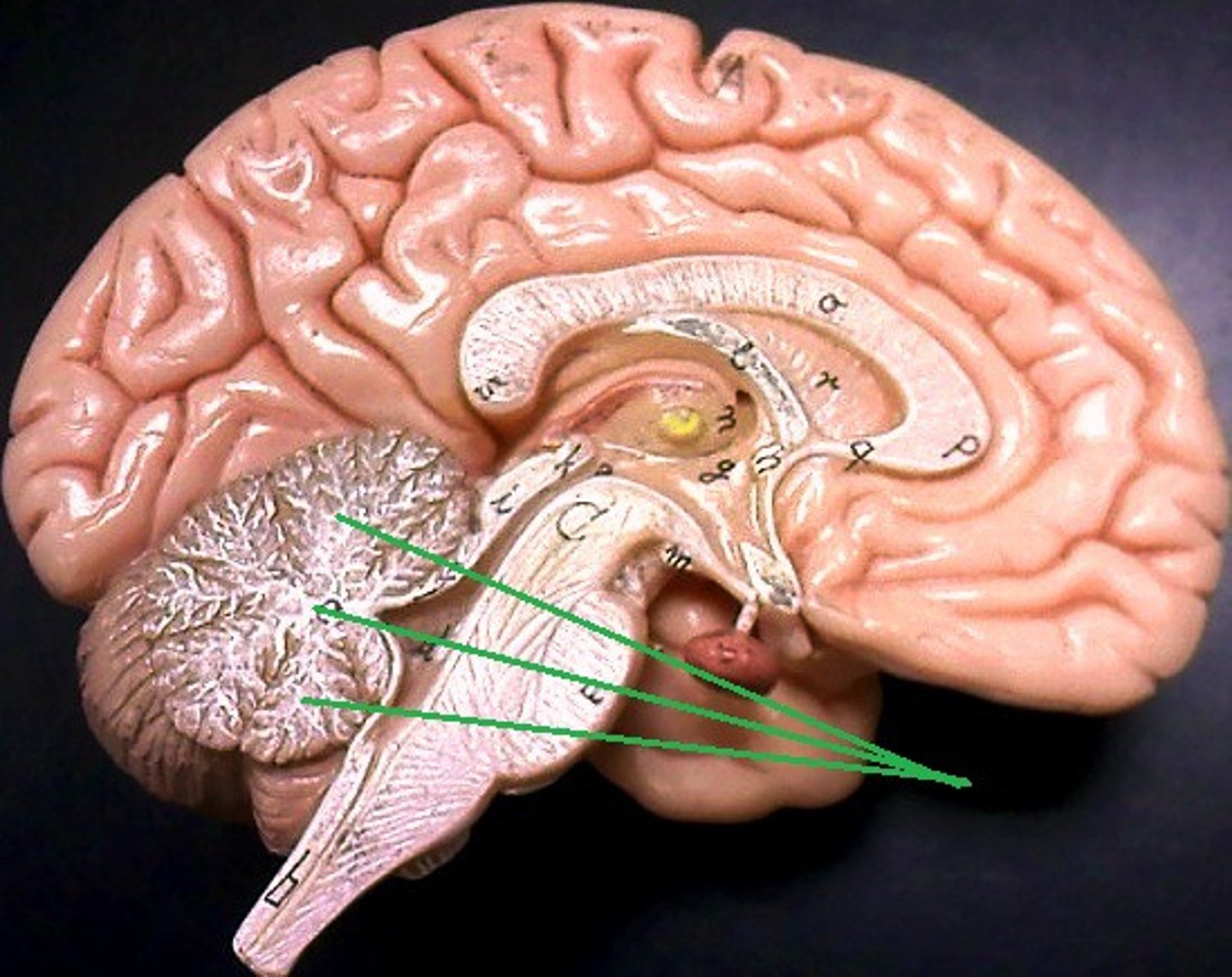
Lateral Ventricle
inferior to rostral end of corpus callosum
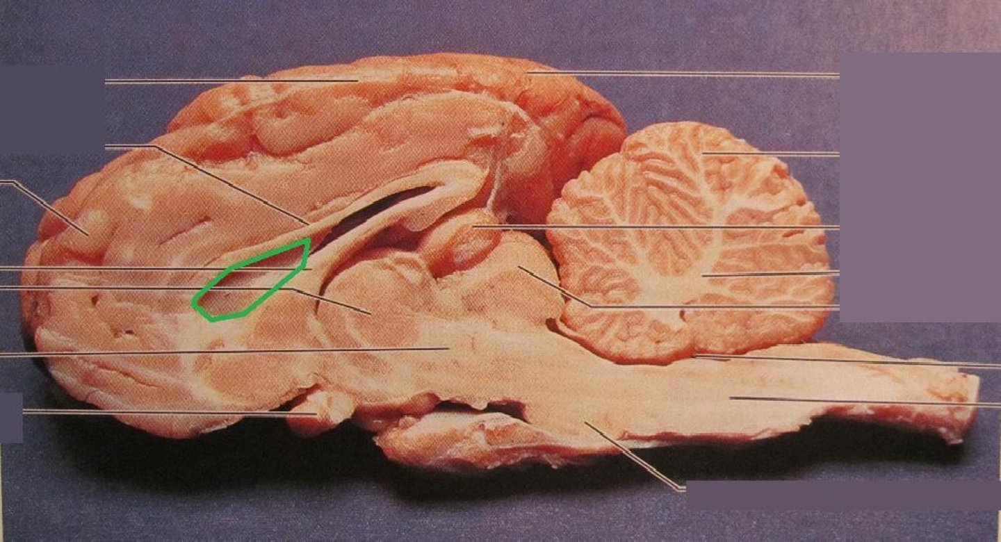
Oculomotor (III)
-eye movement
-inferior to optic, medioanterior to trochlear
-midbrain
Trochlear (IV)
-motor, eye movement
-posteriolateral to oculomotor
-midbrain
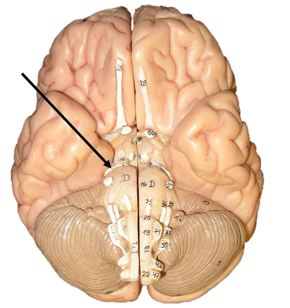
Trigeminal (V)
-head sensation
-posteriorlateral to trochlear
-pons
Glossopharyngeal (IX)
-taste and swallowing
-medulla
-most rostral, rostral to vagus
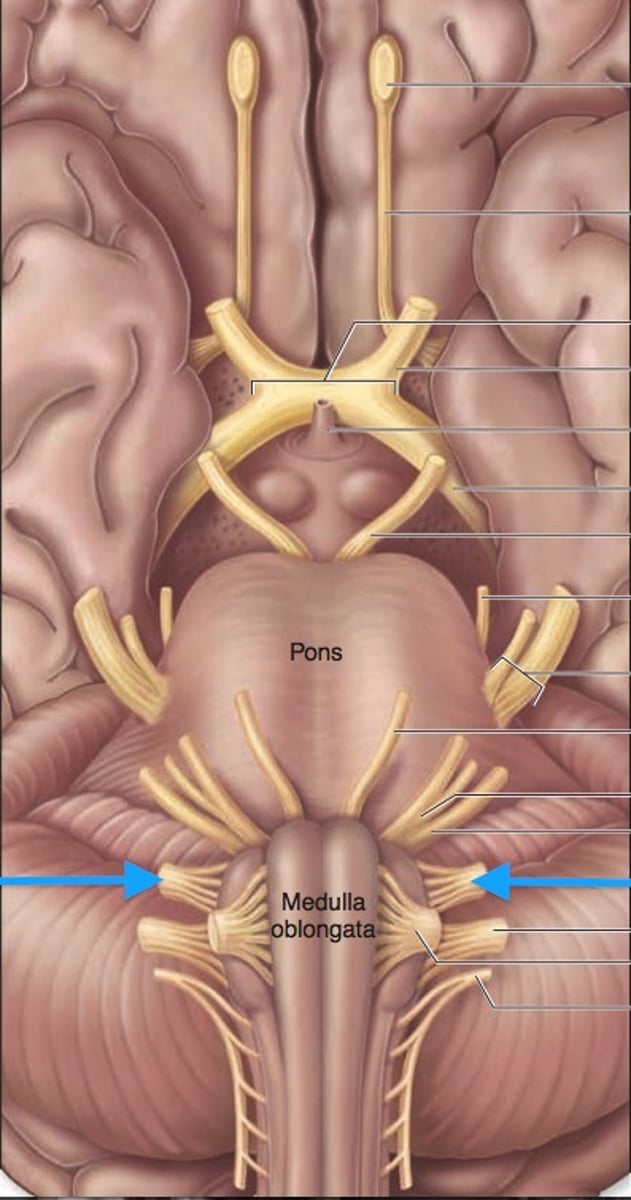
Vagus Nerve (X)
-parasympathetic innveration
-medulla
-caudal to glossopharyngeal
Hypoglossal (XII)
-tongue movement
-below the glossopharyngeal
Conus Medullaris
-end of spinal cord
-L1, L2
Cauda Equina
-"horse's tail", a fan of nerve fibers below the spinal cord
-inferior to conus medullaris
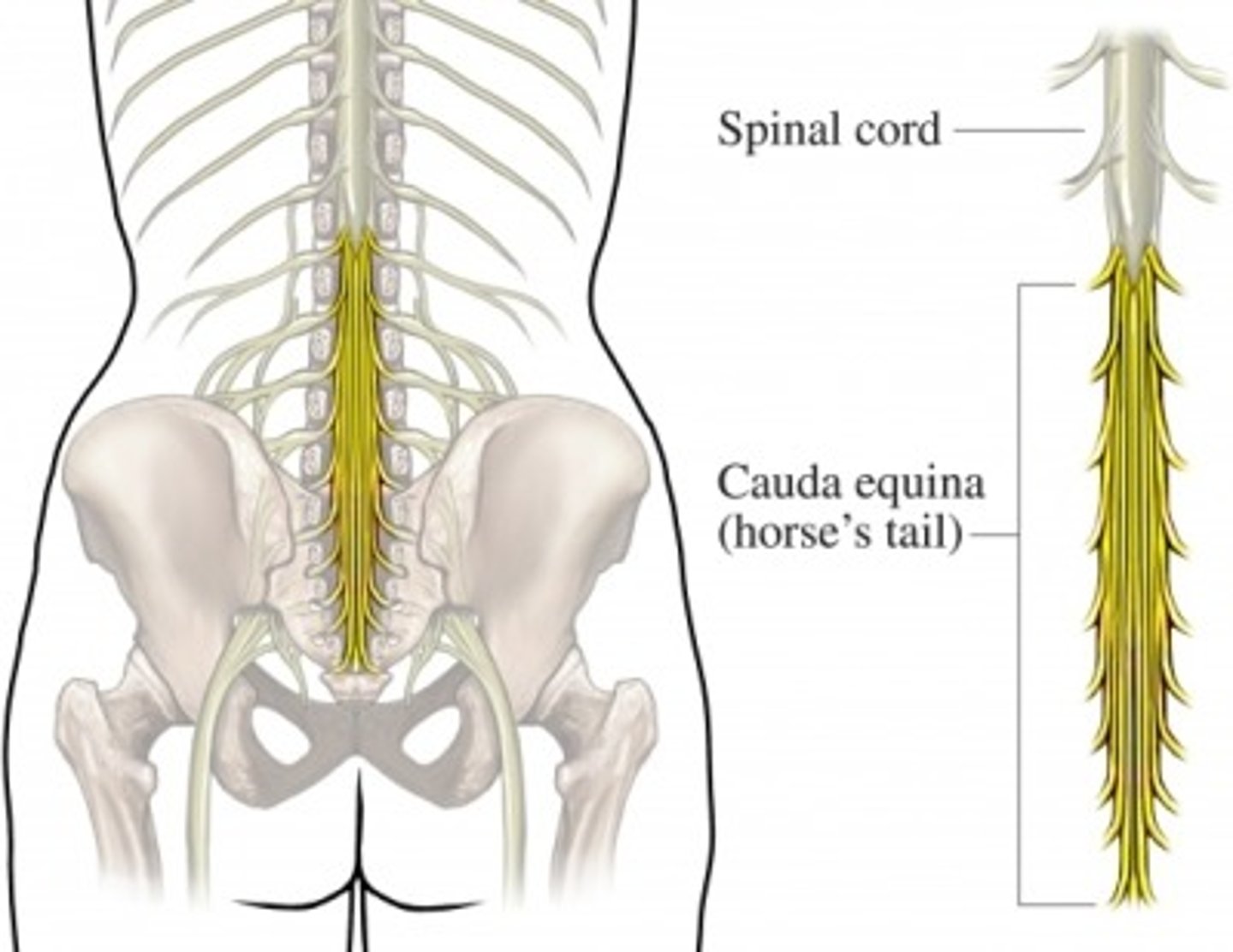
Filum Terminale
-anchors spinal cord to coccyx
-end of cauda equina
Spinal Dural Sheath
-Tough dura mater of the spinal cord
-Does not attach to surrounding bone
Cervical Nerves (8)
-corresponding nerve is above vertebrae
-eg) nerve c3 is below vertebrae c2
-nerve c1 above vertebrae c1
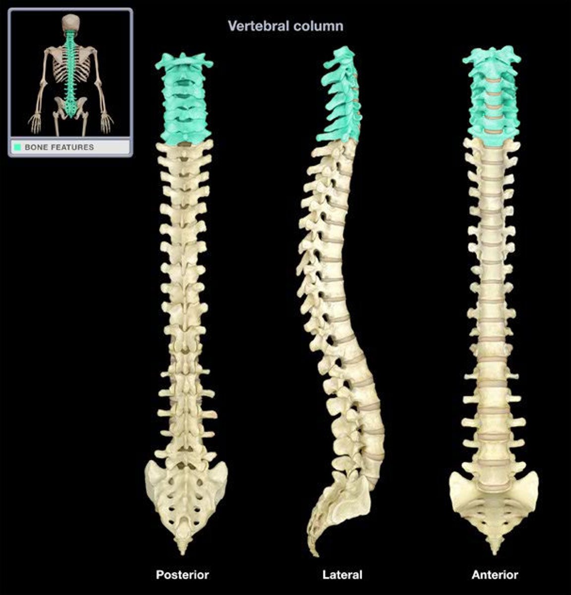
Thoracic Nerves (12)
-corresponding nerve is below the vertebrae
-eg) nerve T10 is below vertebrae T10
Lumbar Nerves (5)
same naming convention as thoracic
Sacral Nerves (5)
same naming convention as thoracic
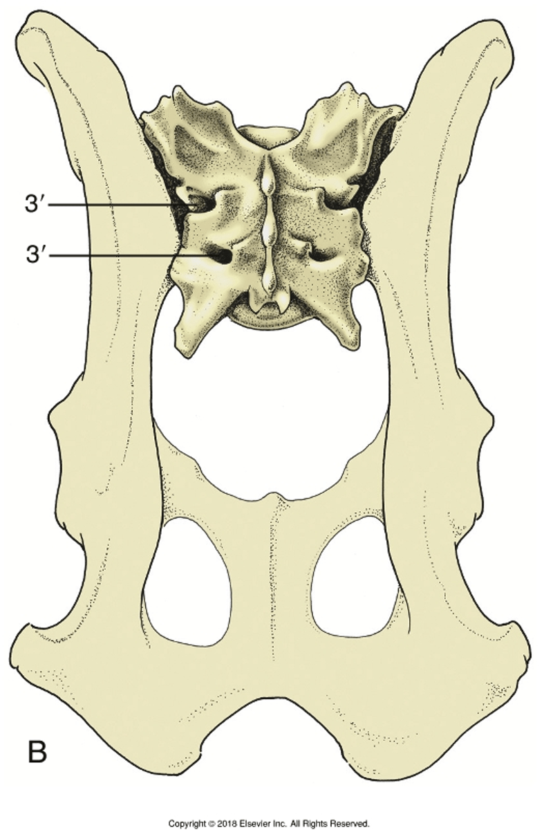
Coccyx Nerve (1 group)
-1 nerve
-butt
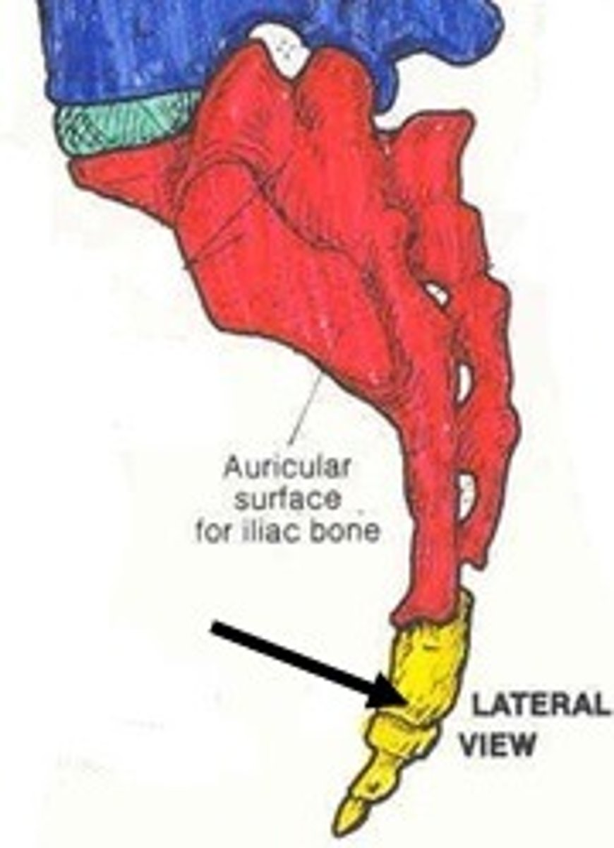
Optic Nerve (innervation)
-posterior of eyeball
-innervates eye
-originates from retina
-passes through optic canal
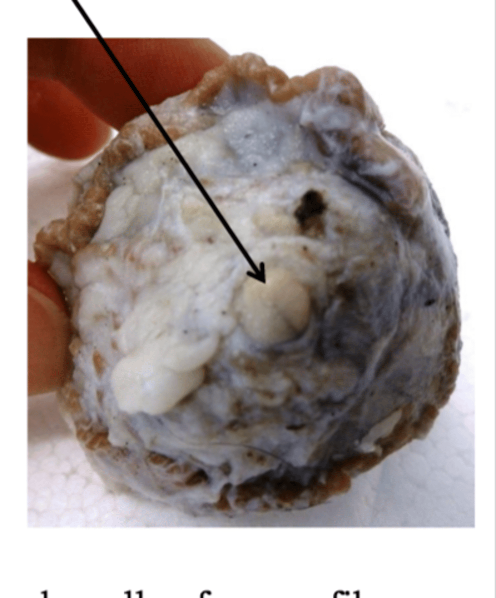
Vagus Nerve (innervation)
-originates from medulla
-passes through jugular foramen, through much of the upper thorax
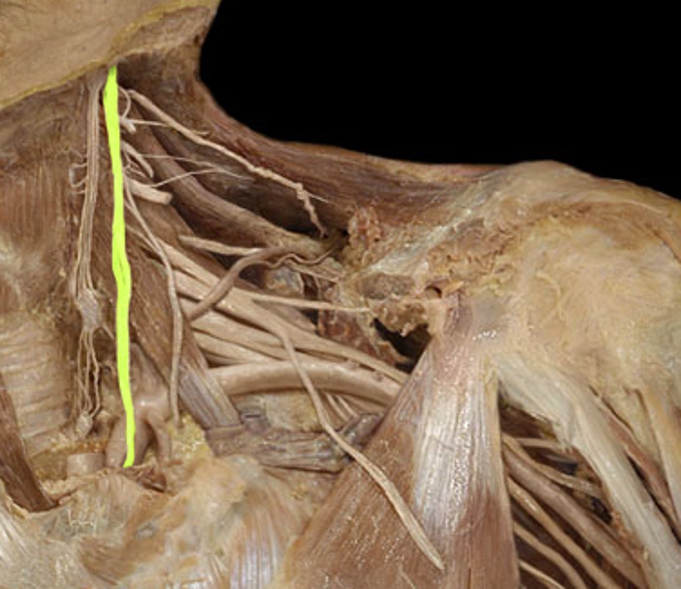
Phrenic Nerve (innervation)
-C3-C5
-Cervical plexus
-Innervates diaphragm, sensory
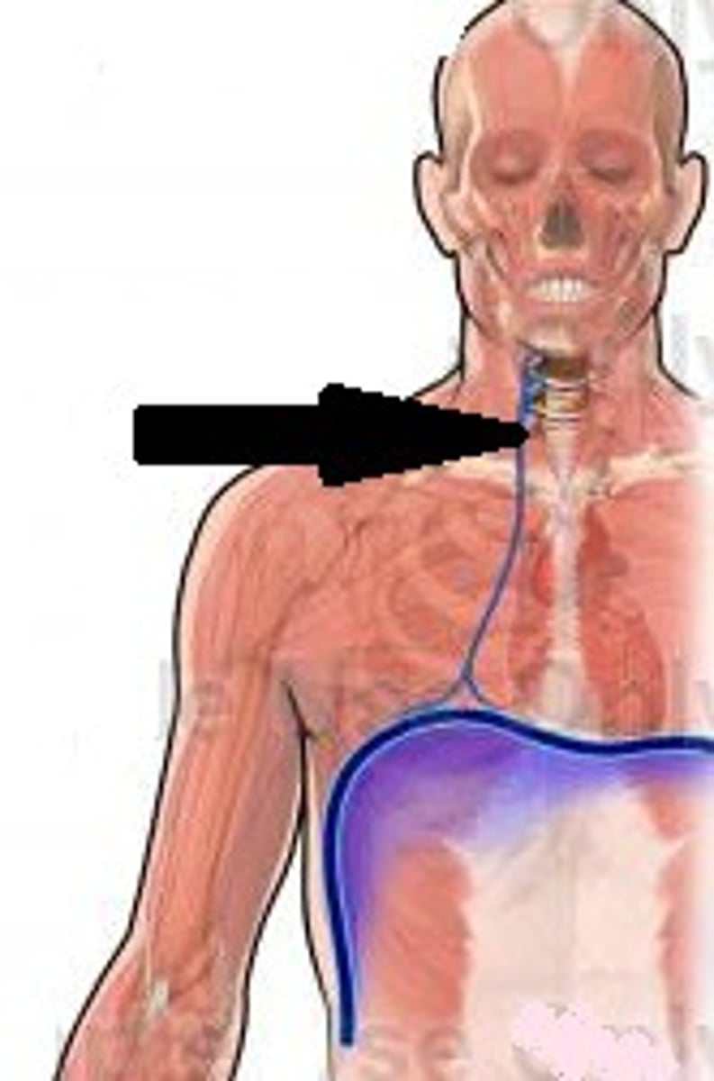
Ulnar Nerve (innervation)
-C8-T1
-Brachial plexus
-innervates medial hand skin
Obturator Nerve (innervation)
-L2-L4
-Lumbar plexus
-innervates inner thigh adductor medially
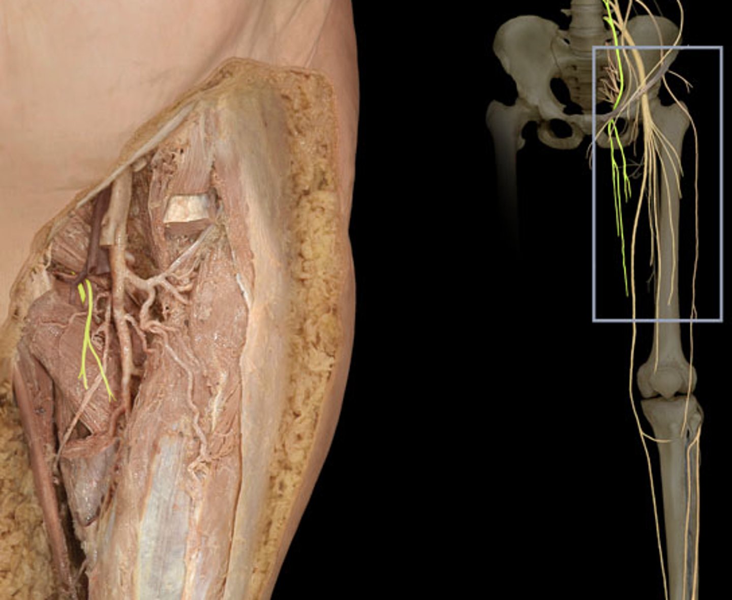
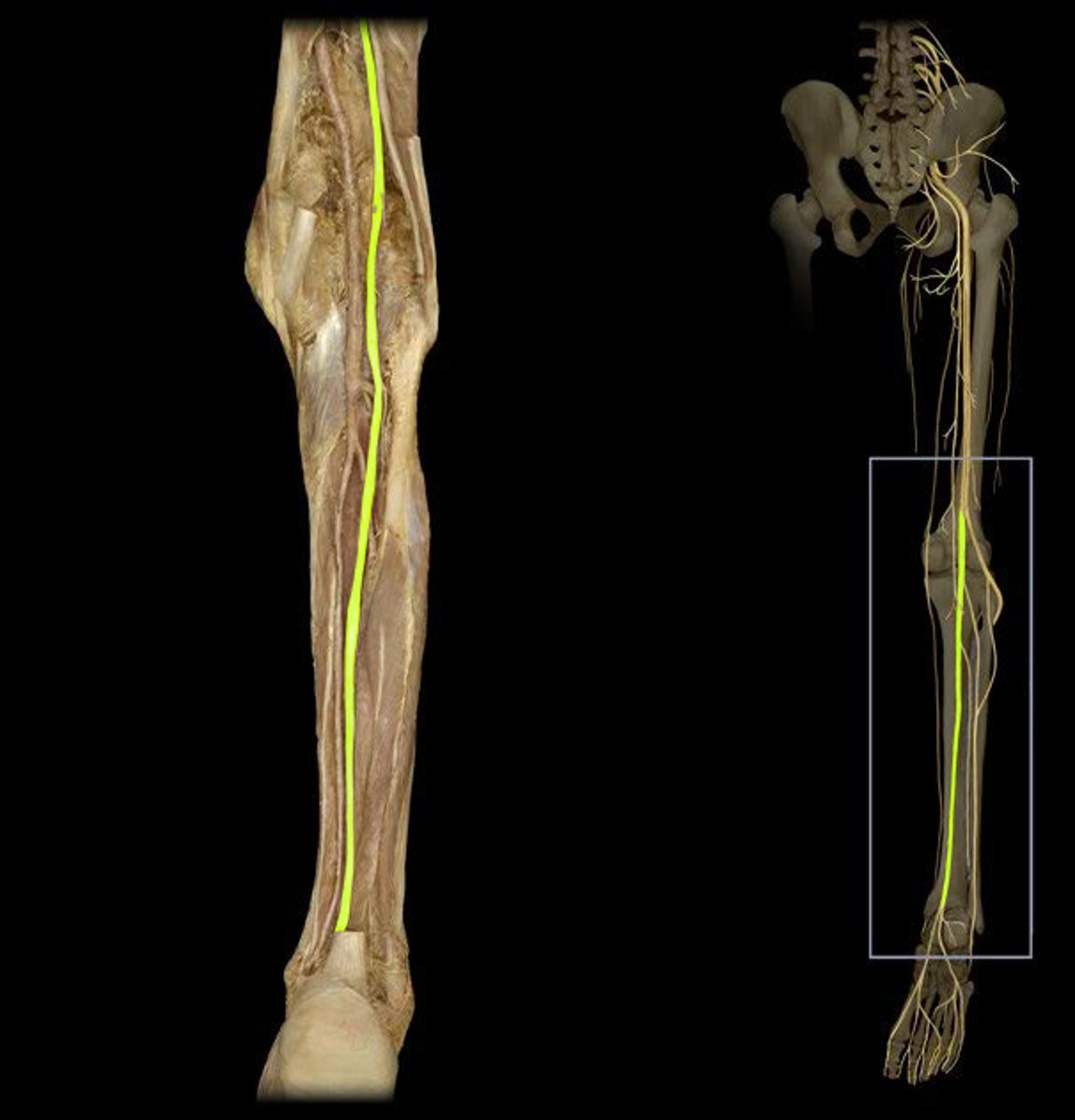
Tibial Nerve
-L4-S3
-Sacral Plexus
-runs through posterior lower limb towards ankle
-innervates posterior lower limb, sole of foot
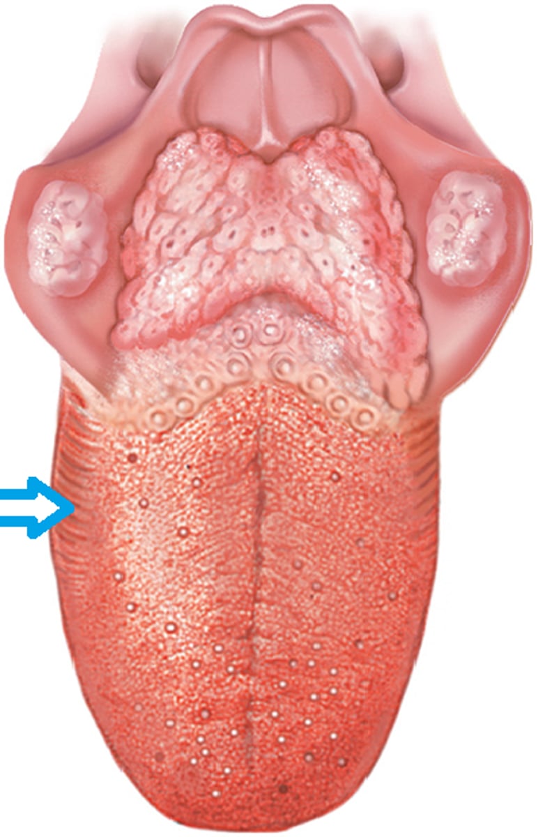
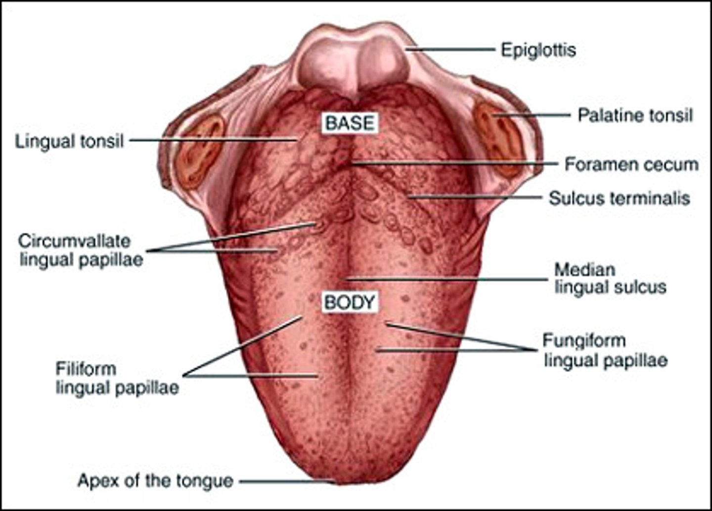
Foliate Papillae
side of tongue
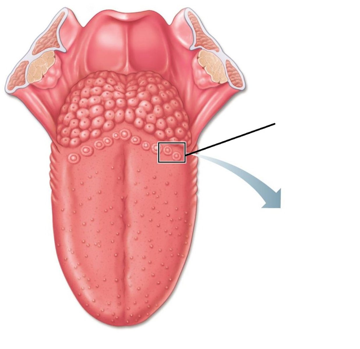
Vallate Papillae
-largest taste buds
-back of tongue, form V
Fungiform Papillae
knoblike projections on apical tongue surface
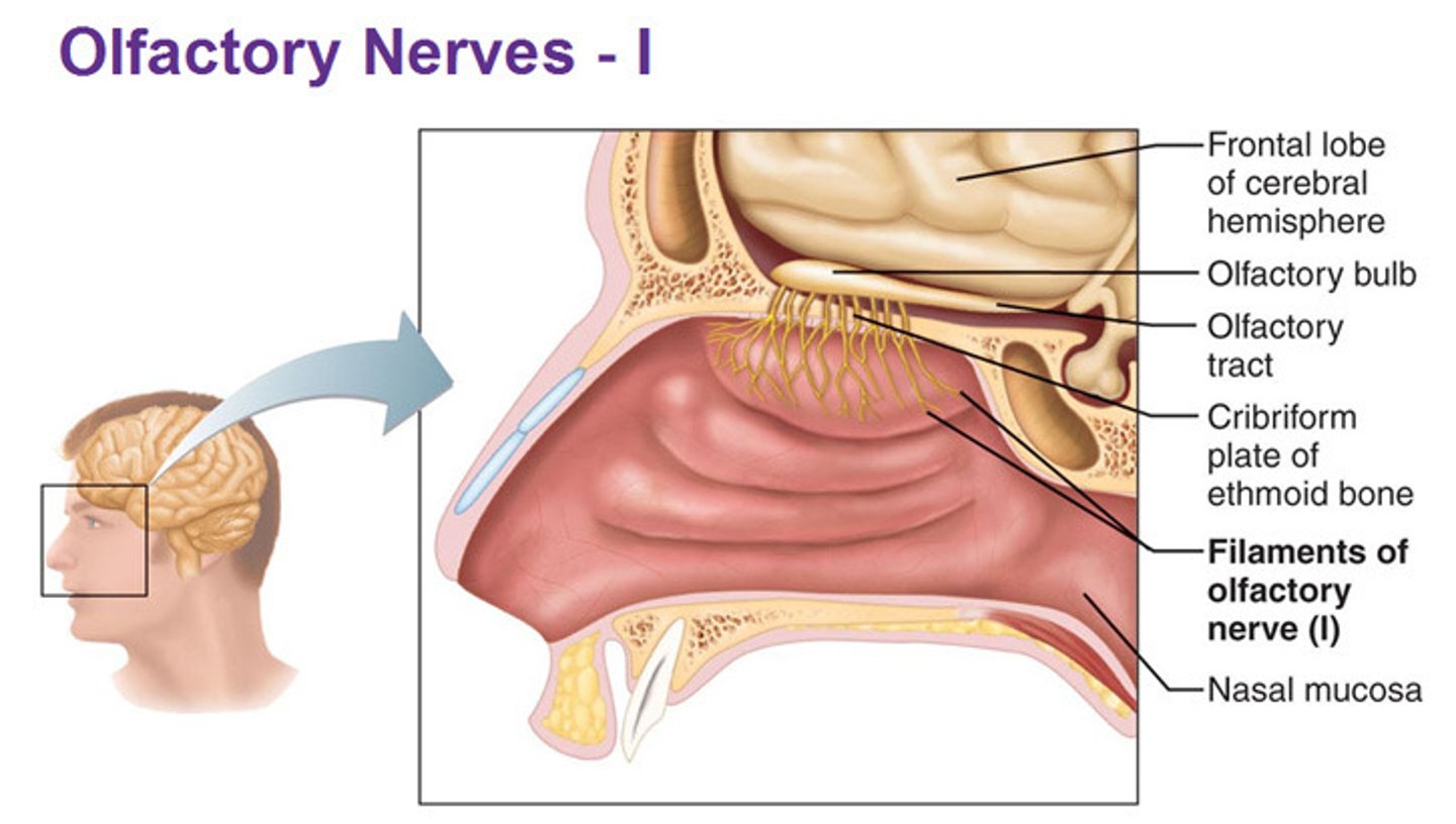
Olfactory Tract
connects olfactory bulb to brain
Olfactory Nerve Filaments
connect olfactory sensory neurons to the bulb
Olfactory Sensory Neurons
receptors for smell
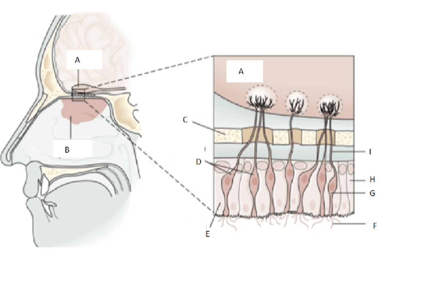
Olfactory Sensory Neurons
receptors for smell
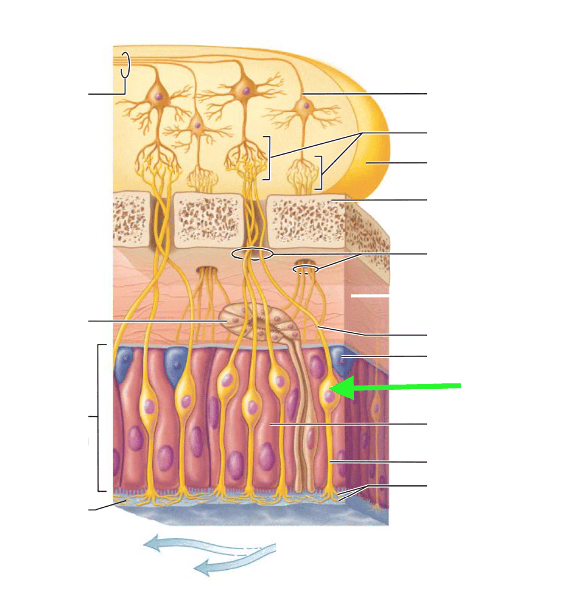
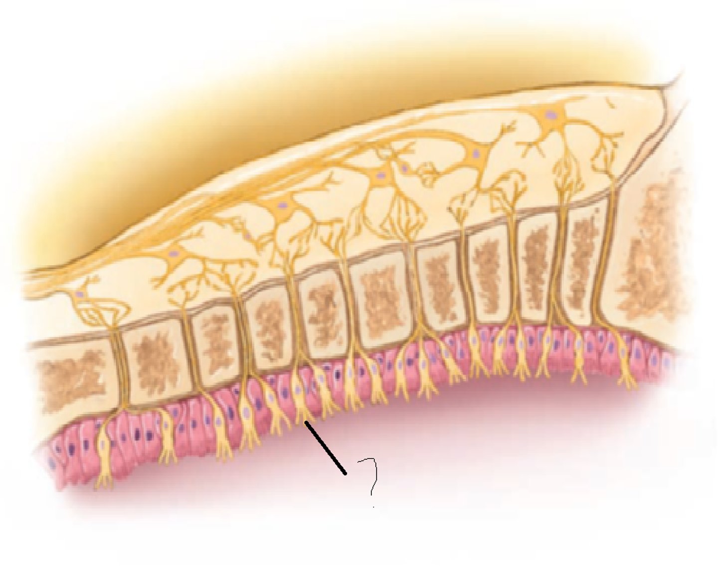
Olfactory Cilia (hairs)
roof of nasal cavity
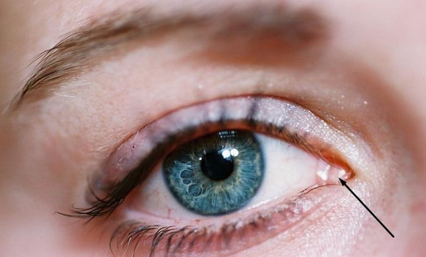
Lacrimal Caruncle
located on the medial commissure - produces oily secretion
Eyelashes
trigger reflex blinking
Eyelids
protect eyes
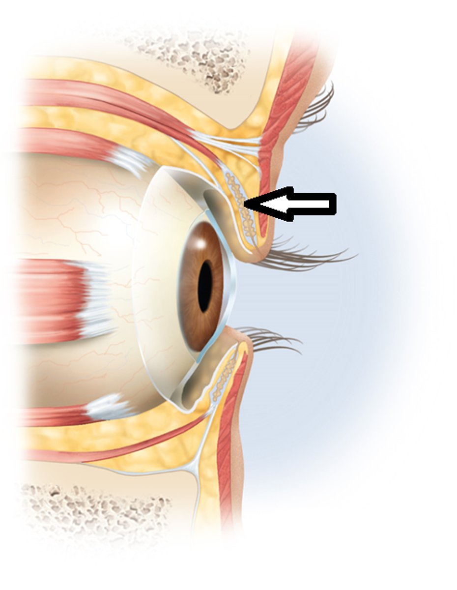
Tarsal Glands
-lubricate eyes
-secrete lipids for tears
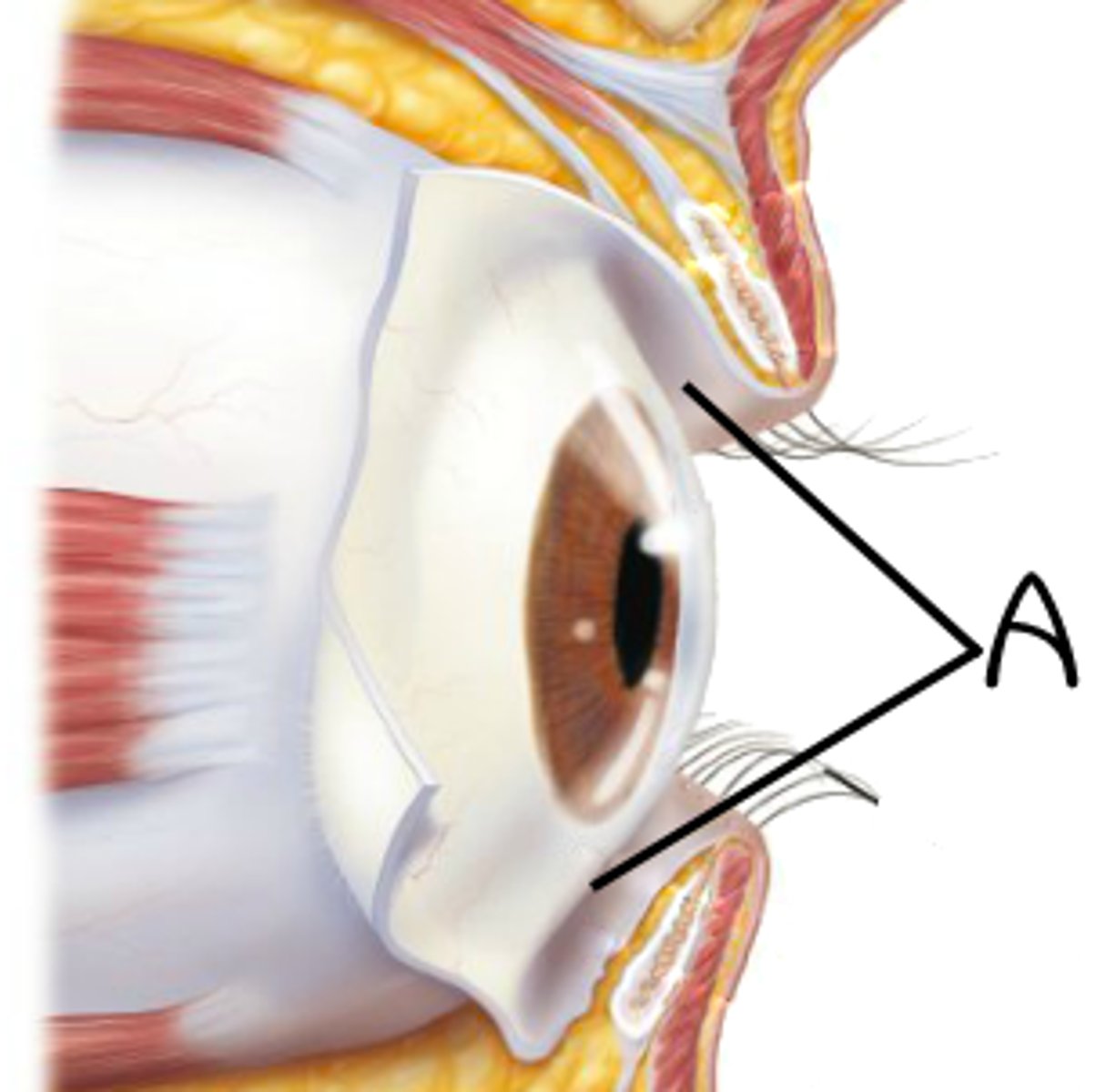
Conjunctiva
-mucus membrane lining eyelids
-keeps eyes wet
Lacrimal Apparatus
system of glands, sacs, ducts, that make new tears and drains lacrimal fluid
Lacrimal Gland
-produces mucus lacrimal fluid
-superolateral orbit of eye
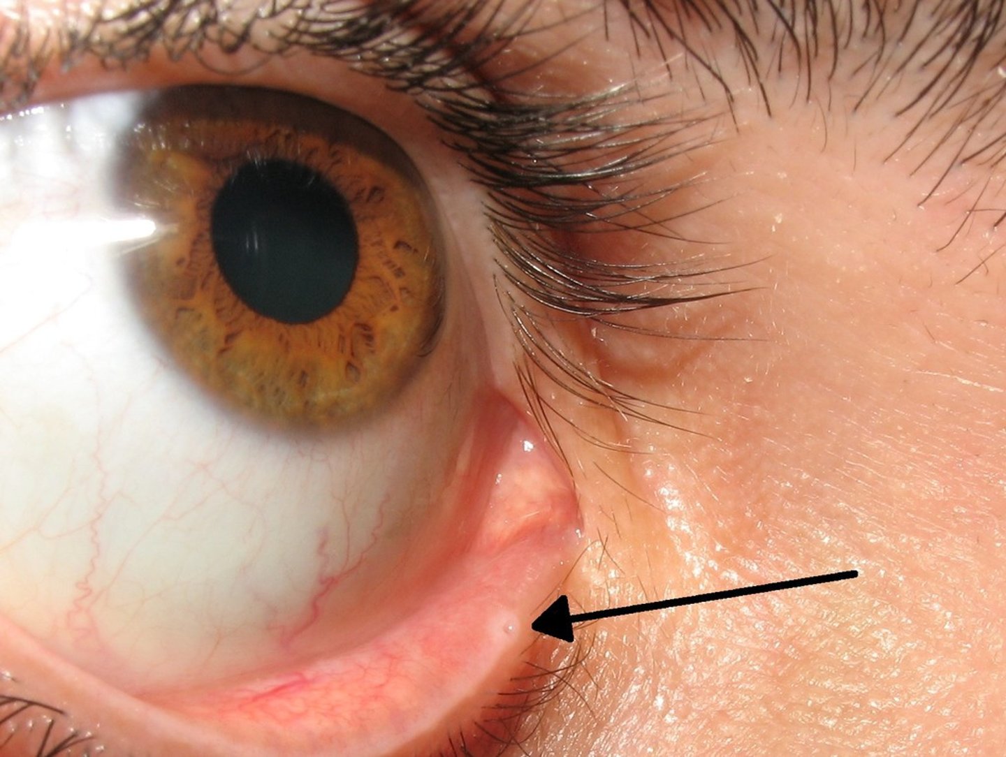
Lacrimal Punctum
-opening, drains fluid
-medial angle of eye, medial to caruncle
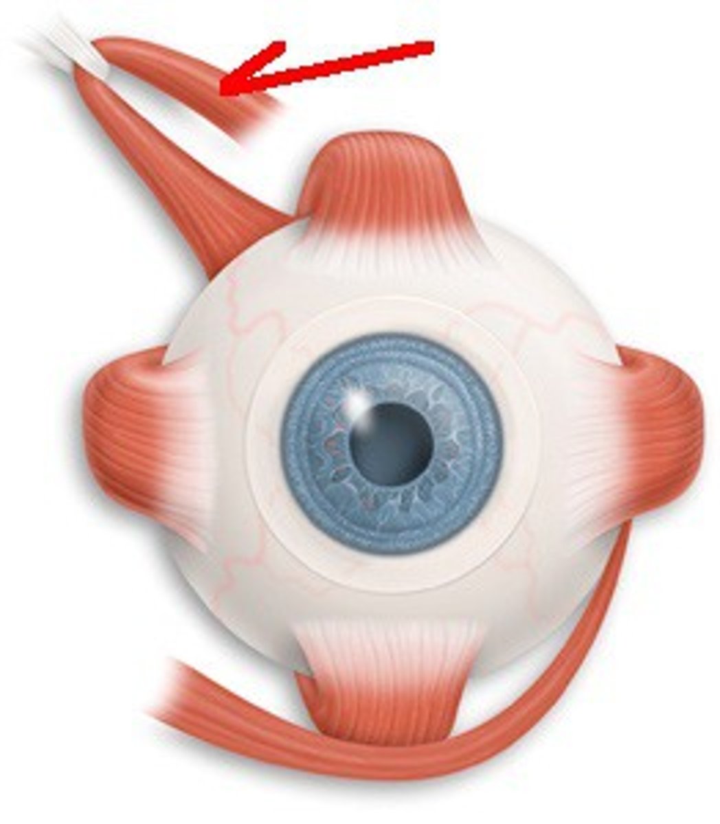
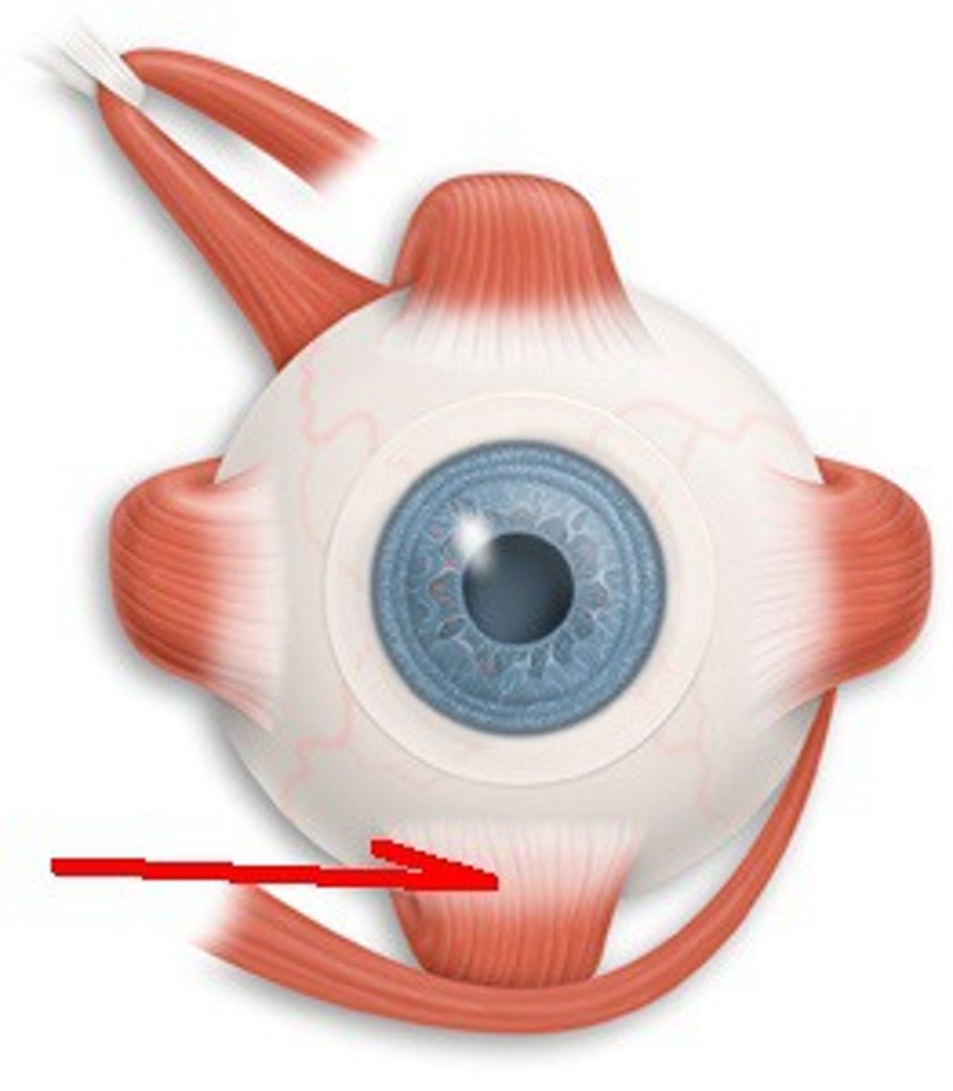
Superior Oblique Muscle
Down and out
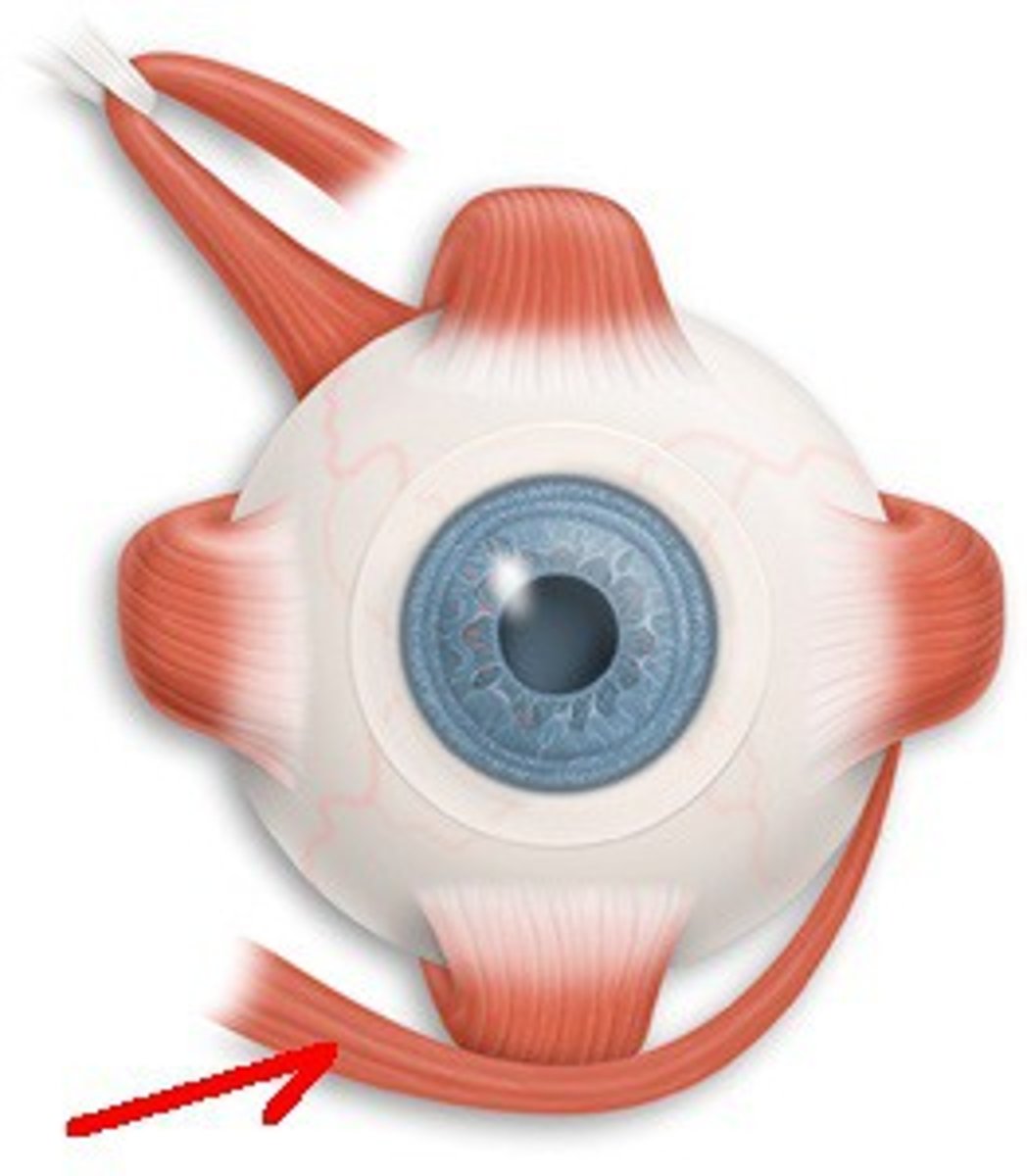
Inferior Oblique Muscle
Up and out
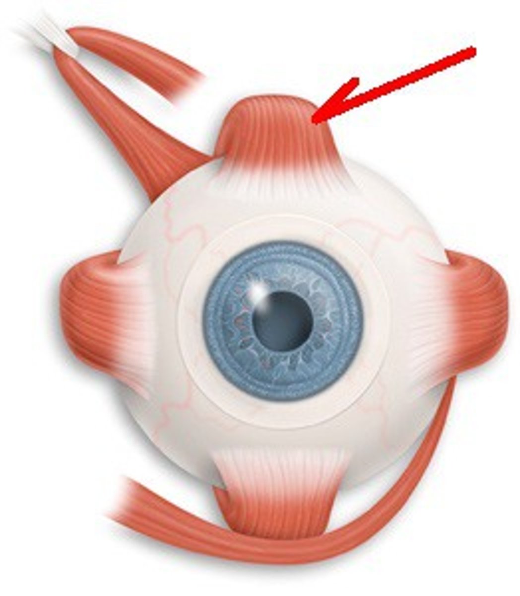
Superior Rectus Muscle
Up and in
Inferior Rectus Muscle
Down and in
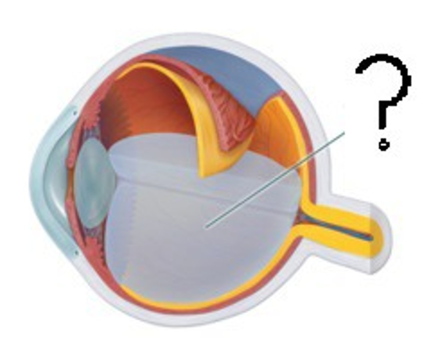
Vitreous Humor
-jellylike substance
-behind the lens in the posterior cavity
Retina
-deep to choroid and sclera
-superficial to vitreous humor
Sclera
-outermost protective layer of eye
-white tissue
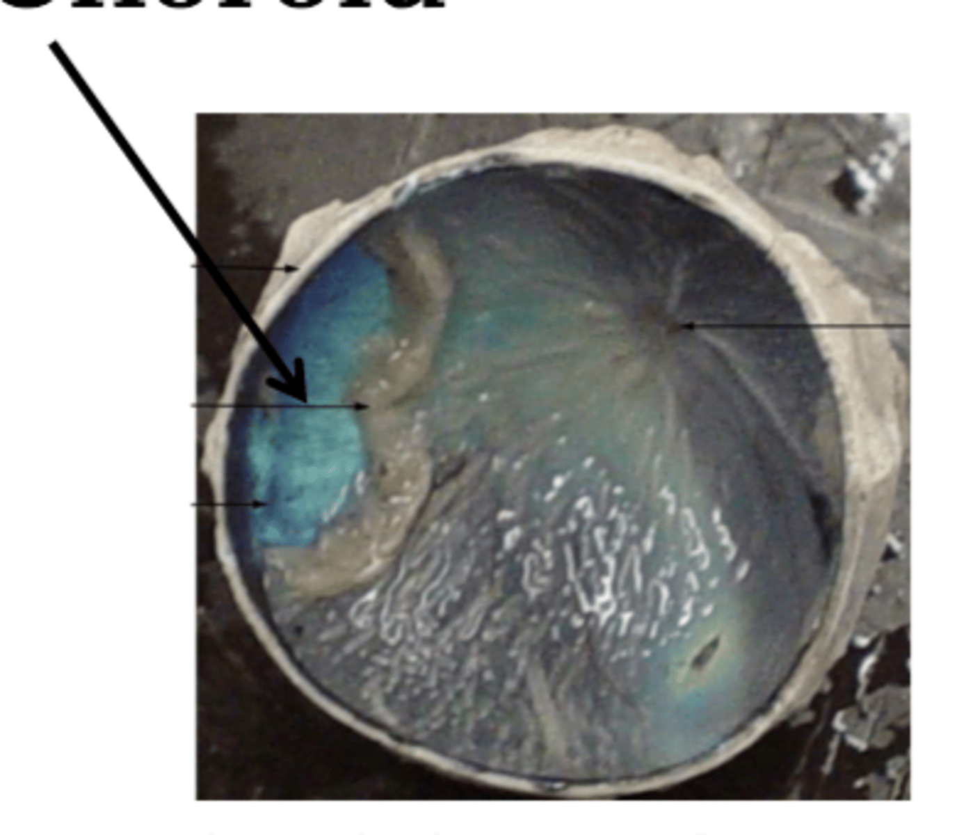
Choroid
-middle vascular layer
-deep to sclera
-superficial to retina
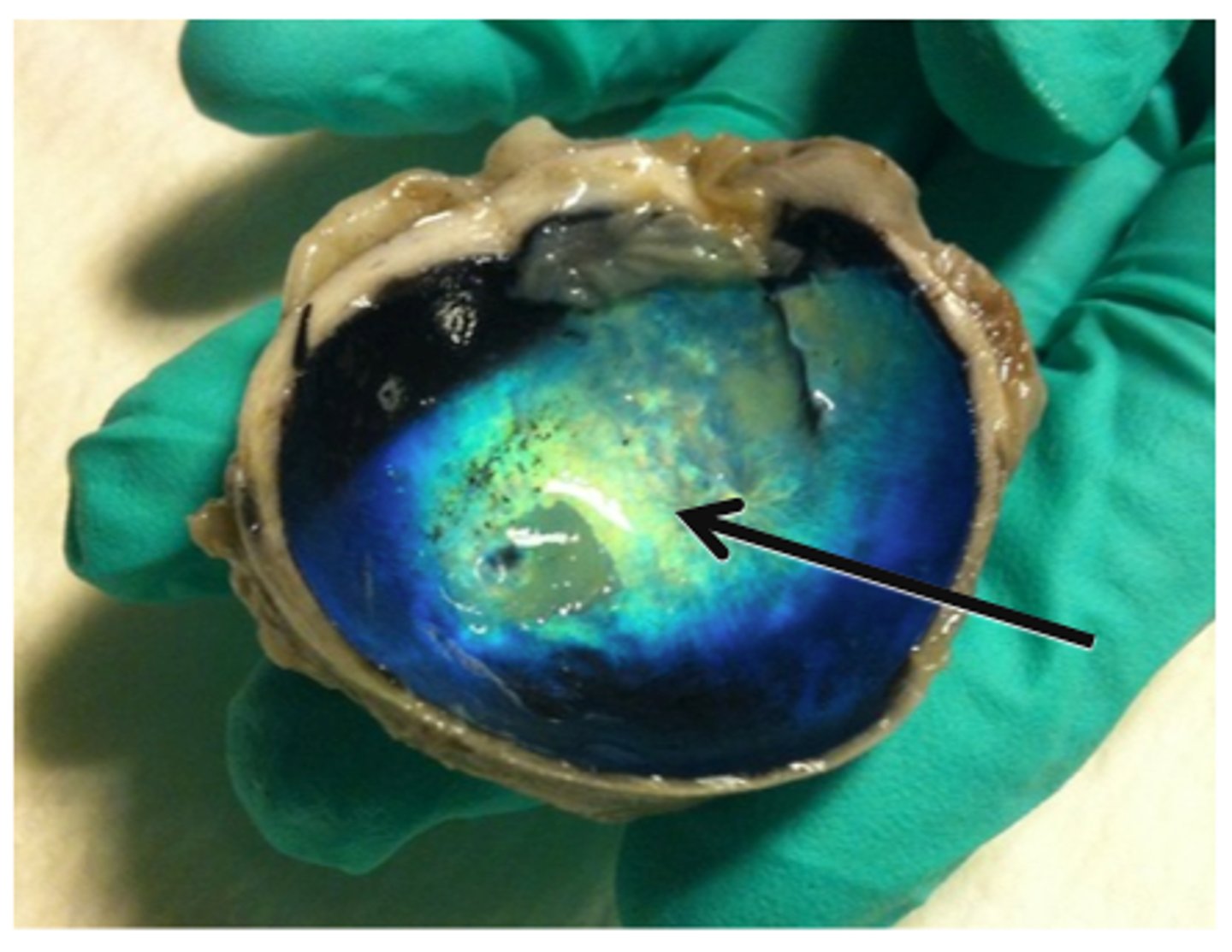
Tapetum Lucidum
-Iridescent layer in posterior wall
-found in nocturnal animals
Ciliary Body
ring of tissue around iris
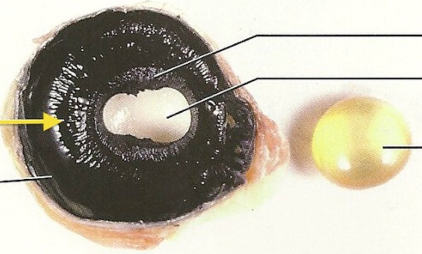
Cornea
-transparent outer covering of the eye
-covers iris and pupil
Lens
the transparent structure behind the pupil that changes shape to help focus images on the retina
Iris
Colored part of the eye

Pupil
opening in the center of the iris
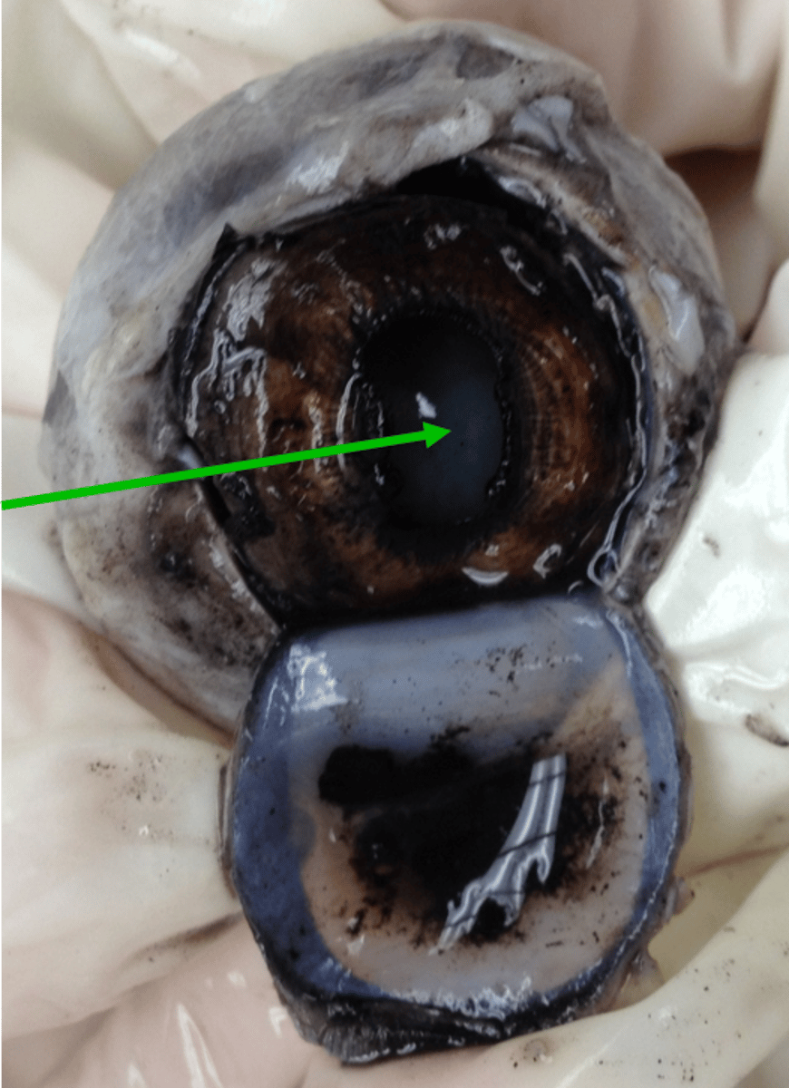
Fibrous Pericardium
tough white outer layer of heart
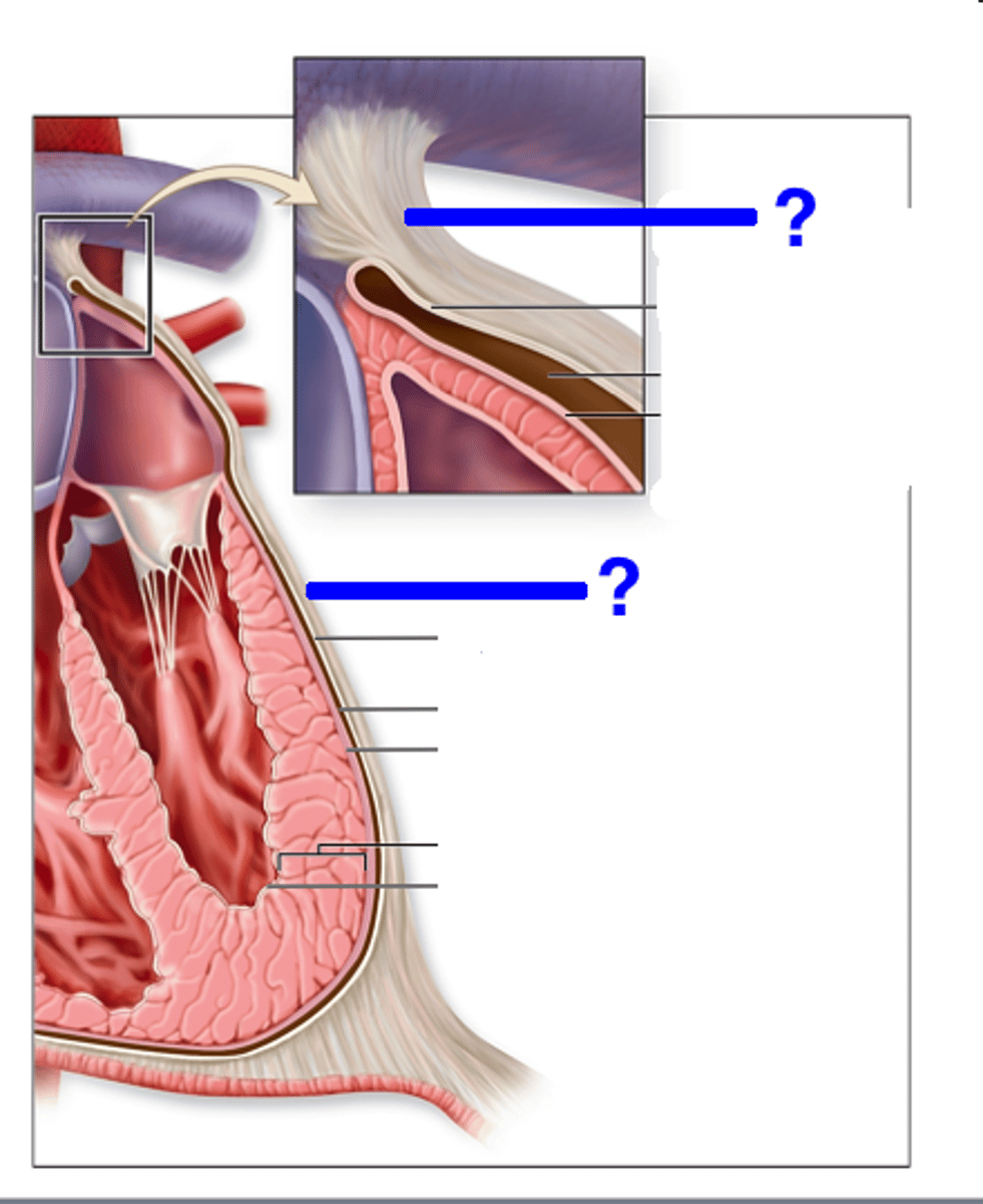
Parietal Serous Pericardium
-lines the inner surface of the fibrous pericardium
-deep to fibrous layer, superficial to visceral layer
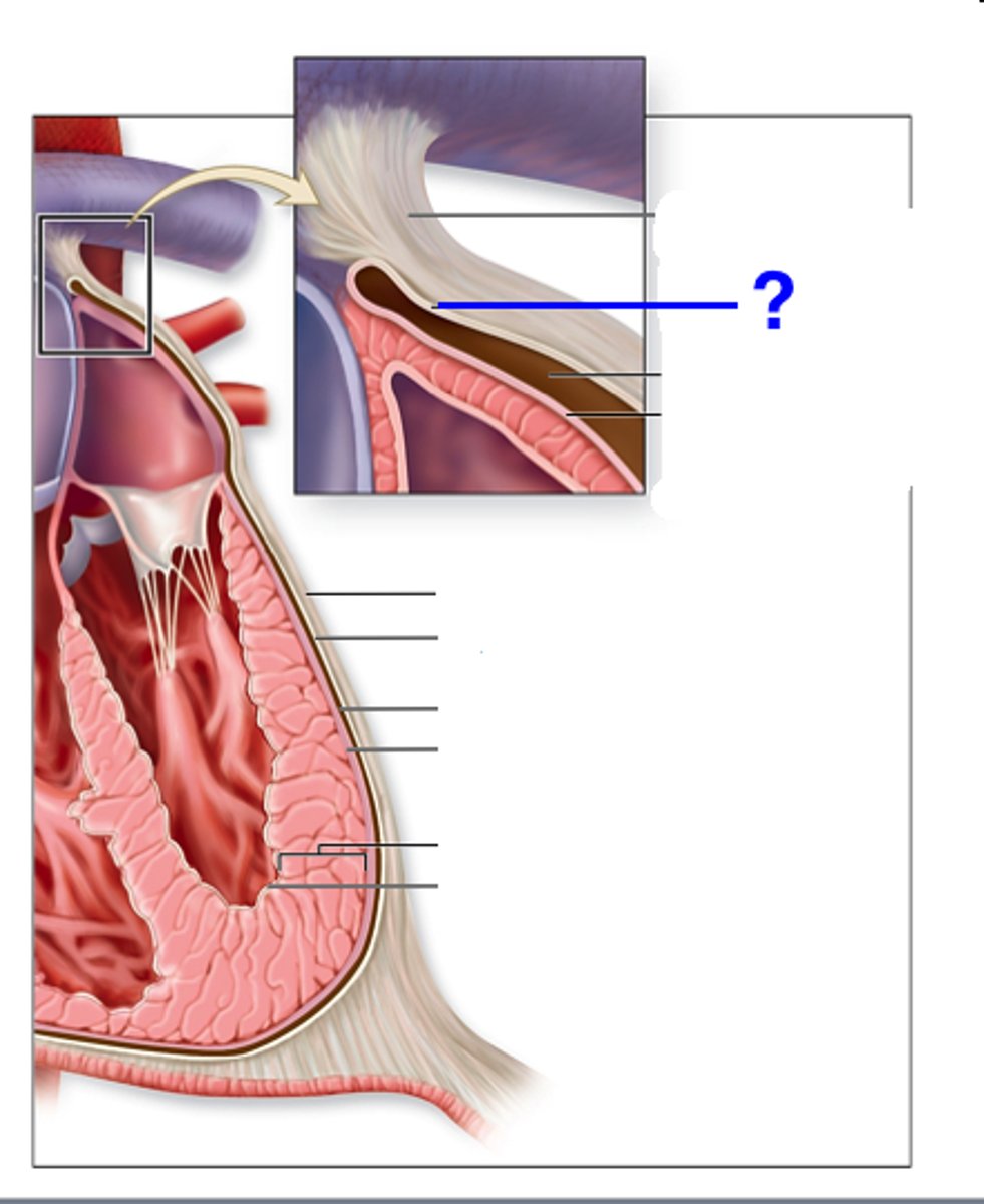
Visceral Serous Pericardium
-covers the heart itself
-deep to parietal layer
Epicardial Adipose Tissue
adipose upon surface of myocardium
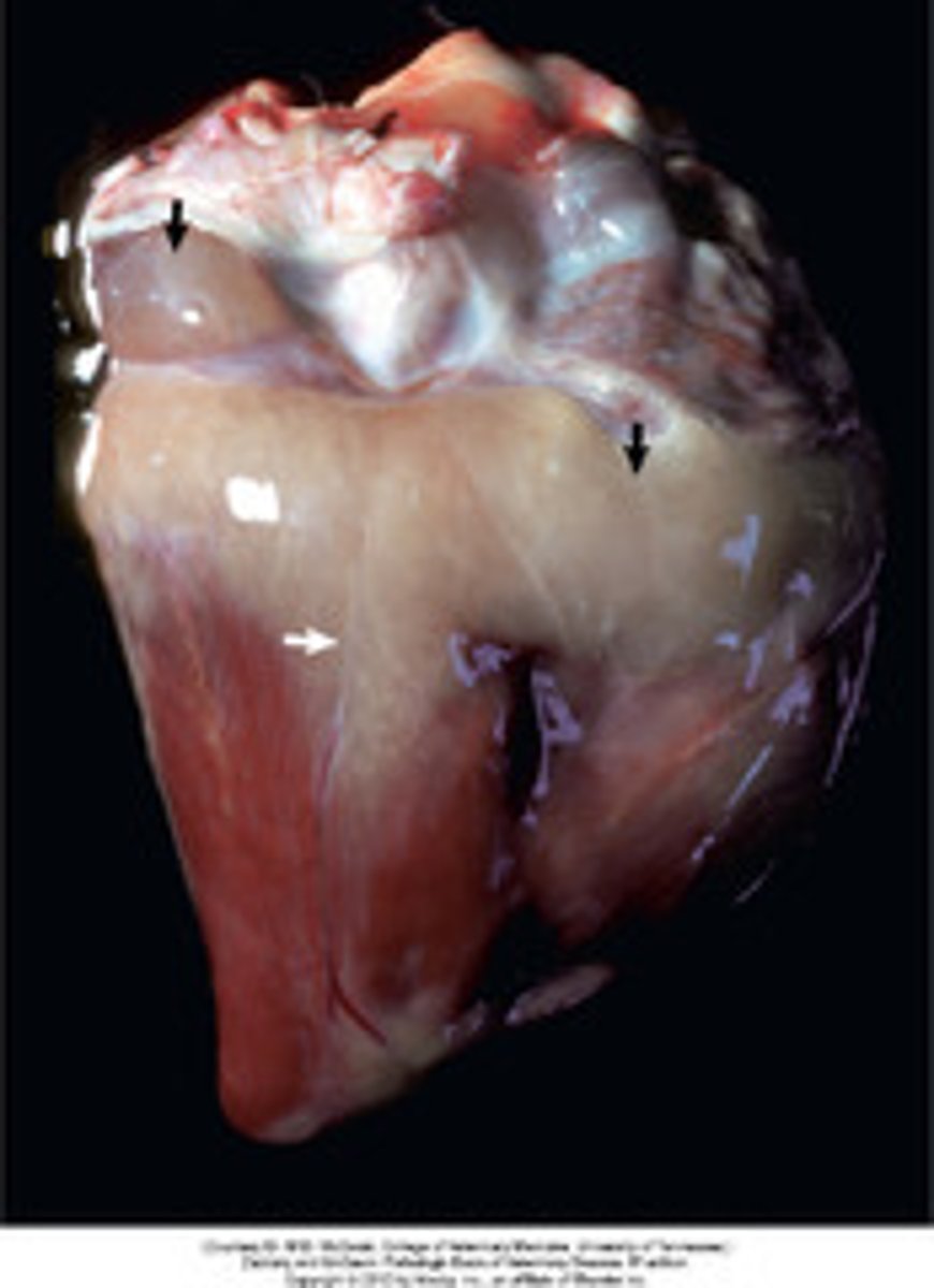
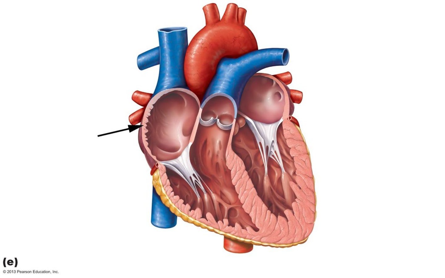
Pectinate Muscles
ridges, lines internal ATRIAL walls
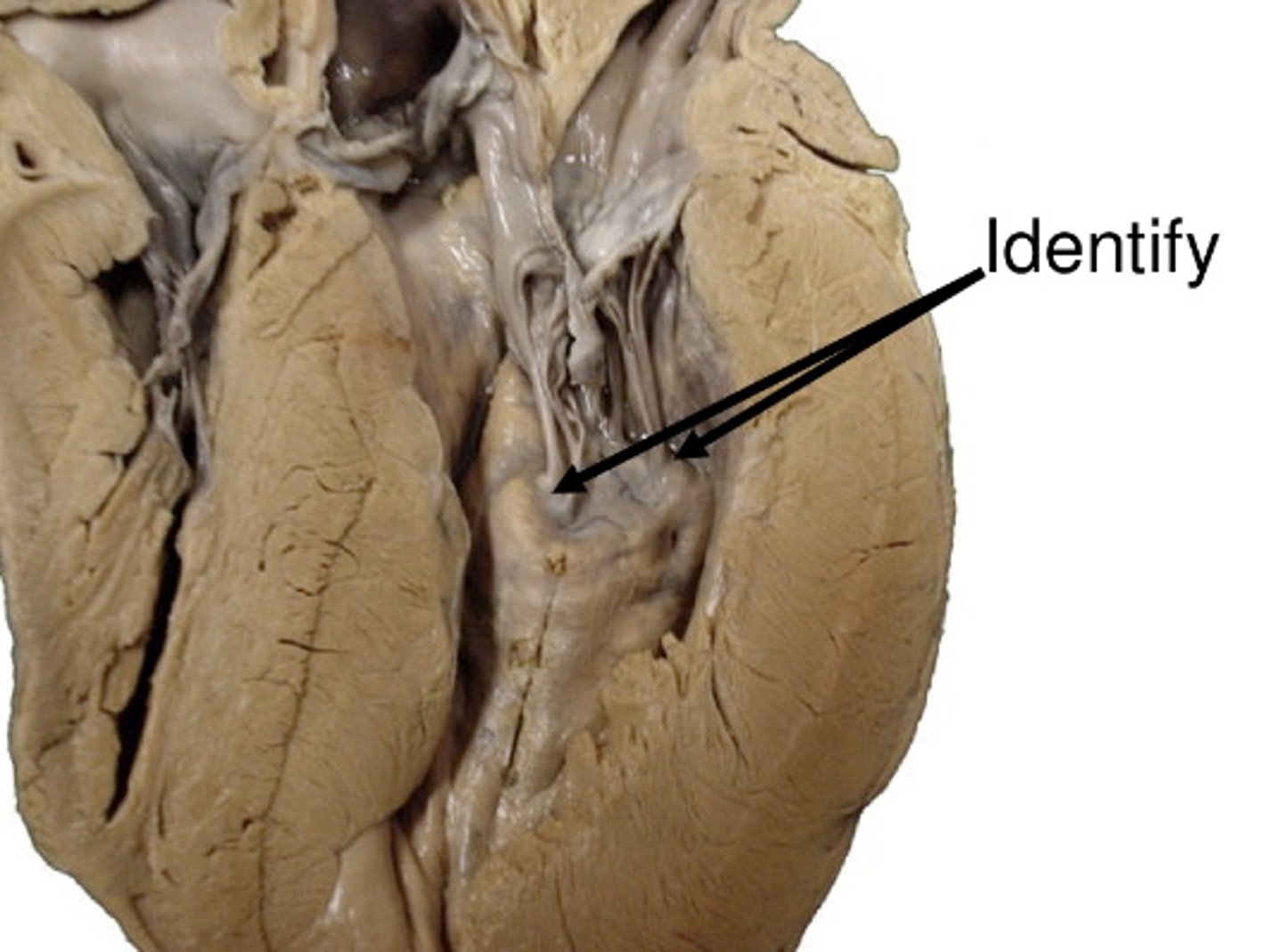
Papillary Muscles
-ventricles
-anchor chordae tendineae
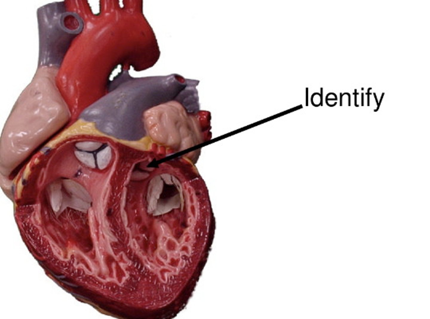
Trabeculae Carneae
ridges, line internal VENTRICLES

Tricuspid Valve
valve between the right atrium and the right ventricle

Bicuspid (Mitral) Valve
valve between the left atrium and the left ventricle

Aortic Valve
-semilunar
-separates LV from aorta
Pulmonary Valve
-semilunar
-separates RV from pulmonary trunk
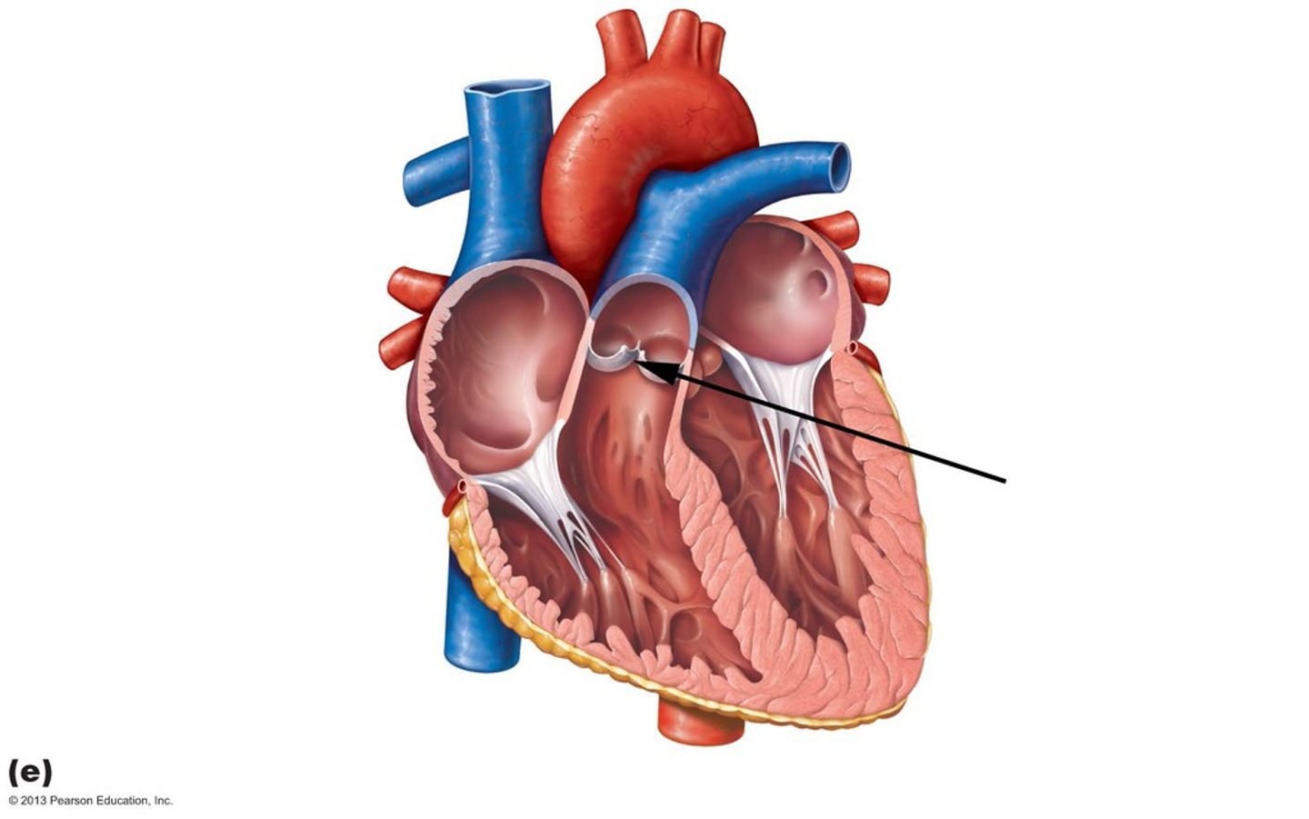
Vena Cava
superior and inferior veins, feed O2 poor blood into RA

Pulmonary Veins
two pairs of veins, feed O2 rich blood into LA

Pulmonary Trunk
-carries blood from the RV to the lungs (pulmonary circuit)
-branches into pulmonary arteries

Aorta
carries blood from the LV to the systemic circuit

Right Coronary Artery
supplies the RA
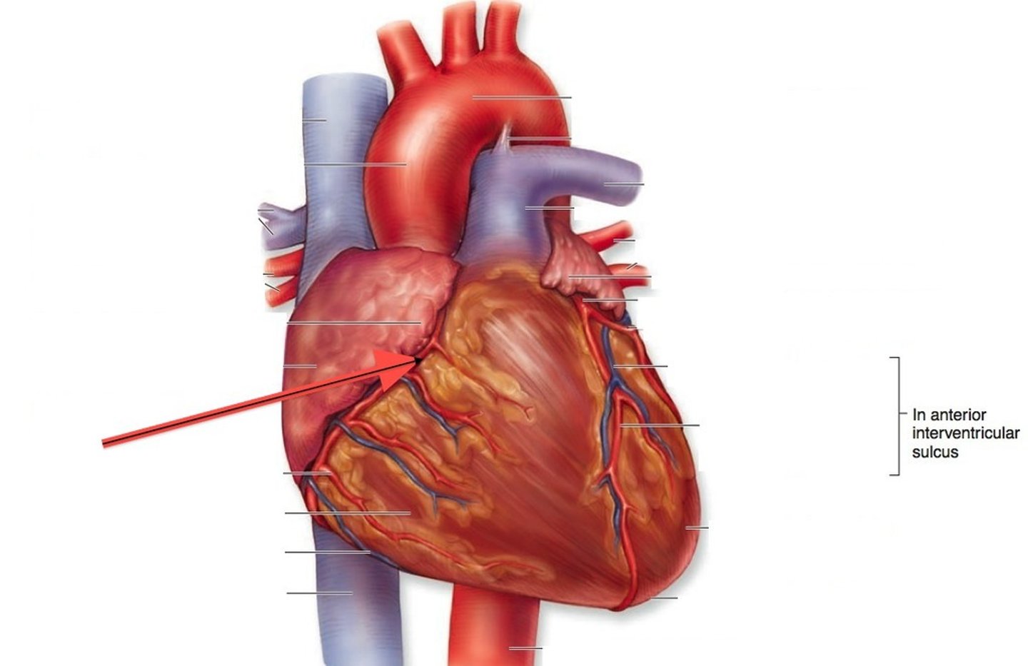
Marginal Artery
-supplies lateral RV
-branches off RCA
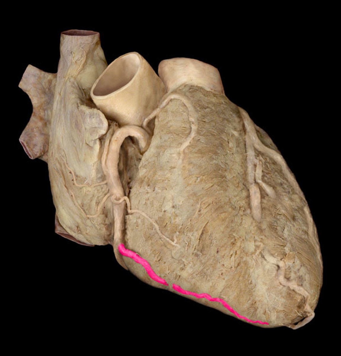
Anterior Interventricular Artery
-lies upon interventricular sulcus
-supplies blood there + both ventricles
-branches off LCA

Left Coronary Artery
-supplies to LA, LV
-branches off circumflex artery

Circumflex Artery
-supplies left atrium and ventricle
-branches off LCA

Small Cardiac Vein
vein, travels along side the right marginal artery.
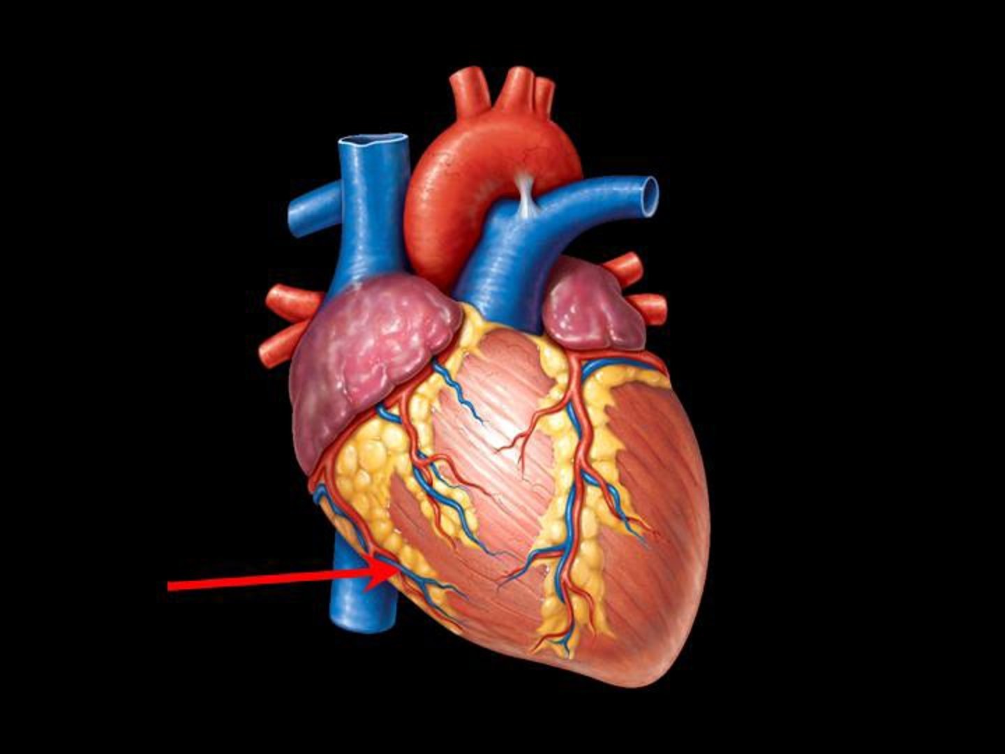
Great Cardiac Vein
-runs alongside the anterior interventricular artery
-originates from posterior/inferior side, coronary sinus

Coronary Sinus
-posterior aspect of heart
-feeds into LA
-connects to great cardiac vein

Posterior Interventricular Artery
-decends inferiorly to apex of heart
-supplies posterior ventricular walls
-branches off RCA

Middle Cardiac Vein
runs alongside the posterior interventricular artery
