Labs for Anemia
 Call Kai
 Learn
Learn Practice Test
 Spaced Repetition
 Match
Match Flashcards
 Knowt Play
1/81

There's no tags or description
Looks like no tags are added yet.
Name | Mastery | Learn | Test | Matching | Spaced | Call with Kai | Chat |
|---|
No analytics yet
Send a link to your students to track their progress
82 Terms
Where does normal erythropoiesis begin?
kidney where peritubular interstitial cells alter erythropoietin production depending on amount of O2 demand
What takes place in the RBC cycle within the bone marrow?
erythropoietin enhances the growth of 'erythroid cells' into reticulocytes
What are causes of WBC abnormalities?
Infection, Inflammation, Neoplasm/malignancy, and Drug reactions
What are causes of Hemoglobin and Hematocrit abnormalities?
Anemia and Polycytehmia
What are causes of Platelet abnormalities?
Bleeding disorders and Hypercoagulable states
What comprises a RBC count?
# of RBCs in a specified volume of whole blood that can be affected by volume status but DOES NOT reflect changes in RBC size
What causes an increased RBC count?
Dehydration, COPD, Smoking, High altitude, and Polycythemia vera
What causes an decreased RBC count?
blood loss, anemia, and fluid overload
What are some causes of anemia?
- Fe, B12, or Folate deficiency
- Hemolytic anemia
- Bone marrow failure
What are some causes of fluid overload?
cirrhosis, pregnancy, and CHF
What is the heme portion of hemoglobin (Hgb)?
contains iron and porphyrin (red pigment)
What is the globin portion?
four amino acid chains -one heme molecule attaches to each of the four globin chains
What is Hematocrit (Hct)?
percentage of the total volume of RBCs relative to the total volume of whole blood (3x that of Hgb)
What can influce hematocrit value?
fluid status and RBC size therefor, if Hgb x3 ≠ Hct, think fluid problem
What causes a high RBC count, Hgb, or Hct?
Polycythemia
What causes a low RBC count, Hgb, or Hct?
Anemia
What is MCV?
measure of average RBC size that can be microcytic (little), normocytic, macrocytic (big)
How is MCV calculated?
Hct / RBC
What causes a high MCV?
Macrocytosis: B12 or folate anemia
What causes a low MCV?
Microcytosis: Fe deficiency anemia, thalassemia common
What is MCH?
weight of Hgb in RBC which tends to mirror MCV and therefore adds little information i
What is MCHC?
Hgb concentration that can be hypochromic, normochromic, and hyperchromic (higher concentration = higher color)
How is MCHC calculated?
Hgb / Hct
What causes high MCHC?
Hyperchromic cells
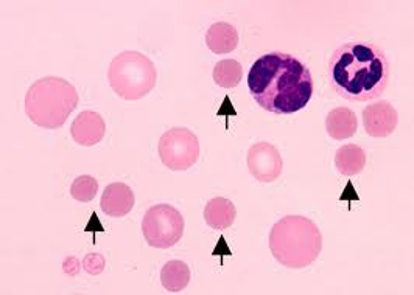
What causes low MCHC?
Hypochromic cells seen in Fe deficiency)
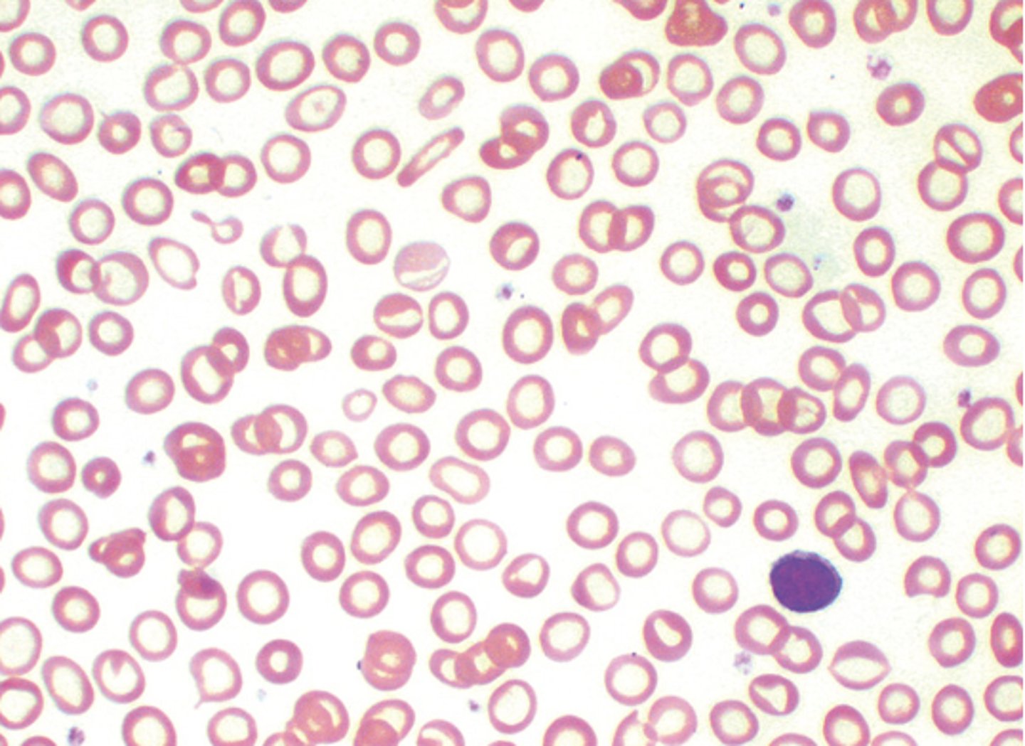
What is RDW?
Measure of variation in RBC size indicates degree of anisocytosis
What is anisocytosis?
abnormal/ increased variability in size MC caused by Fe deficiency
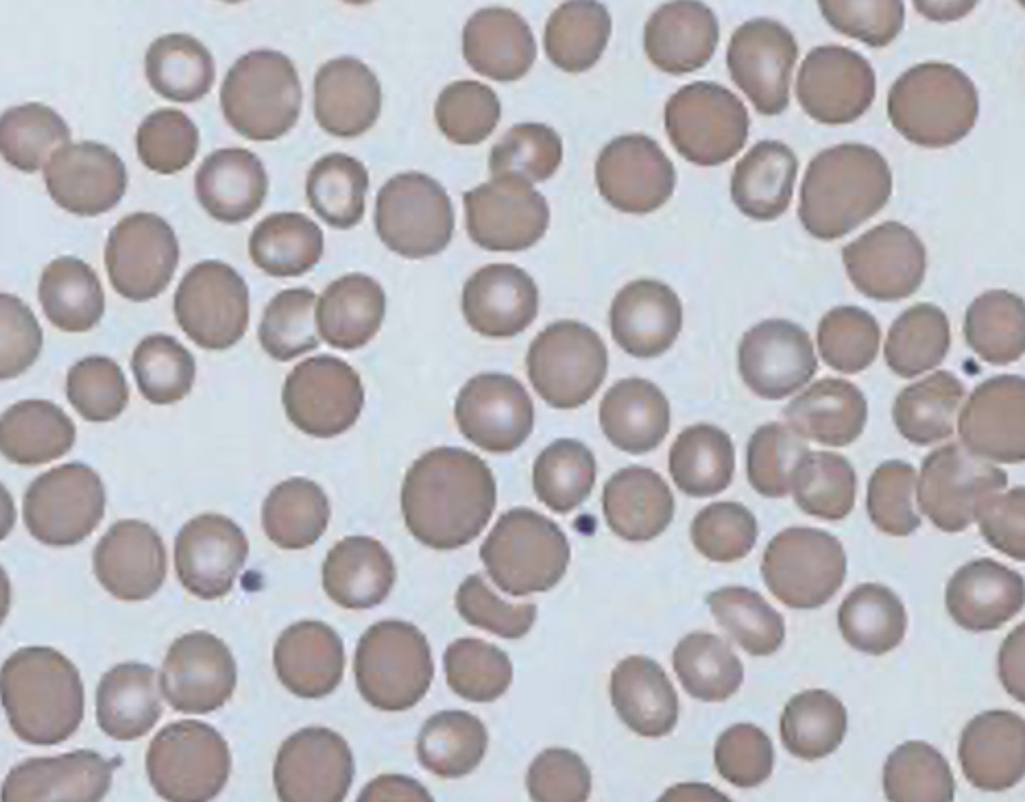
What is a critcal hemoglobin value?
<7 g/dL or >21g/dL
What is a critical hematocrit value?
<21% or >65%
What are peripheral smear used for?
diagnose blood disorders that manifest with changes in blood cell appearance
What conditions prompt a peripheral smear?
Leukemia, Myelodysplastic syndrome, anemias, lymphoma, and malaria
What is a rouleaux formation?
Red blood cells clump together and form what looks like "stacks of coins"
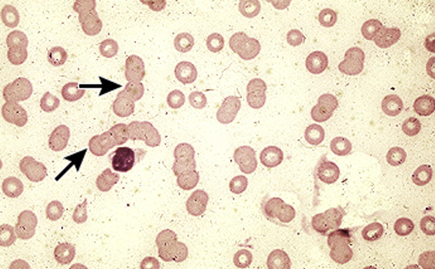
What conditions cause rouleaux formation?
Connective tissue diseases/ inflammatory conditions like multiple myeloma
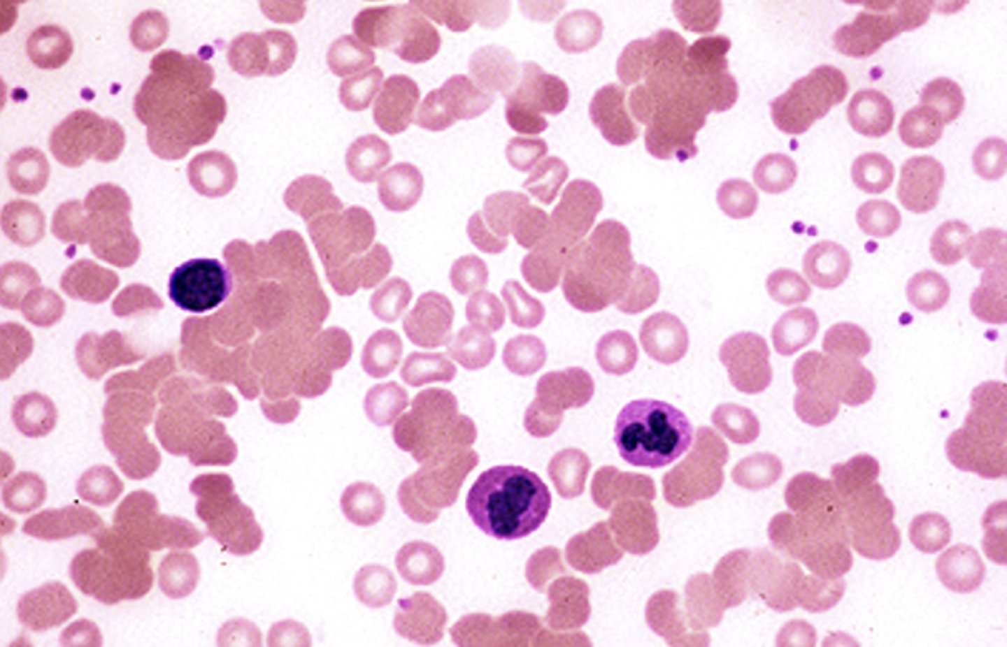
What is RBC Agglutination?
Red blood cells clump together due to antibodies coating RBC surface
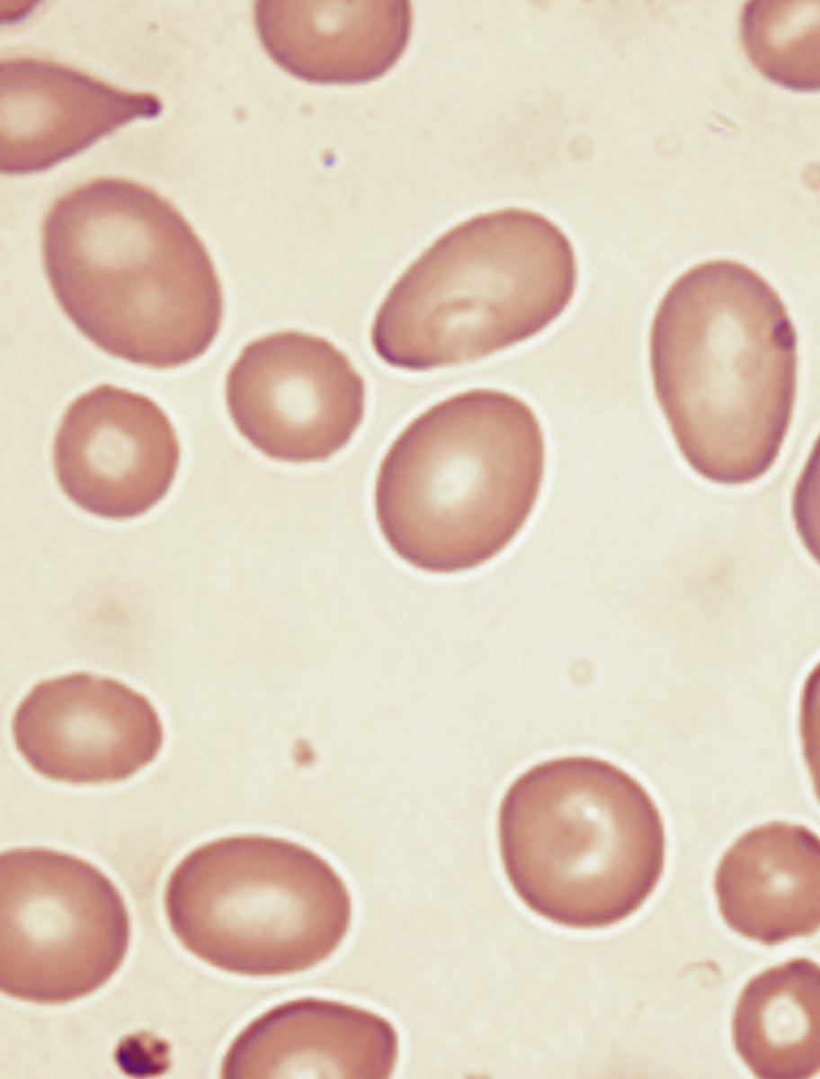
What are Macro-ovalocytes?
Seen with megaloblastic anemia common in B12 and folate deficiency
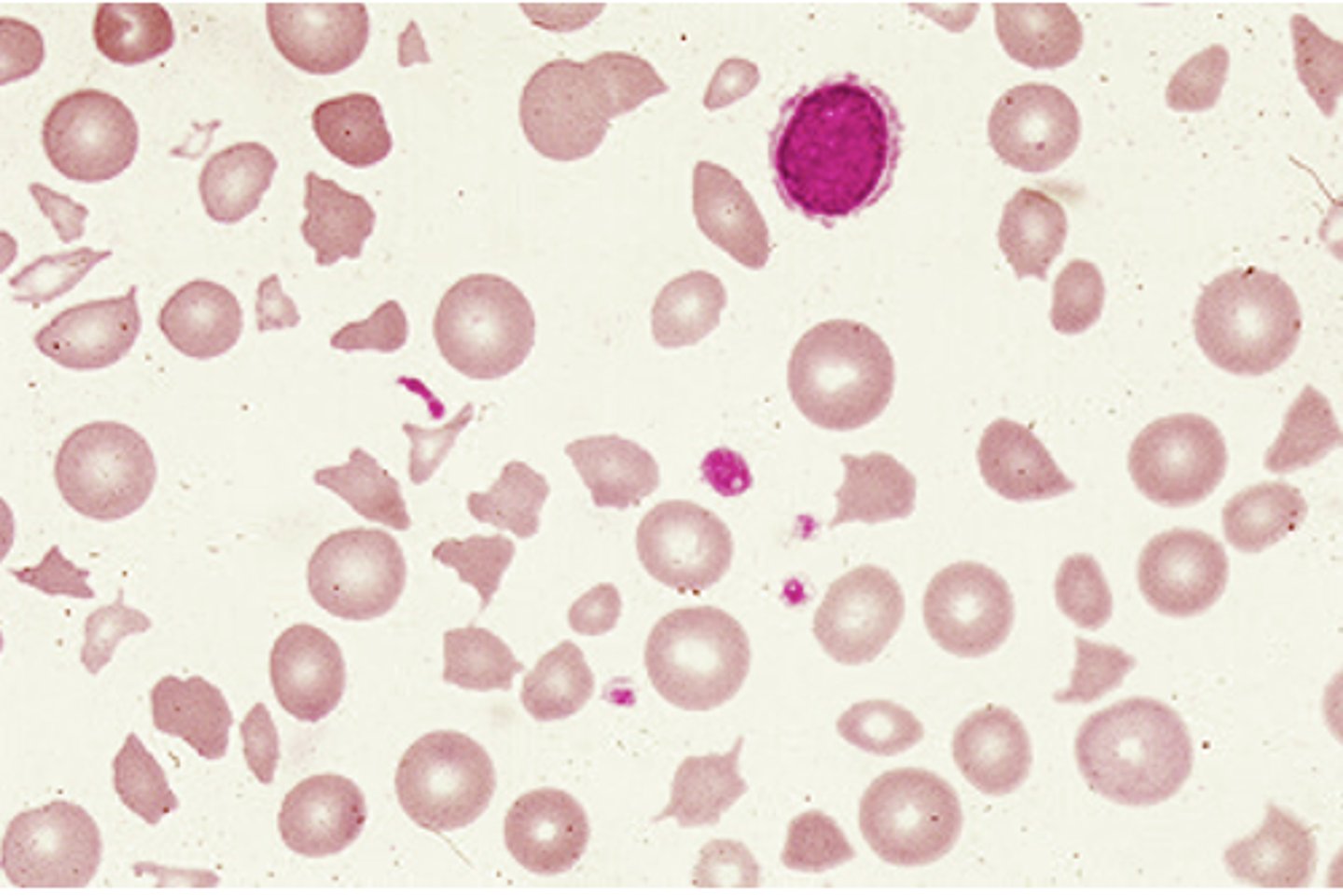
When are Helmet cells and red blood cell fragments (schistocytes) seen?
Fragmented cells seen with DIC, TTP, Heart valves hemolysis, and severe burns
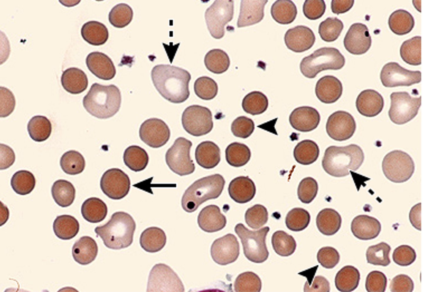
What are spherocytes?
Small, dense, hyperchromic red blood cells lacking the normal central pallor seen in hereditary spherocytosis
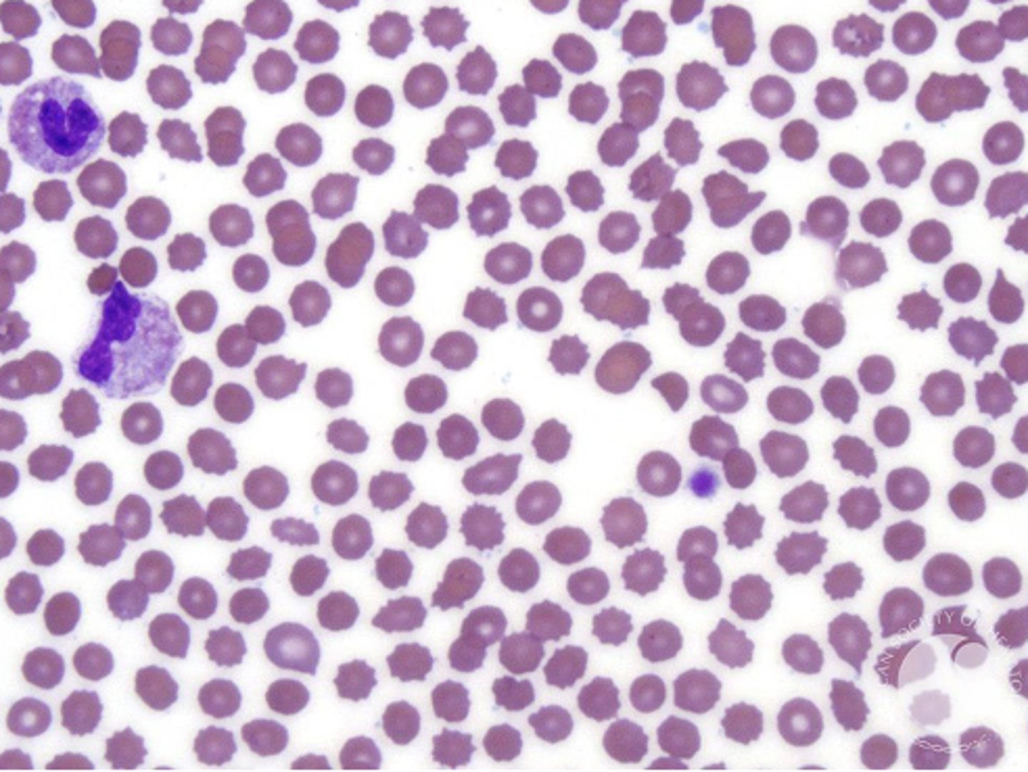
What are burr cells (echinocytes)?
Abnormal RBC membrane seen in metabolic disease like uremia or liver disease (can also be a lab error)
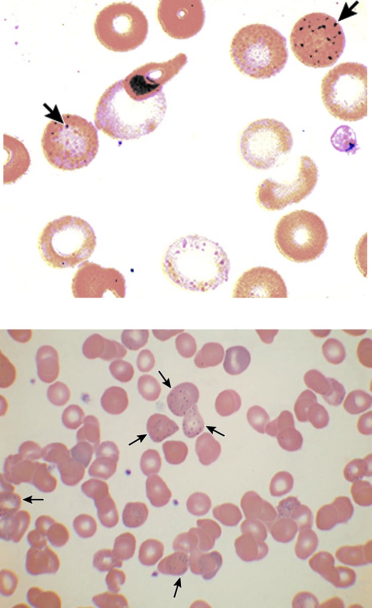
What can cause basophilic stippling?
Lead poisoning, Megaloblastic anemia, Thalassemias, and Alcohol related liver disease
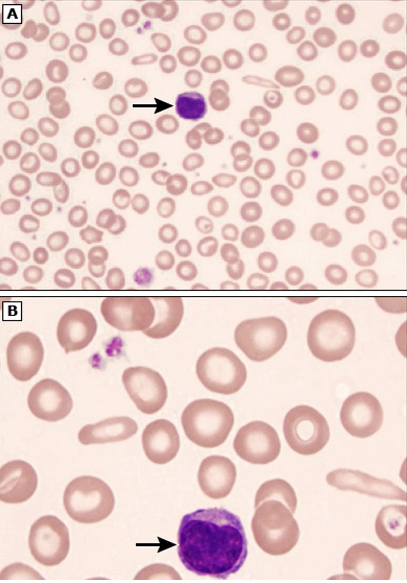
What blood results are seen with iron deficiency anemia?
- Microcytic (small) red blood cells
- Hypochromic (pink not red) RBC
- Anisocytosis (increased RDW)
- Target cells
When are target cells (Codocytes) seen?
Chronic liver disease, Thalassemia, Hemoglobin C, S, or D disease, Iron deficiency, Splenectomy
What are reticulocytes?
Immature RBCs with retained RNA
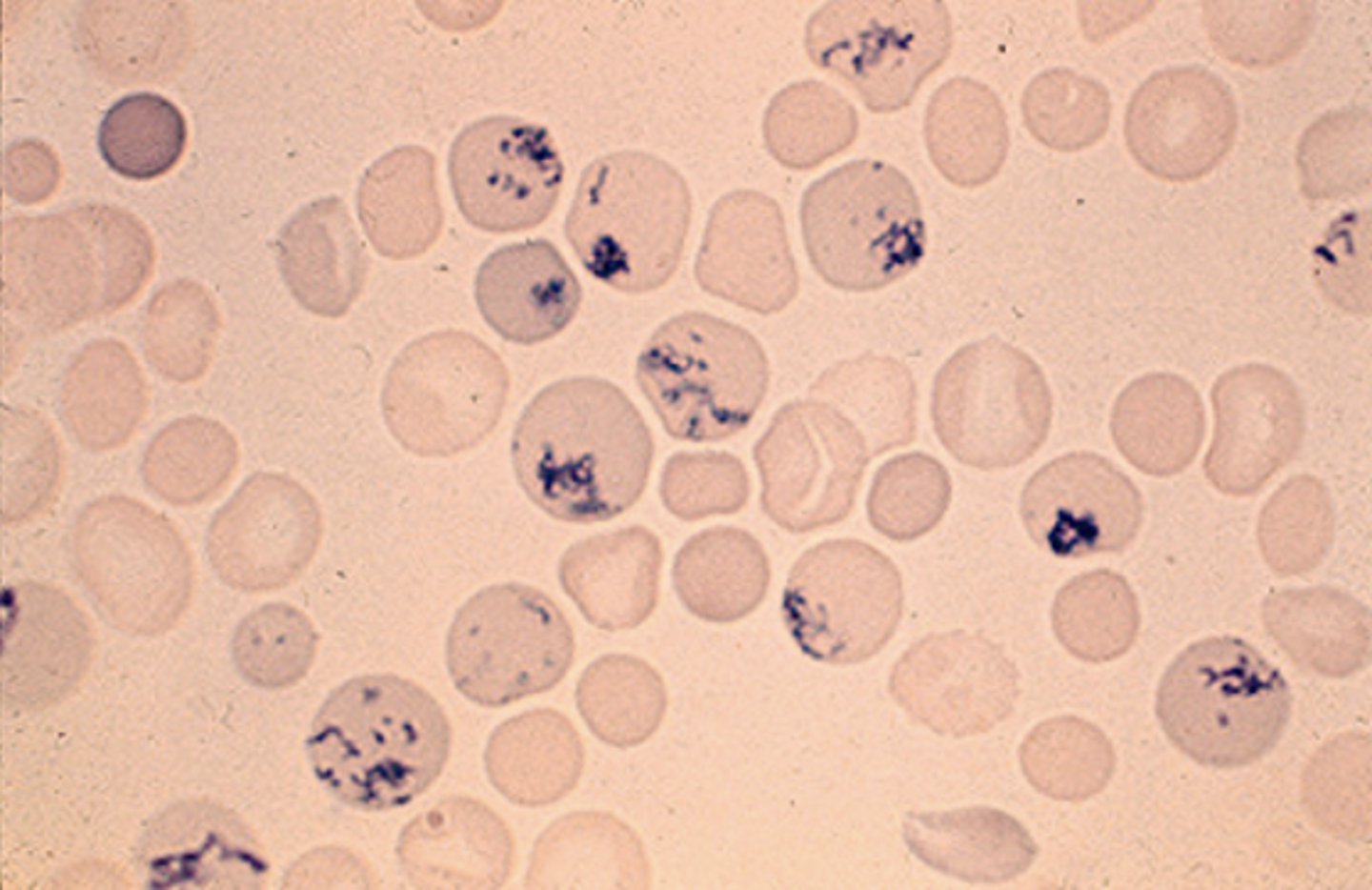
What are reticulocytes used for?
assess the effectiveness of erythropoiesis
When are reticulocytes decreased?
Dysfunctional bone marrow, B12/Folate deficiency, CKD, radiation, malignancy
When are reticulocytes increased?
trauma, chronic blood loss, hemolysis, erythropoietin, supplementation (Fe, B12, Folate), bone marrow transplant
What is an absolute reticulocyte count?
reported as an actual number per unit of blood that does not account for hematocrit or anemia
What is a reticulocyte percentage?
proportion of reticulocytes in circulation calculated by
(# of reticulocytes / RBC count) x 100
What is corrected reticulocyte count?
reticulocyte percentage x (patient's Hct / normal Hct)
What are the severity values for corrected reticulocyte count?
Normal: 1-2%
Mild-Mod: 3-5%
Severe: > 5-6%
What are tear drop cells (Dacrocytes)?
Single spicule with blunt end seen in myeloid metaplasia with myelofibrosis, Myelophthisic process, Thalassemia, Pernicious anemia

What are acanthocytes (spur cells)?
Irregularly spaced spicules of varying lengths seen in liver disease, hypothyroidism, Vit E deficiency, and splenectomy
What are sickle cells (Drepanocyte) seen?
Sickle shape, pointed at both ends seen in sickle cell anemia and other hemoglobinopathies
What are types of iron studies?
serum iron, TIBC, transferrin, transferrin saturation, and ferritin
What is the most sensitive test to detect iron deficiency?
Ferritin
What is Ferritin?
Marker of stored iron unaffected by recent iron intake and can be increased with acute inflammation
What is an important caveat about serum iron?
should NOT be used in isolation to diagnose iron deficiency/overload
What causes iron-deficiency anemia?
Acute/chronic blood loss, Chronic disease, and Low dietary intake
What causes iron overload?
Hemochromatosis and Excessive dietary intake or supplementation
What is total iron binding capacity (TIBC)?
measurement of all proteins available for binding iron (indirect measurement of transferrin and other iron binding proteins)
When is TIBC most commonly increased?
iron deficiency
What is transferrin?
Major iron-binding protein and upregulated when iron stores are low
When is Transferrin most commonly increased?
iron deficiency
What is transferrin saturation?
percentage of transferrin saturated with iron is calculated by dividing serum iron level by TIBC
How is transferrin saturation calculated?
(serum iron level / TIBC) x 100
When is transferrin saturation elevated?
iron overload
What is Hemaglobin electrophoresis used for?
Detects abnormal forms of Hgb (hemoglobinopathies) such as sickle cell anemia
What results are seen with Macrocytic (Megaloblastic) anemias?
- MCV >100 fL
- Mild leukopenia and/or thrombocytopenia
- Low reticulocyte count
- Hypersegmented neutrophils
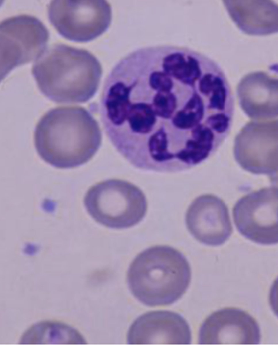
What are hypersegmented neutrophils?
Neutrophils with > 5 lobes
What are the varying cobalamin levels?
Normal: > 300 pg/mL
Borderline: 200 – 300 pg/mL
Low: < 200 pg/mL
What causes a low cobalamin level?
low vitamin B12 levels
What confirms a low cobalamin level due to low vitamin B12?
an elevated serum methylmalonic acid (MMA)
What is the most common cause of a folic acid deficiency?
inadequate dietary intake
What are the varying serum folate levels?
Normal: > 4 ng/mL
Borderline: 2 – 4 ng/mL
Low: < 2 ng/mL
What confirms a low serum folate level due to B12 and folate deficiency?
an elevated Homocysteine level
What causes low haptoglobin levels?
Hemolyzed Hgb is bound to haptoglobin intravascularly
What causes increased Lactate Dehydrogenase (LDH)?
Released with RBC destruction
What causes increased indirect (unconjugated) bilirubin?
Released with RBC destruction
What is a direct Coombs'/Direct antiglobulin test?
A positive test provides evidence for an immune etiology for the hemolysis
What is Porphyrias?
Multiple disorders based on enzyme defects in porphyrin metabolism (heme synthesis)
What are the most common porphyrias?
Porphyria cutanea tarda, Acute Intermittent Porphyria, Erythropoietic protoporphyria
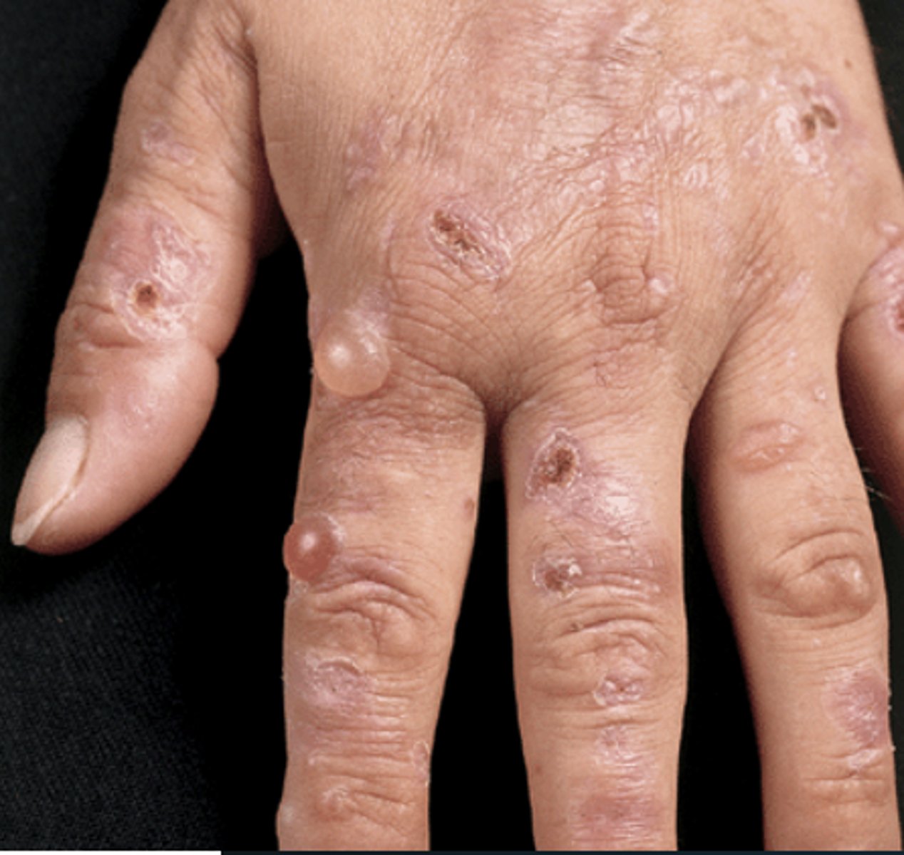
What is Porphyria Cutanea Tarda (PCT)?
Autosomal dominant disorder causing a deficiency of urobilinogen decarboxylase and associated with Hep C, AUD, and HIV
What are the symptoms of PCT?
Vesicles and bulla in dorsa of hands, scarring and small milia, hypertrichosis, mottled pigmentation