Key figures 2
1/7
There's no tags or description
Looks like no tags are added yet.
Name | Mastery | Learn | Test | Matching | Spaced |
|---|
No study sessions yet.
8 Terms
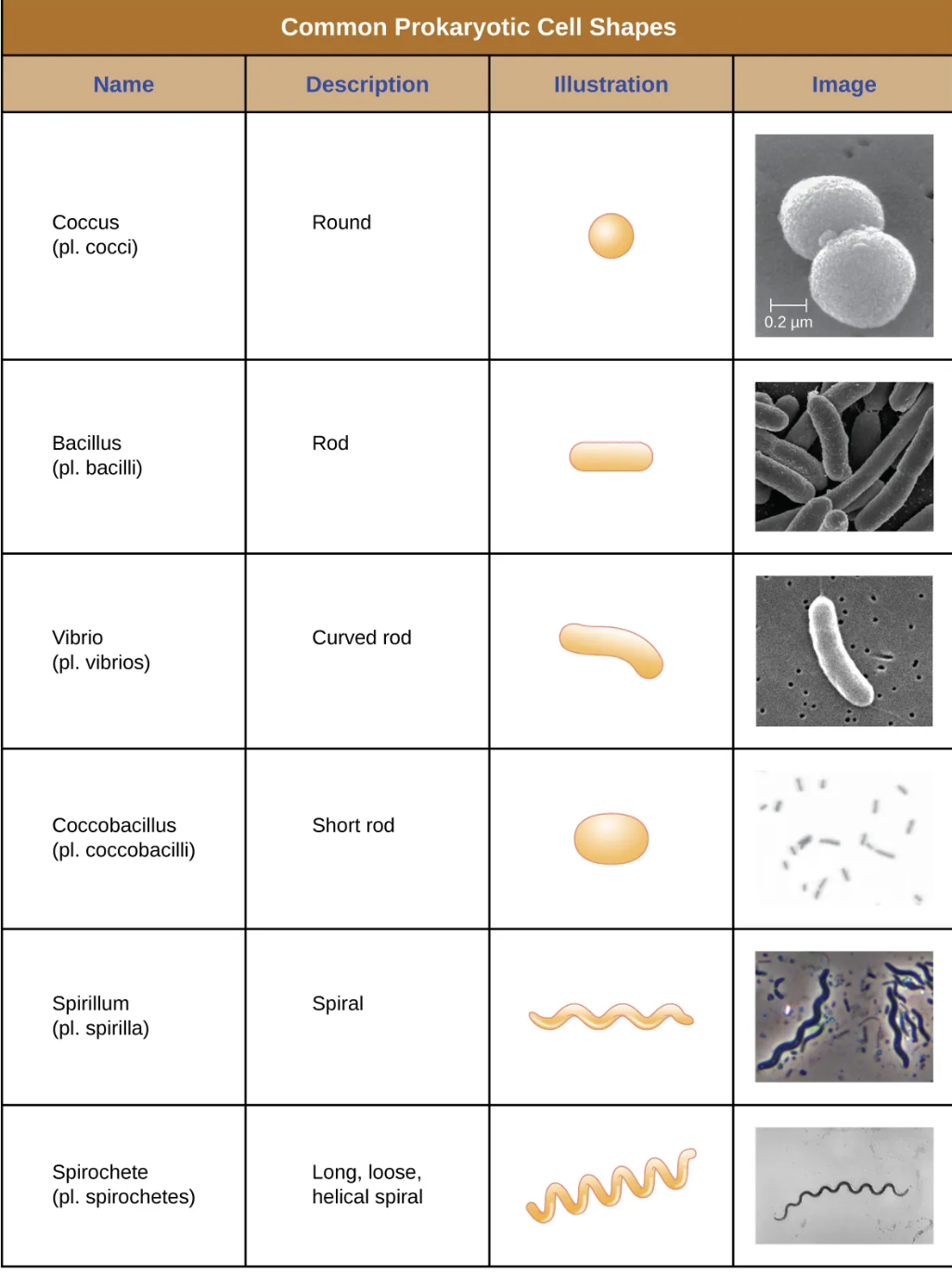
common prokaryotic cell shapes
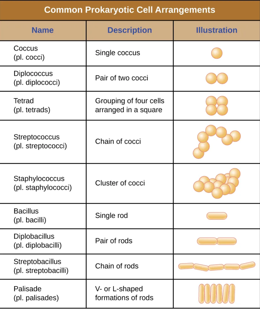
common prokaryotic cell arrangements
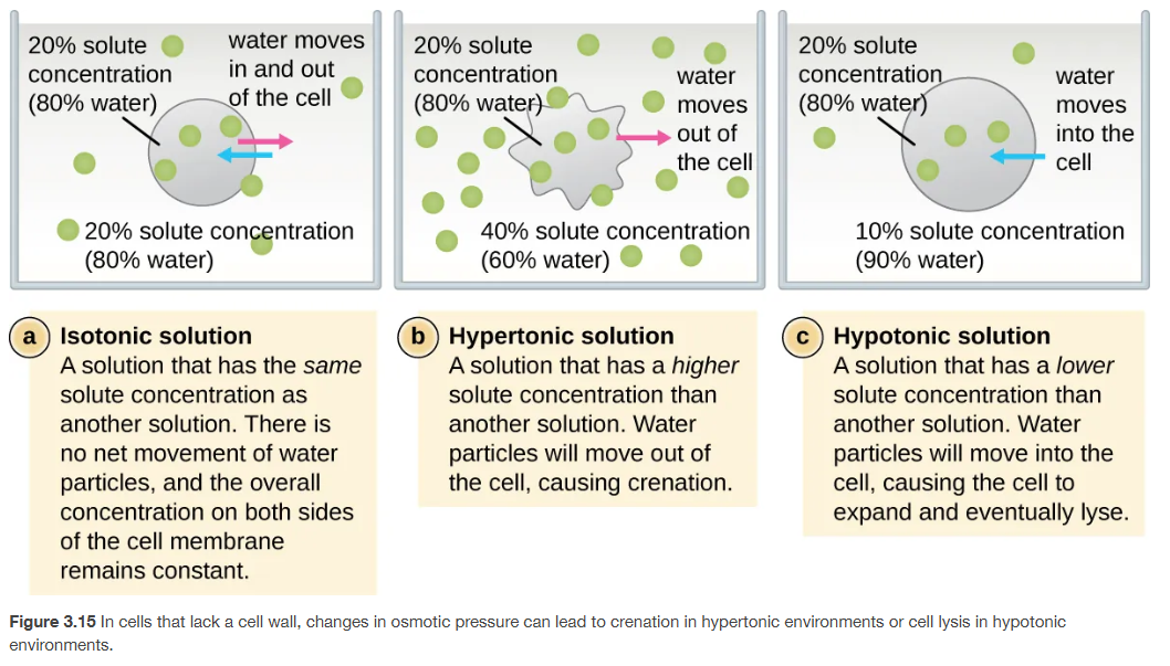
changes in osmotic pressure:
isotonic solutions, hypertonic solutions, and hypotonic solutions
Osmosis = diffusion of water across a semipermeable membrane.
Water moves from high water concentration (low solute) → low water concentration (high solute).
isotonic solution:
Definition: Solute concentration is the same inside and outside the cell.
Water movement: Equal movement in and out → no net movement.
Effect on cell: Cell stays the same size.
hypertonic solution
Definition: Solute concentration is higher outside the cell than inside.
Water movement: Water moves out of the cell.
Effect on cell: Cell shrinks (crenation in animal cells, plasmolysis in plant cells).
Example: Saltwater environment.
hypotonic solution
Definition: Solute concentration is lower outside the cell than inside.
Water movement: Water moves into the cell.
Effect on cell: Cell swells and may burst (lysis) if no cell wall is present.
Example: Freshwater environment.
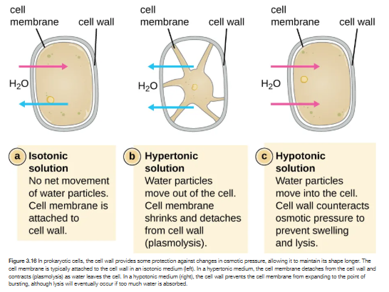
osmosis in prokaryotic cells (with cell walls)
The cell wall provides extra protection against osmotic pressure.
Unlike animal cells, bacteria (and plants) don’t easily burst in hypotonic solutions because the rigid cell wall counteracts swelling.
solutions in cells with cell walls:
isotonic Solution
Definition: Solute concentration is equal inside and outside.
Water movement: No net movement of water.
Effect on cell:
Cell membrane remains attached to the cell wall.
Cell maintains normal shape
Hypertonic solution
Definition: Solute concentration is higher outside the cell.
Water movement: Water moves out of the cell.
Effect on cell:
Cell shrinks.
Cell membrane pulls away from cell wall → plasmolysis.
Cell wall stays intact.
Hypotonic Solution
Definition: Solute concentration is lower outside the cell.
Water movement: Water moves into the cell.
Effect on cell:
Cell swells, but cell wall prevents bursting (lysis) by counteracting osmotic pressure.
If extreme, lysis may still occur if too much water is absorbed.
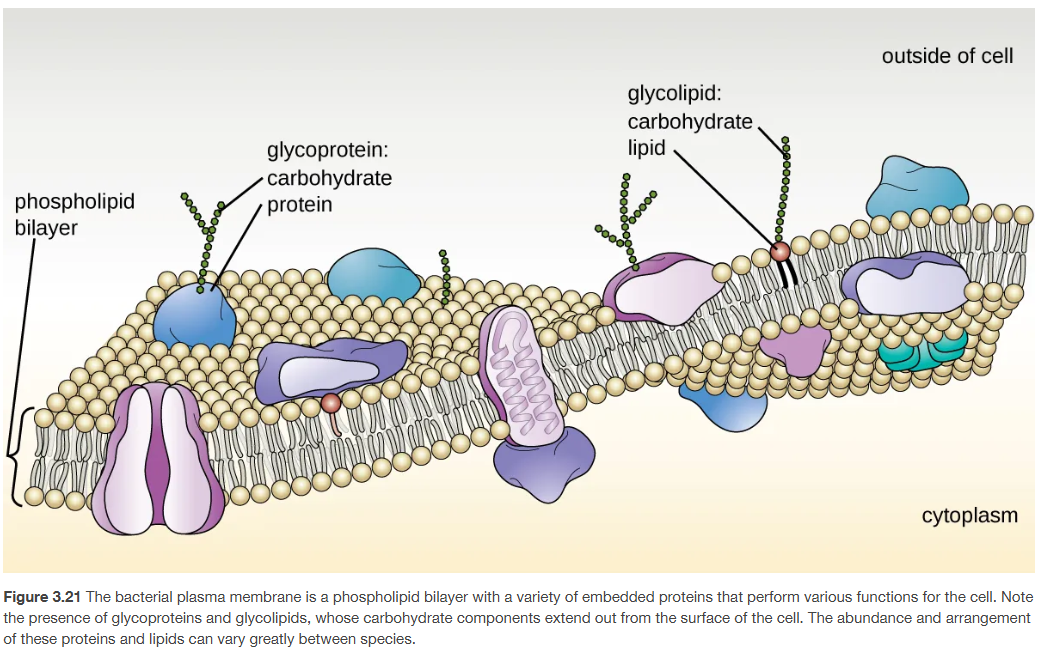
Bacterial plasma membrane
1. Structure: Fluid Mosaic Model
Phospholipid bilayer:
Hydrophilic heads (face outward).
Hydrophobic tails (face inward).
Creates a selective barrier between inside (cytoplasm) and outside environment.
Proteins:
Integral proteins: Span the membrane (transport channels, pumps).
Peripheral proteins: Loosely attached (signaling, structural support).
Carbohydrate attachments:
Glycoproteins (carbohydrate + protein).
Glycolipids (carbohydrate + lipid).
Extend outward from the cell surface.
2. Functions of the Plasma Membrane
Barrier: Separates cytoplasm from the external environment.
Selective permeability: Controls movement of molecules (nutrients in, waste out).
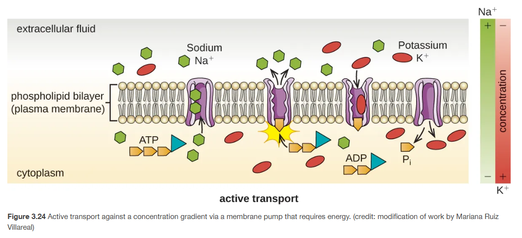
Transport:
Passive (diffusion, osmosis, facilitated diffusion).
Active (ATP-driven pumps, proton motive force).
Group translocation (chemical modification during entry).
Energy generation: Site of electron transport chain and ATP synthesis in bacteria (since they lack mitochondria).
Cell recognition & communication: Carbohydrate chains (glycolipids/glycoproteins) act as ID tags and receptors.
Anchoring site: For flagella, pili, and cytoskeletal elements.
Special Notes for Prokaryotes
Bacteria use the plasma membrane for many processes that eukaryotes perform in organelles (e.g., respiration, photosynthesis).
Archaea have unique membrane lipids (ether-linked, branched hydrocarbons) that provide stability in extreme environments.
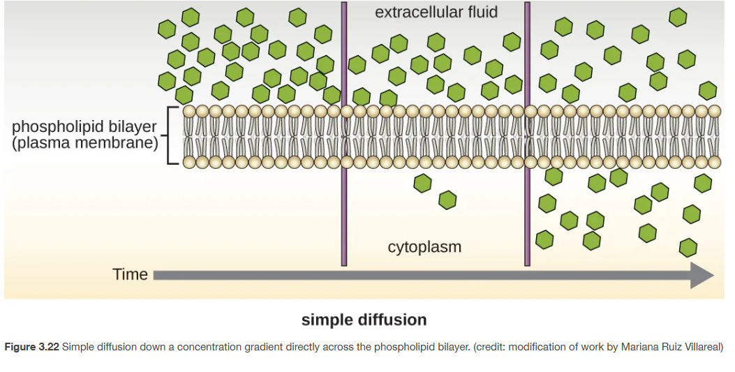
simple diffusion across plasma membrane
Simple diffusion: Passive movement of molecules directly across the phospholipid bilayer from an area of high concentration → low concentration.
Does not require energy (ATP).
Does not require transport proteins.
characteristics:
Driven by the concentration gradient until equilibrium is reached.
Only works for molecules that can freely cross the lipid bilayer:
Small, nonpolar molecules (O₂, CO₂).
Small lipid-soluble molecules.
Limited small uncharged polar molecules (H₂O, sometimes).
Large, charged, or polar molecules (e.g., glucose, ions) cannot pass by simple diffusion → they need facilitated diffusion or active transport.
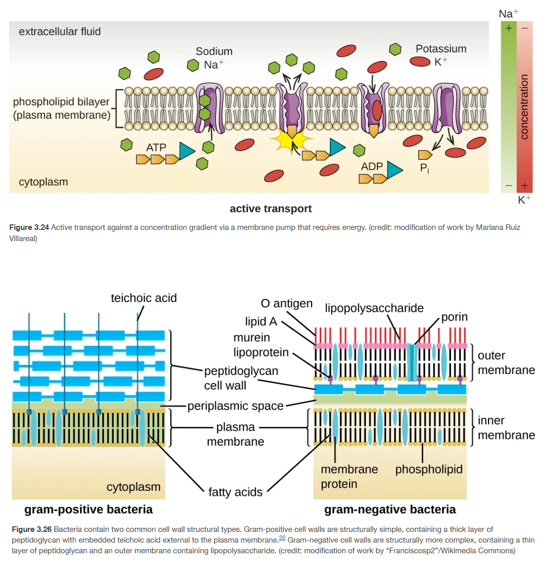
Gram-positive and gram-negative
Gram-positive:
thick peptidoglycan layer
contains teichoic acids and lipoteichoic acids (help with stability and ion regulation, also antigenic).
no outer membrane
staining - purple
periplasmic space - minimal/absent
antibiotic susceptibility - more sensitive to penicillin
gram-negative:
thin peptidoglycan layer
Surrounded by an outer membrane containing:
Lipopolysaccharides (LPS)
Porins (allow passage of small molecules)
periplasmic space - Prominet
staining - pink/red
antibiotic susceptibility - more resistant