Lecture 4
 Call Kai
 Learn
Learn Practice Test
 Spaced Repetition
 Match
Match Flashcards
 Knowt Play
1/14

There's no tags or description
Looks like no tags are added yet.
Name | Mastery | Learn | Test | Matching | Spaced | Call with Kai | Chat |
|---|
No analytics yet
Send a link to your students to track their progress
15 Terms
Expression Profiling Using Microarrays
A rapid, high-throughput technique used to measure the simultaneous expression levels of thousands of known genes across the genome.
Microarray Chip (Probes): Contains an arrayed grid of known single-stranded reference DNA sequences (probes) spotted by a robotic microarrayer. Probes represent standard genome sequences, not specific tissues.
Experimental Sample (Target cDNA): mRNA extracted from a tested tissue (e.g., brain or liver) and converted into single-stranded cDNA labelled with fluorescent dyes.
Hybridization: Labelled cDNA is applied to the chip, where complementary strands selectively hybridize to their target probe spots via standard base pairing.
Fluorescence Detection:
High Fluorescence Signal: Confirms high expression levels of that specific known gene in the tested tissue.
Low / No Signal: Confirms the gene is minimally expressed or silent in the tested tissue
Expression Profiling Using Microarrays (Two-colour comparison)
Two-Color Comparative Microarray (Dual Labelling): Directly compares expression between two samples (e.g., Green = Brain cDNA, Red = Liver cDNA).
Green Spot: Gene expressed primarily in Brain.
Red Spot: Gene expressed primarily in Liver.
Yellow Spot: Equal expression in both tissues (mix of Green and Red cDNA).
Shades of Orange/Lime: Expressed in both, but slanted toward whichever tissue has higher cDNA abundance.
Black Spot: Silent gene; not expressed in either tissue sample.
Global Genome/Proteome Regulation using Microarrays
Types of Microarray Probes:
ESTs (Expressed Sequence Tags): Short cDNA fragments corresponding to expressed genes.
Full ORFs (Open Reading Frames): Complete protein-coding gene sequences amplified by PCR; ideal for smaller genomes (e.g., yeast).
Oligo Chips: Short, chemically synthesized oligonucleotides (up to 70-mers) representing individual genes; ideal for complex, large genomes.
Yeast Expression Profiling Example:
Array Design: 6,116 yeast ORFs spotted onto a coated glass slide.
Dual-Color Labeling: Strain 1 cDNA labeled with Cy3 (Green); Strain 2 cDNA labeled with Cy5 (Red).
Readout: Green spots indicate genes upregulated in Strain 1; Red spots indicate genes upregulated in Strain 2; Yellow spots indicate equal expression across both strains.
Affymetrix Oligo Chips (GeneChips)
High-density silicon chips containing hundreds of thousands of short 25-mer oligonucleotide probes synthesized in situ.
Probe Design & Control (PM vs. MM):
Probe Cell: Contains ≈40×107 copies of a unique 25-mer single-stranded DNA probe.
Probe Pair (PM / MM): Consists of a Perfect Match (PM) probe and a Mismatch (MM) probe (single-base alteration in the center) to control for non-specific hybridization background.
Probe Set: Uses 11 probe pairs scattered across a single gene to yield robust signal measurement.
Glass Slide vs. Affymetrix Comparison:
Glass Slide Microarrays: Measure relative expression ratios between two comparative samples (Cy3 vs. Cy5) on a single slide.
Affymetrix GeneChips: Measure absolute gene expression levels for a single sample per chip using biotin-labelled cRNA stained with fluorescent streptavidin-PE.
Serial Analysis of Gene Expression (SAGE)
A digital, sequencing-based method used to profile global gene expression without pre-designed probes or prior gene sequence knowledge.
Key Concept: Isolates short 10–14 bp sequence tags from near the $3'$ poly(A) tail of mRNAs, which uniquely identify specific transcripts.
Concatemerization: Short tags are linked tail-to-tail into long DNA molecules (concatemers) so a single sequencing reaction reads dozens of transcript tags simultaneously.
Enzymatic Process:
Anchoring Enzyme (AE): A 4-base cutter that restricts cDNA into fragments anchored to streptavidin beads.
Tagging Enzyme (TE): A Type IIS restriction enzyme that cleaves 10–14 bp away from its binding site to release the signature tag.
Ditags & PCR: Tags are paired into ditags, PCR-amplified, released from linkers, and ligated into long chains for plasmid cloning and sequencing.
Quantification: Provides a direct digital count of transcript abundance, sensitive enough to detect low-abundance genes.
RNA Interference (RNAi)
A post-transcriptional gene silencing mechanism where short single-stranded RNAs guide protein complexes to degrade target mRNA or repress its translation.
Key Terms
miRNA (microRNA): Short (21–23 nt) non-coding RNAs.
siRNA (short interfering RNA): Short (21–27 bp) double-stranded RNAs.
Endogenous: Originating inside the cell/genome (e.g., miRNA genes).
Exogenous: Originating outside the cell (e.g., viruses or lab-introduced constructs).
Transient vs. Stable Knockdown: Transient is temporary (dilutes out as cells divide); Stable is permanent/long-term (DNA integrates into host genome).
Core Biological Machinery
Drosha: Cleaves primary miRNA (pri-miRNA) into pre-miRNA hairpins inside the nucleus.
Exportin 5: Translocates pre-miRNA from the nucleus to the cytoplasm.
DICER: Cytoplasmic RNase III enzyme that cuts dsRNA, pre-miRNA, or shRNA into 21–27 bp duplexes.
RISC (RNA-Induced Silencing Complex): Complex containing Argonaute (AGO) protein that discards the passenger strand and uses the guide strand to locate target mRNA.
miRNA vs. siRNA Comparison
Here is the standalone Markdown table for easy copying and pasting:
Feature | microRNA (miRNA) | Small Interfering RNA (siRNA) |
Origin | Endogenous (encoded in host genome) | Exogenous (viral or synthetic lab constructs) |
Binding Match | Partial Match (usually targets mRNA 3' UTR) | Extensive / Exact Match (100% complementary) |
Main Outcome | Translational Repression (blocks ribosomes) | mRNA Cleavage (Argonaute slices mRNA) |
Specificity | Broad (1 miRNA can regulate 100+ genes) | High (targets 1 specific gene) |
Experimental Formats & Delivery
Long dsRNA (200–500 bp): Processed naturally by Dicer in insect cells; must be cut in vitro into esiRNA for mammalian cells to avoid immune activation.
Synthetic siRNA: Short, chemically modified duplexes used for short-term (transient) knockdown.
shRNA (short hairpin RNA): Vector-expressed hairpins under U6/H1 promoters delivered via viruses (lentivirus) for long-term (stable) knockdown. Often paired with GFP or Puromycin selection markers.
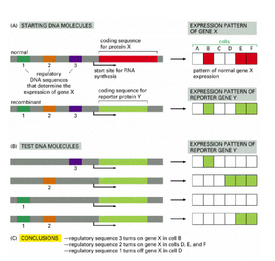
Reporter gene assays
A technique used to map out regulatory DNA elements (promoters and enhancers) and determine where, when, and how a specific gene is expressed.
Core Concept: Instead of tracking the original protein (Protein X), you swap out its coding region for a reporter gene (Reporter Y, such as GFP or LacZ) that produces an easily visible or measurable signal. Because Reporter Y is placed downstream of Gene X's regulatory DNA sequences (1, 2, and 3), it will be expressed in the same spatial pattern as Gene X.
How the Mapping Analysis Works (Section B & C)
By systematically deleting or testing individual regulatory regions, you can figure out what each specific regulatory sequence does:
Test Region 3 Only: Reporter Y turns ON only in Cell B.
Conclusion: Regulatory sequence 3 acts as an activator that turns on Gene X in cell B.
Test Region 2 Only: Reporter Y turns ON in Cells D, E, and F.
Conclusion: Regulatory sequence 2 acts as an activator that turns on Gene X in cells D, E, and F.
Test Region 1 Only: Reporter Y does not turn on anywhere.
Conclusion: Regulatory sequence 1 by itself cannot activate expression.
Test Region 1 + 2 Combined: Reporter Y turns ON in Cells E and F, but stays OFF in Cell D.
Conclusion: Regulatory sequence 1 acts as a repressor that turns off Gene X specifically in cell D.
DNA Sequencing (Sanger method)
Developed by Frederick Sanger in 1977; uses chain-terminating nucleotides to determine DNA sequence base-by-base.
Reaction Components:
Single-stranded denatured DNA template & complementary primer.
DNA Polymerase.
dNTPs (dATP, dCTP, dGTP, dTTP): Standard building blocks with a 3'-OH group required for strand elongation.
ddNTPs (ddATP, ddCTP, ddGTP, ddTTP): Chain-terminating nucleotides lacking an OH group at the 3’ end, instead having just an H. Cannot form a phosphodiester bond with the next nucleotide, causing immediate chain termination.
Classic 4-Reaction Protocol:
Reaction mixture split into 4 separate tubes (G, A, T, C), each receiving a small amount of one specific ddNTP alongside standard dNTPs.
Random incorporation of ddNTPs yields DNA fragments of every possible single-nucleotide length ending at that specific base.
Fragments are separated by size on a high-resolution polyacrylamide gel.
How to Read Sanger Sequencing Gels:
Direction: Read from bottom to top (5' to 3' direction of the newly synthesized strand).
Smallest Fragment: Positioned at the very bottom of the gel; represents the first nucleotide added after the primer (5' end).
Template Sequence: Deduced by converting the gel's 5' to 3' synthesized strand into its complementary sequence (3' to 5’).
DNA Sequencing Using Universal Vector Primers
Solves the problem of sequencing unknown DNA inserts without needing prior sequence knowledge to design primers.
Universal Binding Sites: Modern cloning plasmids (e.g.,
pCR2.1-TOPO) contain standardized, known sequences built into the plasmid backbone flanking the insertion site (e.g., M13 Forward and M13 Reverse primer sites).Mechanism:
An unknown DNA insert is cloned into the plasmid vector's Multiple Cloning Site (MCS).
Standard off-the-shelf primers (M13 Forward/Reverse) bind to the known plasmid backbone sequences outside the insert.
DNA polymerase exte
Automated Sanger Sequencing (Fluorescent Automated Sequencing)
Four Dyes, One Tube: Instead of running four separate reaction tubes (G, A, T, C), each of the four ddNTPs is tagged with a distinct fluorescent color (e.g., ddATP, ddTTP, ddGTP, ddCTP each glow a unique color). This allows all termination reactions to happen in a single tube.
Single-Lane Capillary Electrophoresis: The resulting DNA fragments are loaded into a single lane or capillary tube. As the fragments pass through, a laser excites the fluorescent tag at the end of each fragment.
Chromatogram Output: A detector (photomultiplier) reads the emitted light color as each fragment passes, generating a chromatogram with distinct color peaks representing the 5' to 3 DNA sequence.
Runs can produce sequences of up to 1000 bp.
Purpose of Primers in DNA Sequencing
Starting Anchor: DNA polymerase cannot synthesize DNA de novo; it requires a short double-stranded region with a free 3'-OH group provided by the primer to initiate synthesis.
Positioning: Directs DNA polymerase to begin reading precisely at the border of the target sequence.
Dye-Primer Method (Fluorescently Labeled Primers)
Mechanism: The 5' end of the primer is tagged with one of 4 distinct fluorescent dyes.
Reaction Setup: 4 separate tubes (G, A, T, C) are prepared, each containing a specific colour-tagged primer and one unlabeled ddNTP.
Pooling & Detection: After synthesis, all 4 reactions are combined into a single gel lane or capillary tube. Because the fragment's colour identifies which reaction tube it originated from, the laser reads the sequence directly by observing the colour order of passing bands.
Pyrosequencing
A real-time "sequencing-by-synthesis" method that detects light emitted every time DNA polymerase incorporates a complementary nucleotide.
Genomic DNA Fragmentation: Genomic DNA is sheared into short pieces (approx 400-500 bp) matching the machine's read length limit.
Emulsion PCR (Bead Prep):
DNA fragments are ligated to adaptors and bound to microscopic beads at a 1:1 ratio.
Beads are isolated inside individual water-in-oil droplets (micro-reactors containing PCR reagents).
PCR amplifies the DNA on the bead, coating it with 1 million identical copies of that single fragment.
Beads are deposited into individual micro-wells on a PicoTiter plate (one fragment per well).
Enzymatic Light Cascade:
Nucleotide IncorporationPolymerasePPiSulfurylase + APSATPLuciferase + LuciferinLight Flash
Role of Apyrase: Phosphatase enzyme that degrades unreacted nucleotides and excess ATP between washes to reset the well for the next flow.
Pyrogram Output:
1x Peak Height: Single base incorporation.
2x/3x Peak Height: Consecutive identical bases (homopolymers, e.g.,
GGorGGG).Flat Line: Non-complementary nucleotide flush (no binding).
Reconstructing the Genome (Assembly): Because millions of genome copies are randomly broken at staggered points, computer algorithms compare the overlapping 5' and 3' ends of short reads from all wells to align and stitch them back into the original genomic sequence (de novo assembly or reference mapping).
Shotgun Sequencing
A strategy where long target DNA or entire genomes are randomly sheared into small fragments, sequenced individually, and computationally reassembled using overlapping sequence ends.
Definition: A continuous, reconstructed stretch of genomic DNA formed by merging overlapping sequence reads.
Assembly Process: Because millions of genome copies are randomly sheared at different locations, the fragments break with staggered, overlapping ends. Bioinformatic algorithms scan the 5' and 3' ends of all short reads to identify matching overlaps and stitch them back together (de novo assembly or reference mapping).
Hierarchical Shotgun Approach (Clone-by-Clone):
Strategy: "Map-first, sequence-later".
The genome is first broken into large, mapped library clones (such as BACs).
Each large clone is individually fragmented, sequenced, and assembled before ordering them across chromosomes.
Highly organized and accurate for repetitive DNA, but slower.
Whole-Genome Shotgun Approach (WGS):
Strategy: "Blow it all up at once".
Skips physical mapping entirely; shears the entire genome into millions of small fragments simultaneously.
All fragments are sequenced in parallel and assembled globally using high-power bioinformatic algorithms.
Faster and more cost-effective, but requires heavy computational power.