Dental Radiology
1/112
There's no tags or description
Looks like no tags are added yet.
Name | Mastery | Learn | Test | Matching | Spaced |
|---|
No study sessions yet.
113 Terms
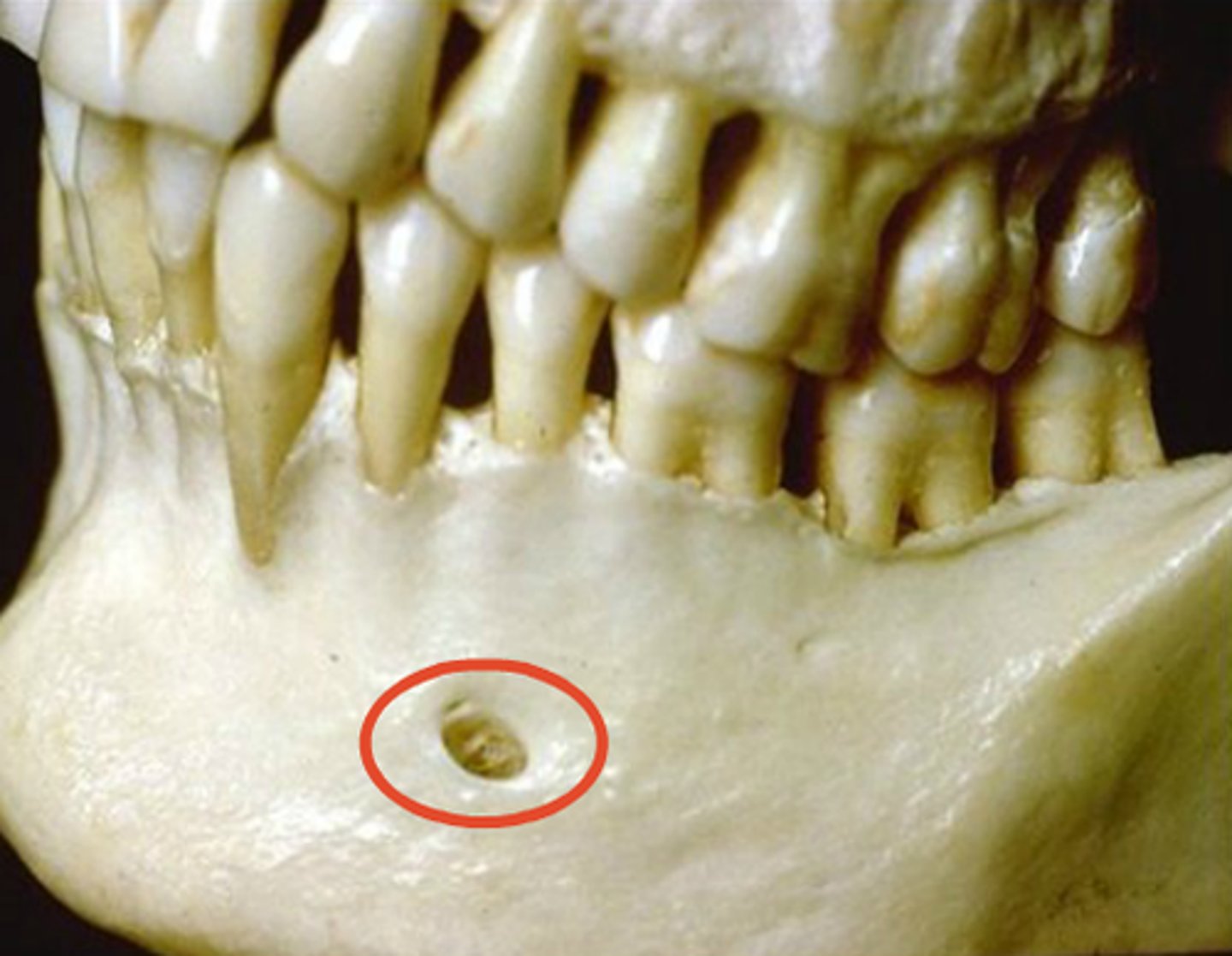
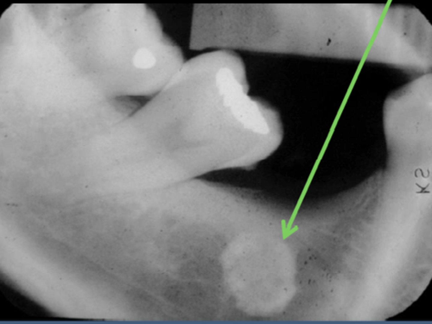
Genial Tubercles
Radiopaque
Lingual surface of mandible at mid-line
Note: Small dark spot, radiolucency, is the LINGUAL FORAMEN
Name structure that arrow is pointing too and what the radiolucent dot is inside?
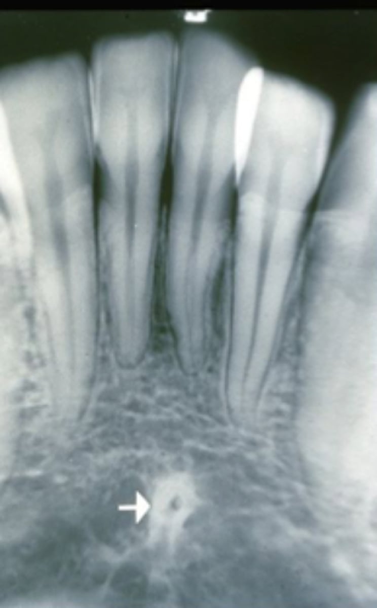
Genial Tubercles aka Mental Spines
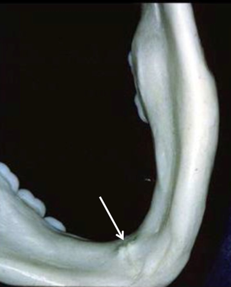
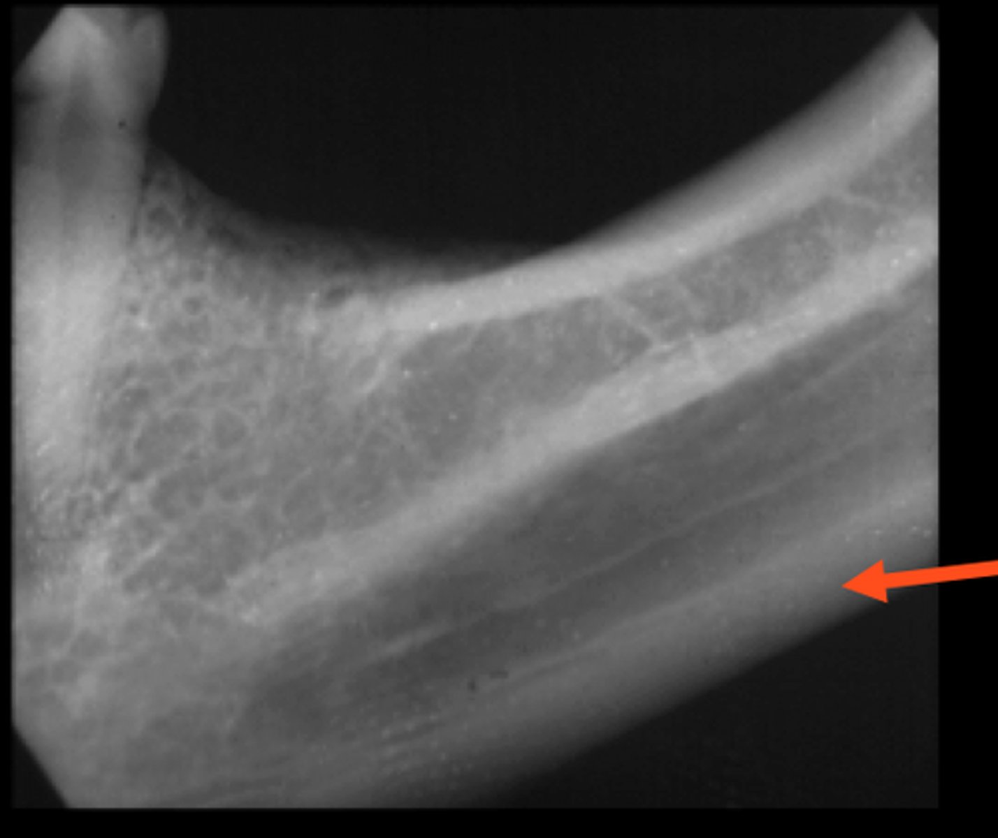
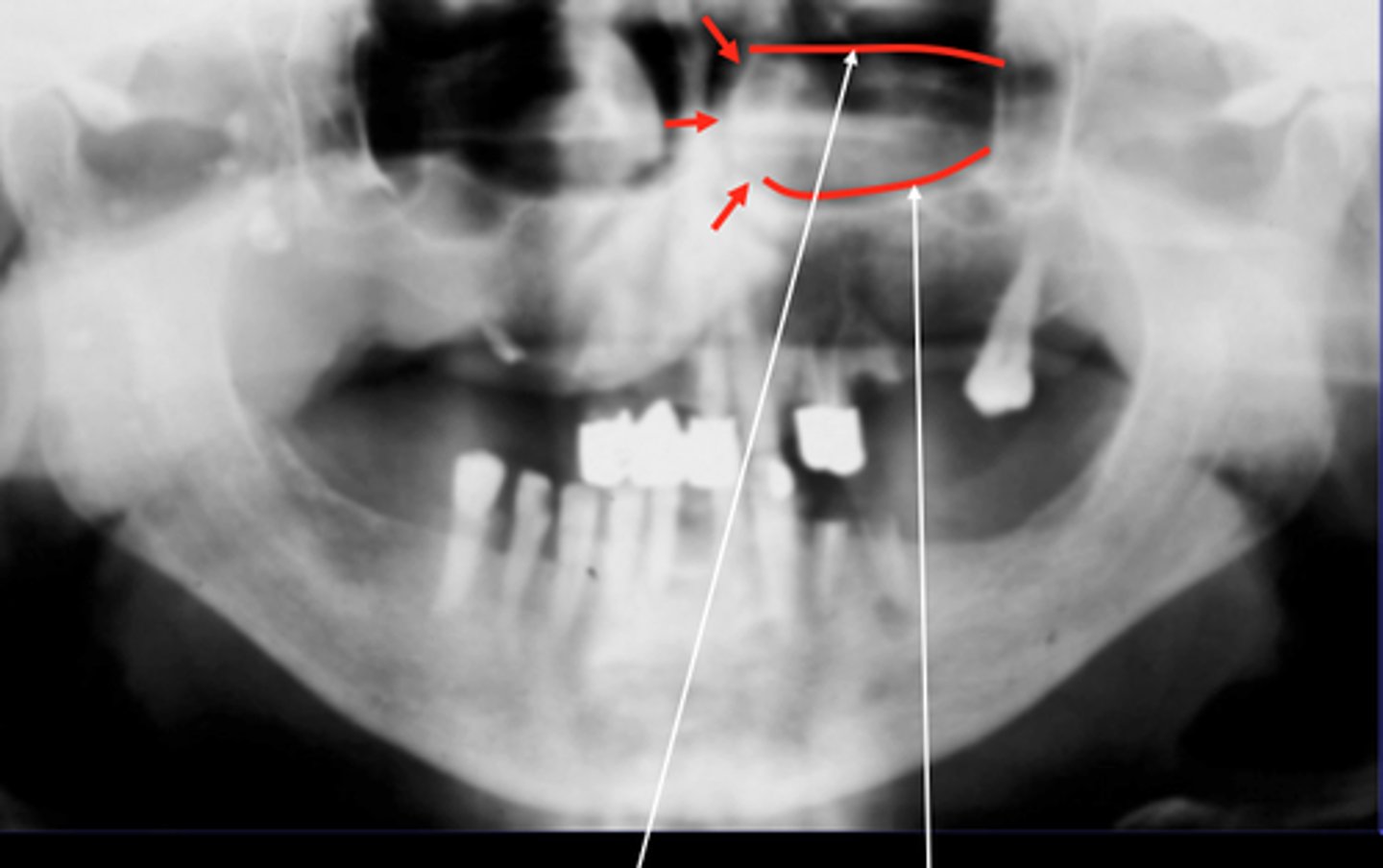
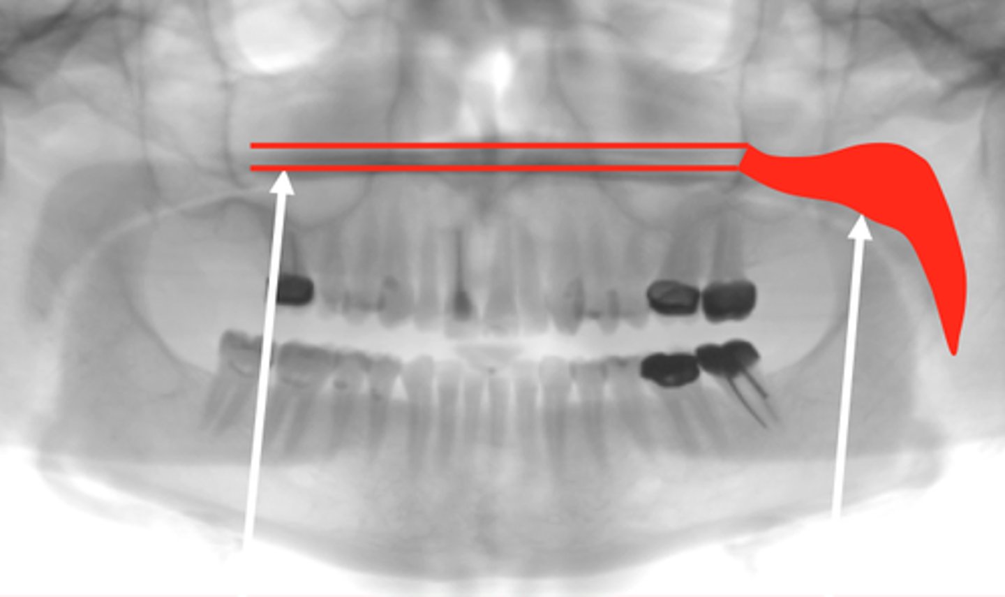
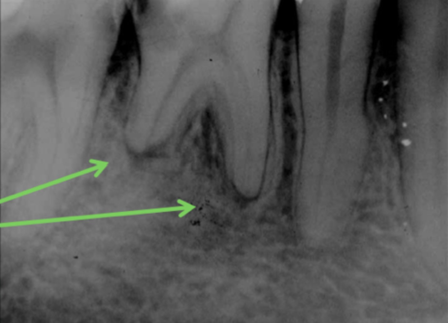
Mylohyoid Ridge
Landmark for molars.
Blends into internal oblique ridge.
Radioopaque
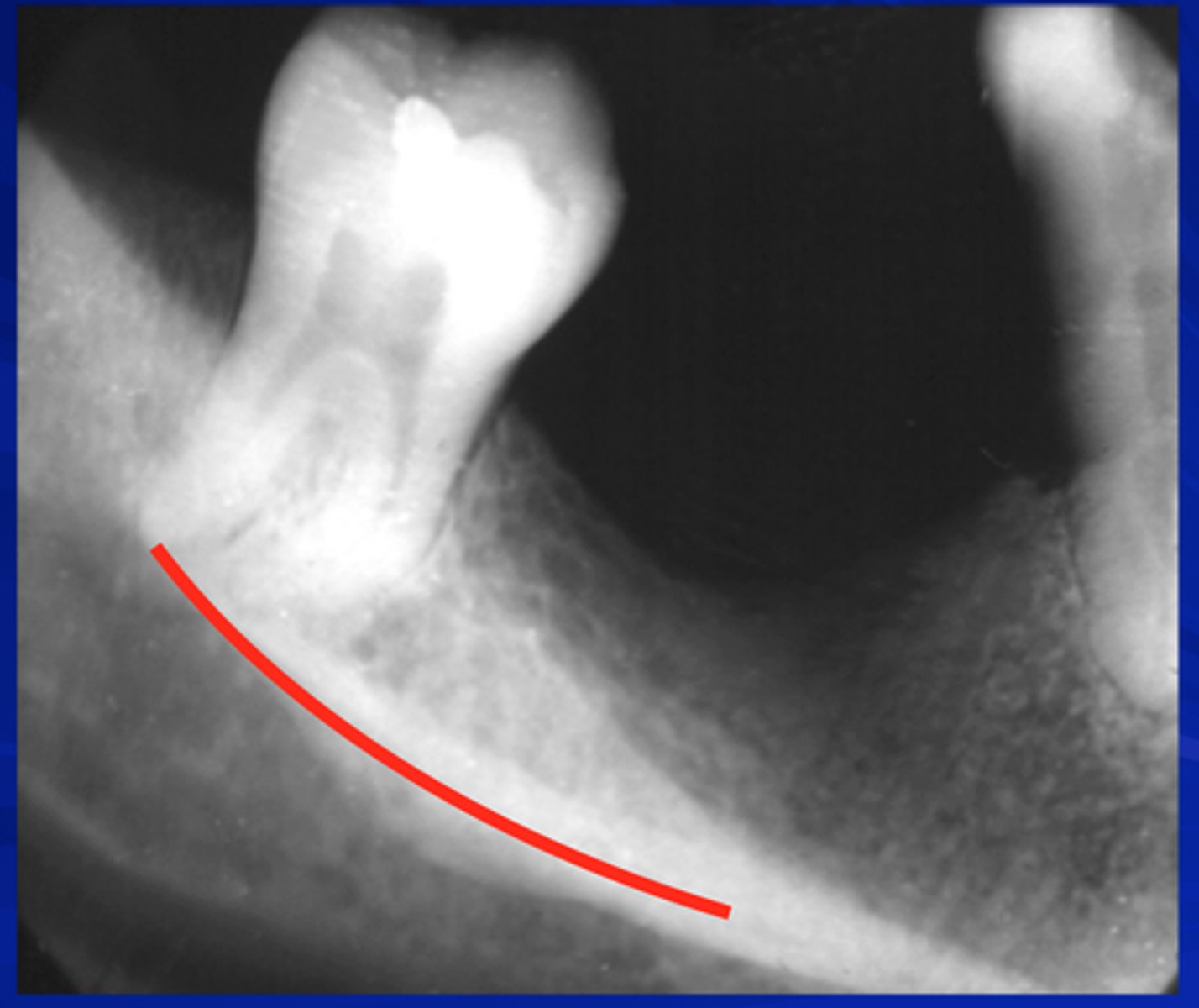
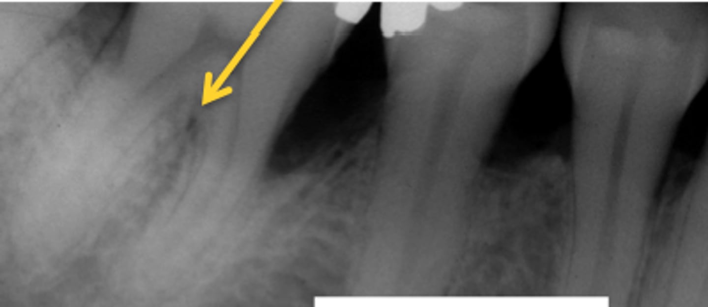
Submandibular Gland Fossa
Radiolucent space below the mylohyoid ridge.
Name structure that is boxed by the dotted red line.
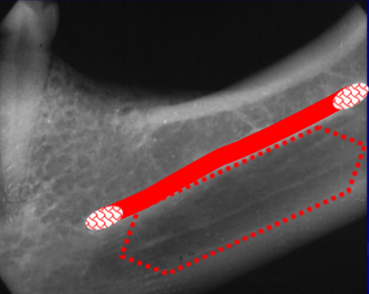
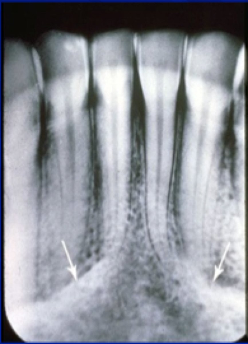
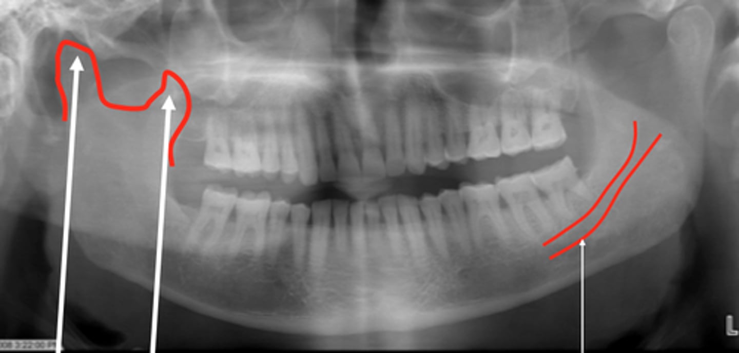
The smaller arrow: Mylohyoid Ridge
The larger arrow: Submandibular Gland Fossa
Arrow on left (small)?
Arrow on right (large)?
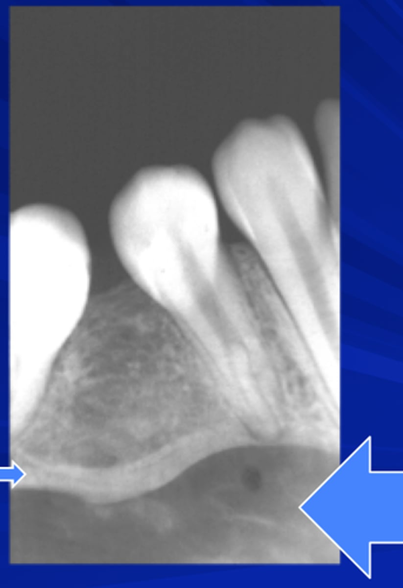
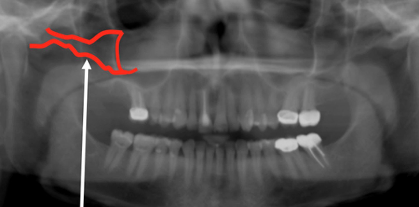
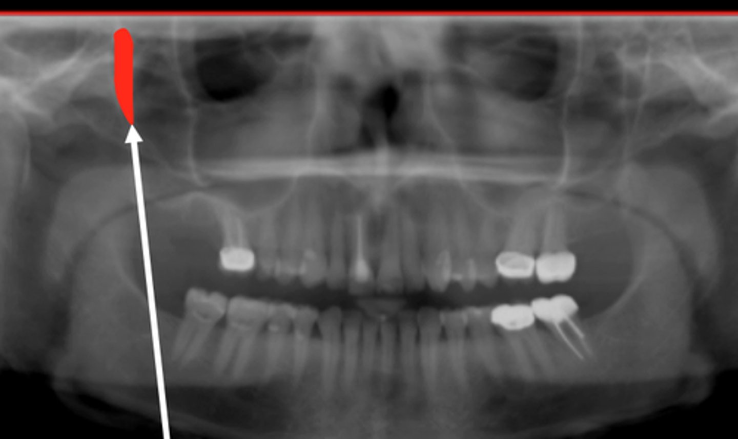
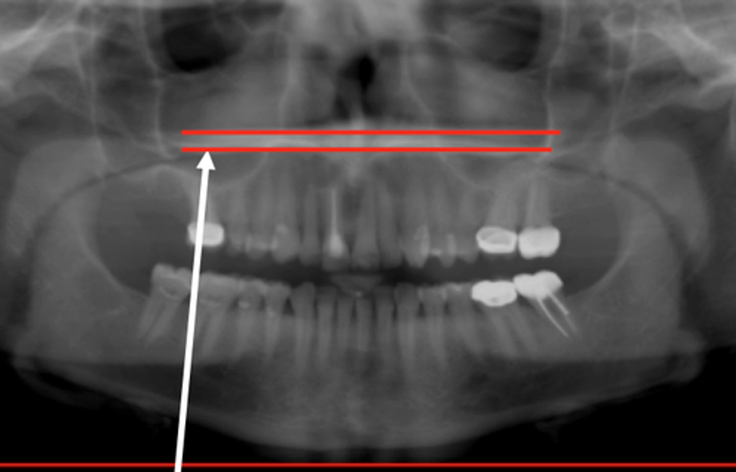
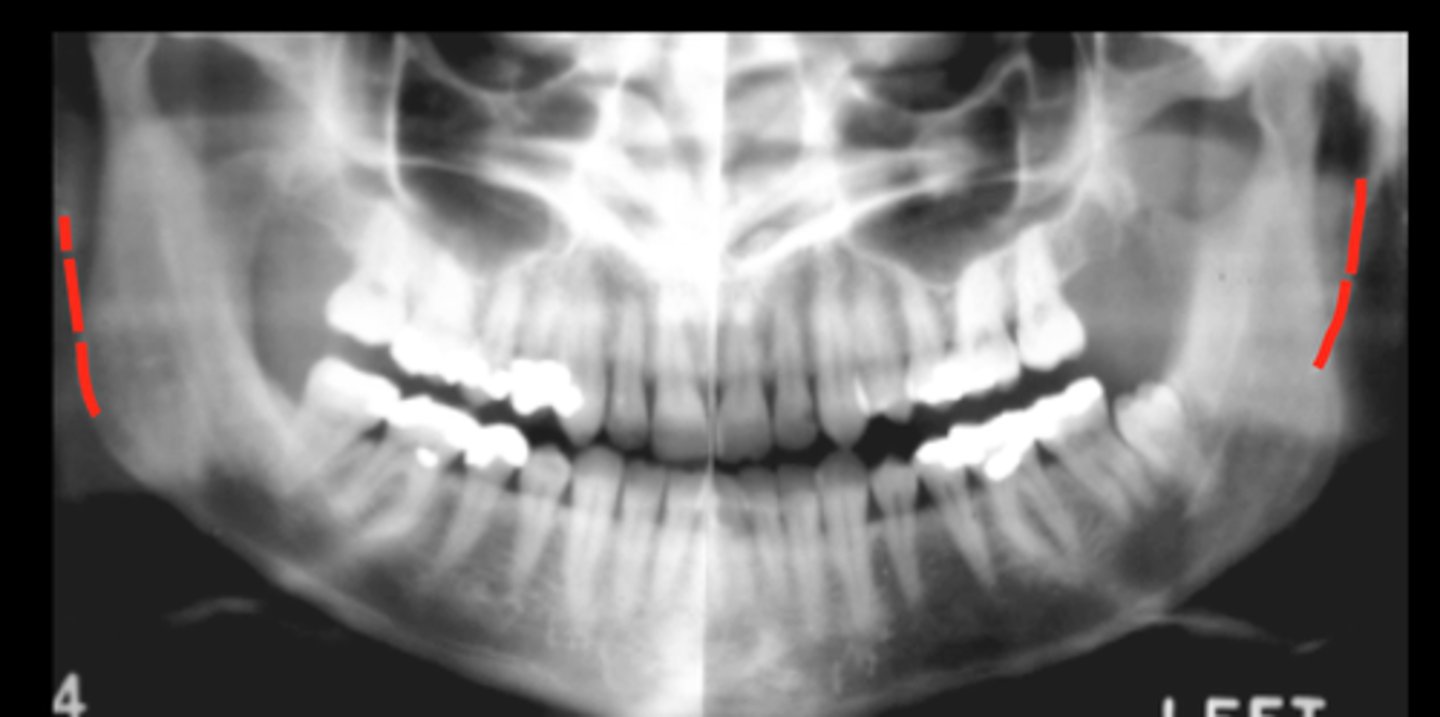
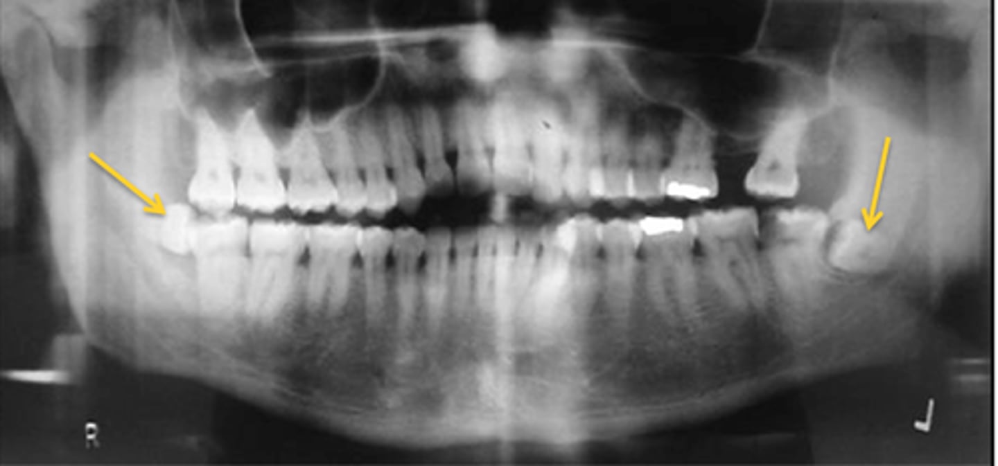
External Oblique Ridge
External Oblique Ridge
Looks like a Chisel going anteriorly
Mylohyoid Ridge
Top line (white arrow)?
Bottom line (Blue arrow)?
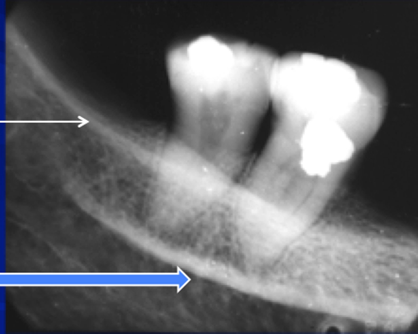
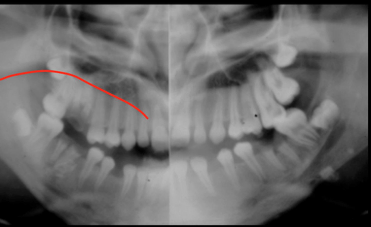
Inferior Alveolar Canal
Extends from mandibular foramen on medial a sect of mandible to mental foramen.
Note: If one border missing, its the Sup Border

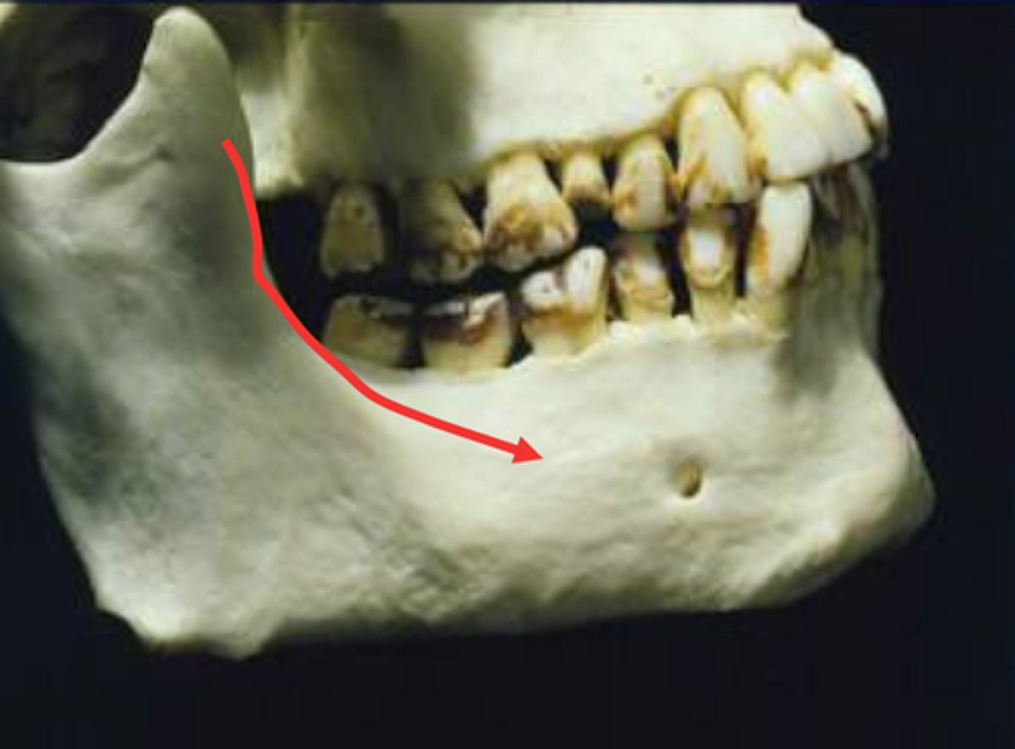
Nutrient Canals
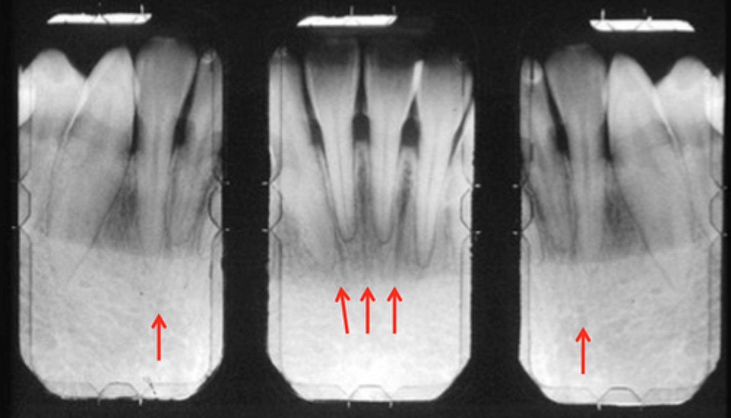
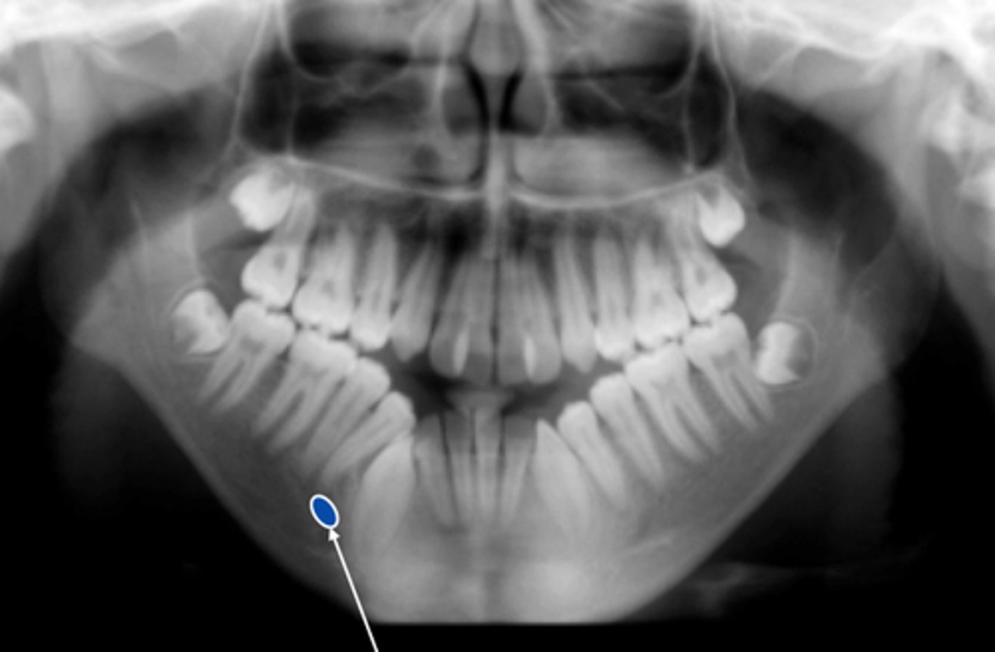
Mental Triangle
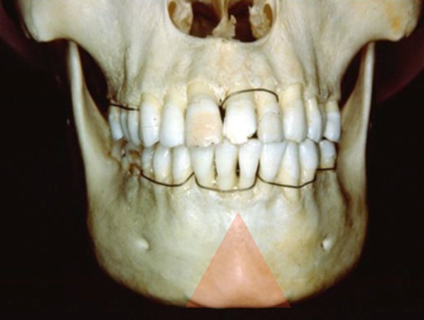
Mental Triangle
Mental Formen
Below the 2nd premolar tooth.
Opens in a post/sup direction
Mental Formen
Below the 2nd premolar tooth on buccal surface.
Inferior Border of Mandible
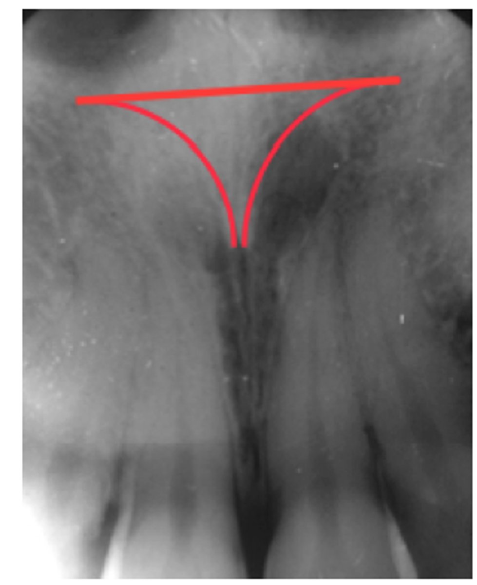
1) Incisive Foramen
2) Midpalantine Suture
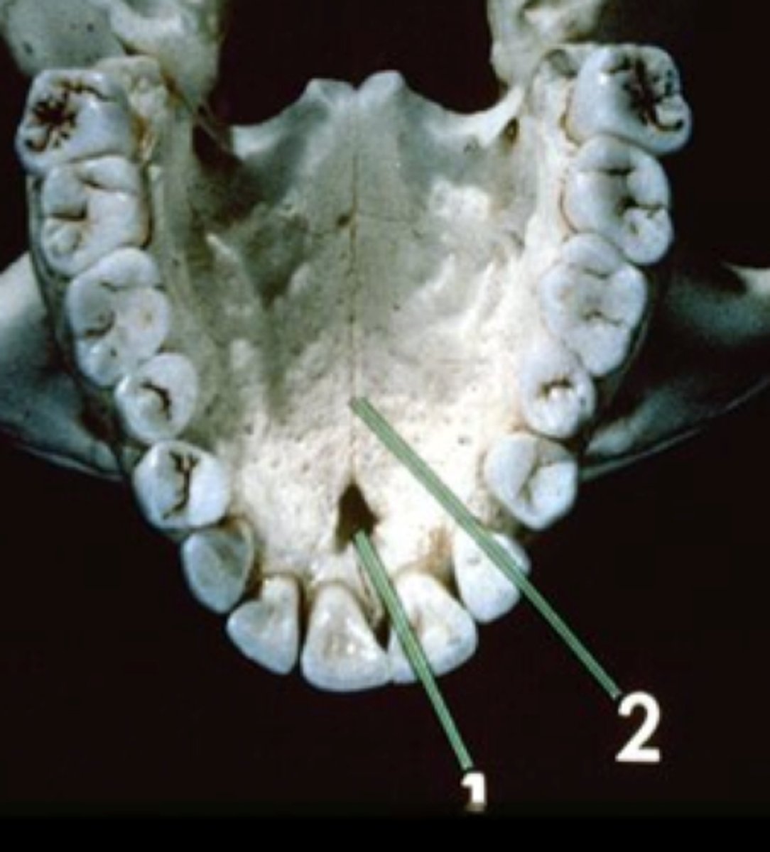
Midpalantine Suture
Suture between 2 palatal processes of Maxilla
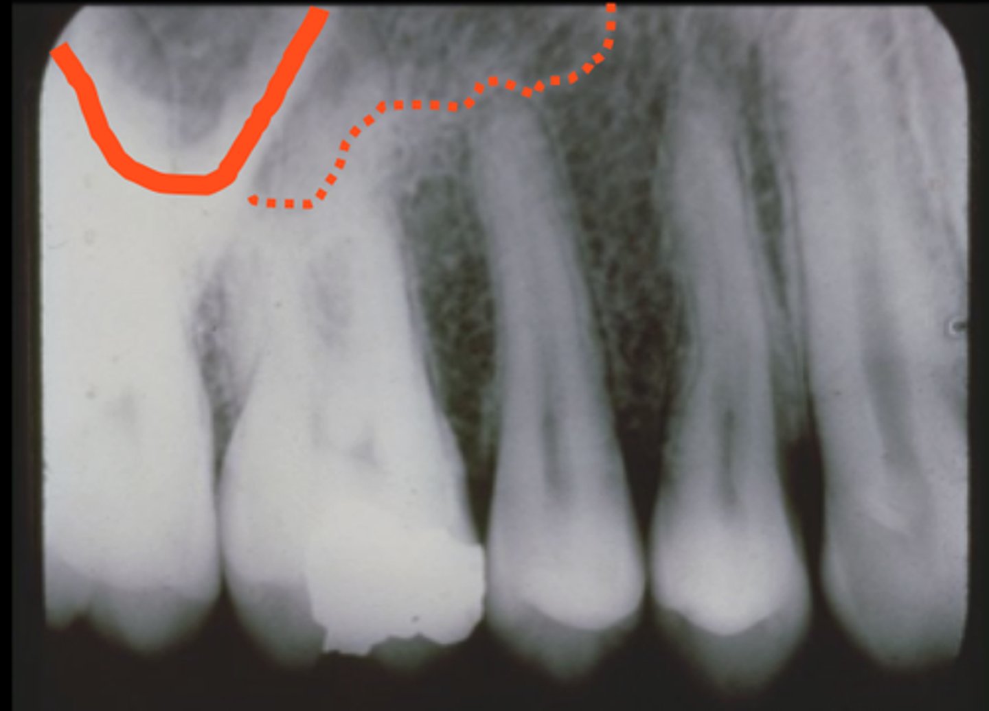
Zygomatic Process of Maxilla (Buttress)
Identify the radiopaque structure
Zygomatic Bone
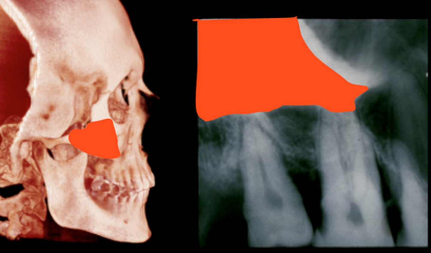
Dotted Line: Floor of Maxillary Sinus
Thick Line: Zygomatic Process of Maxilla
The difference b/w the 2 is the thickness!!!
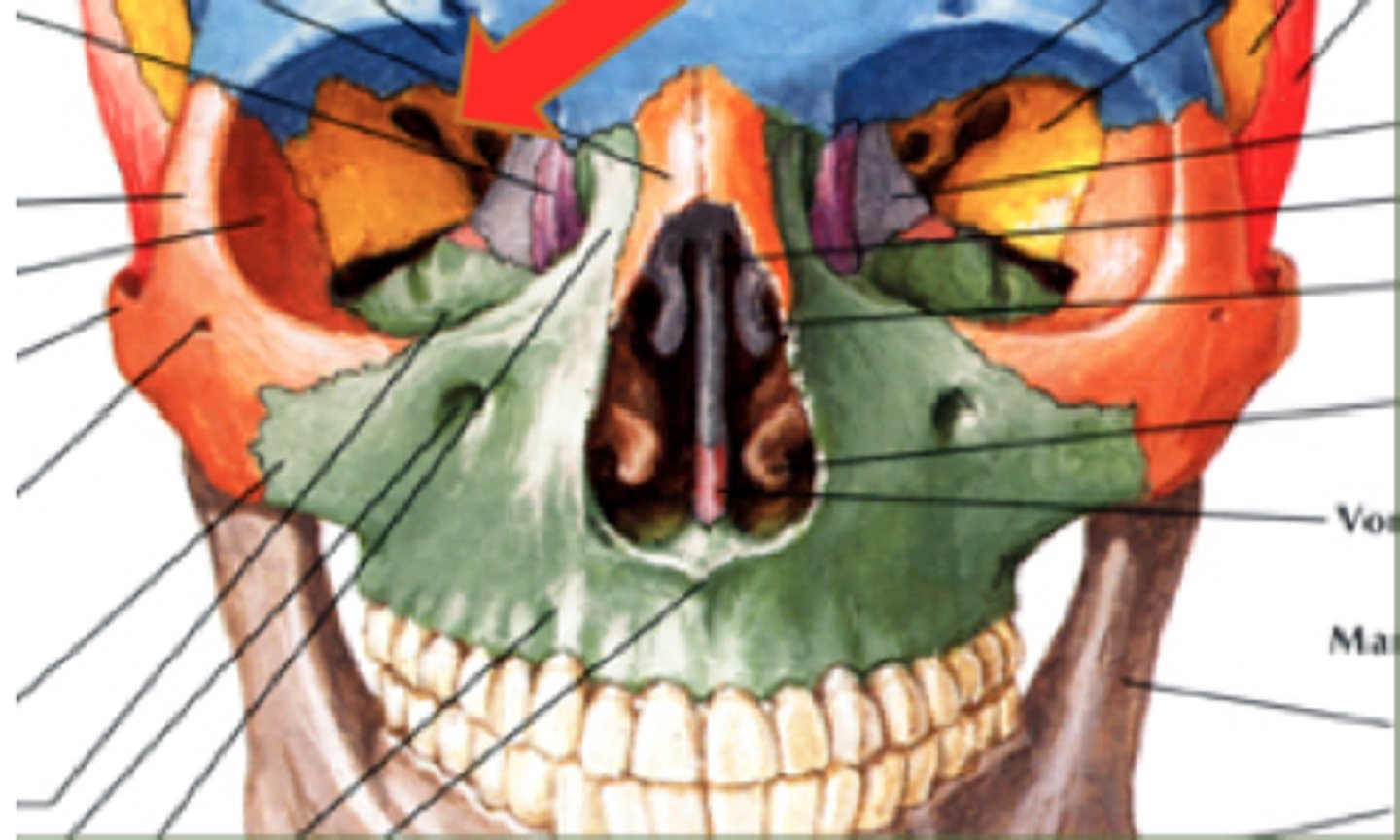
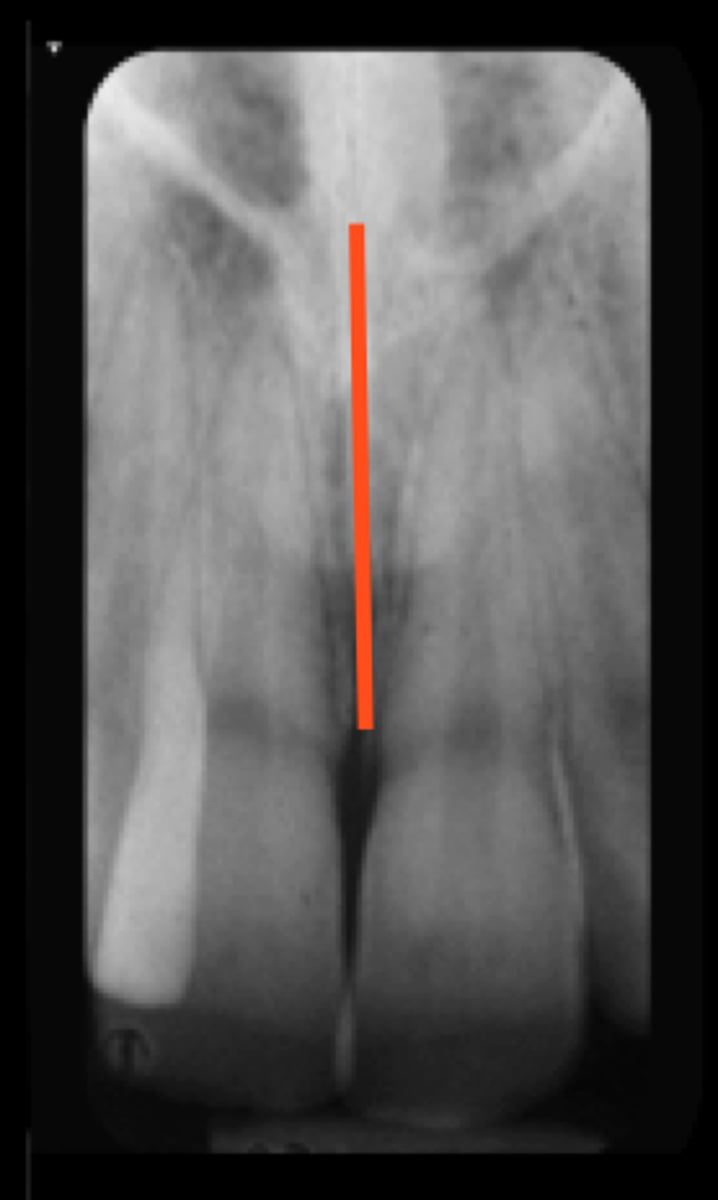
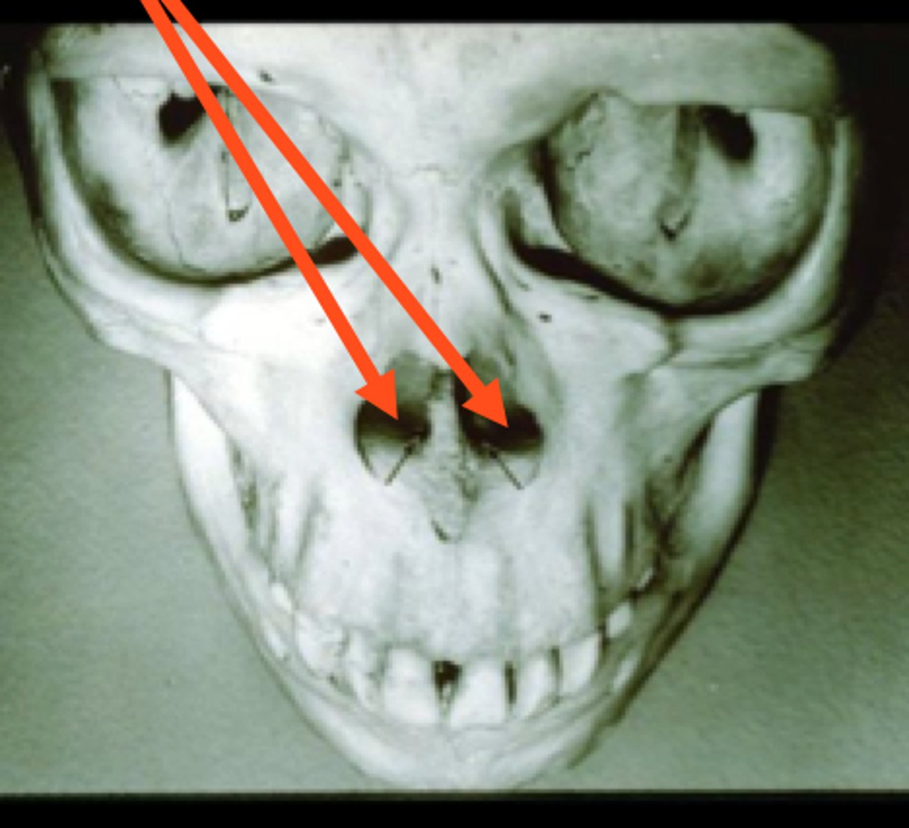
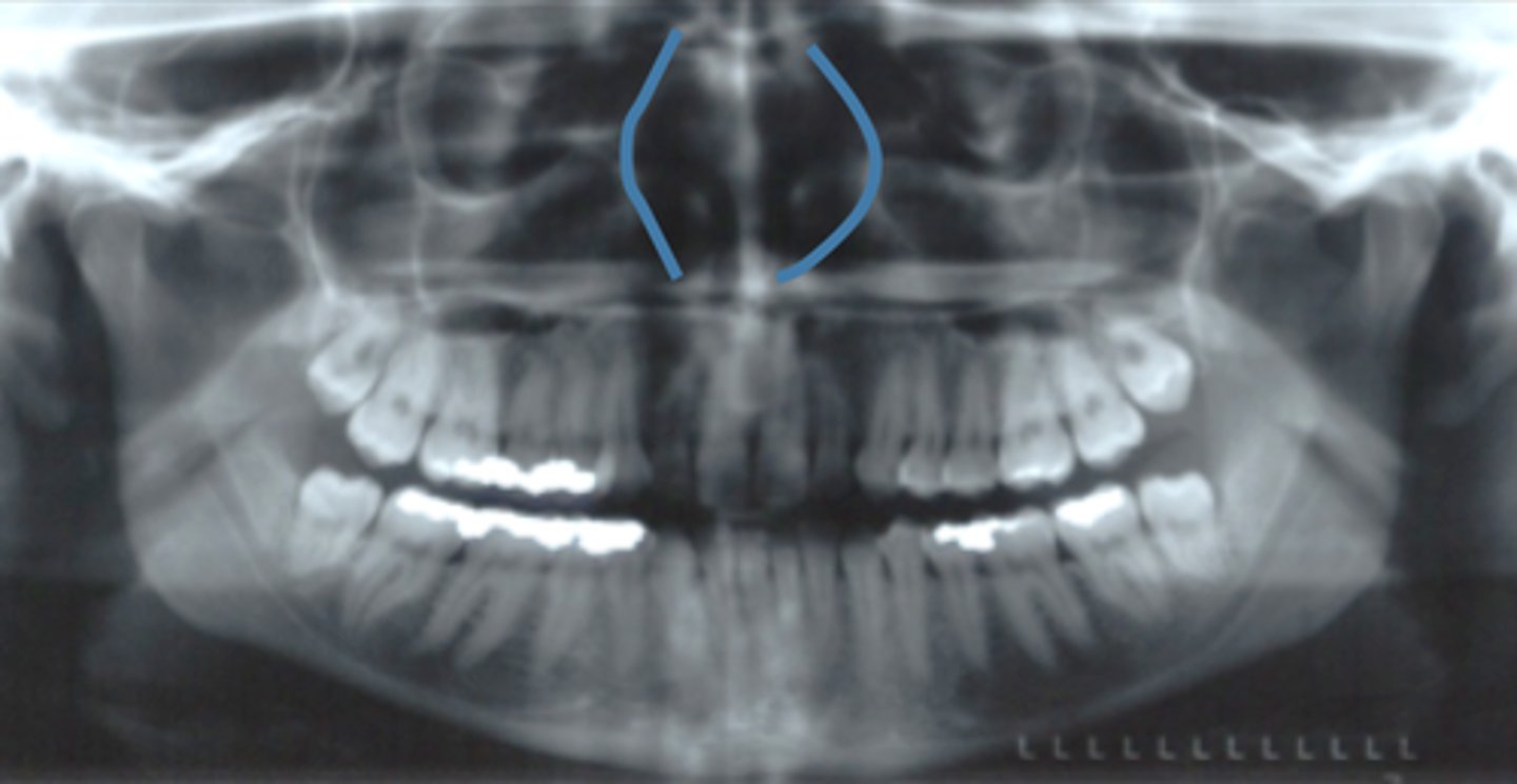
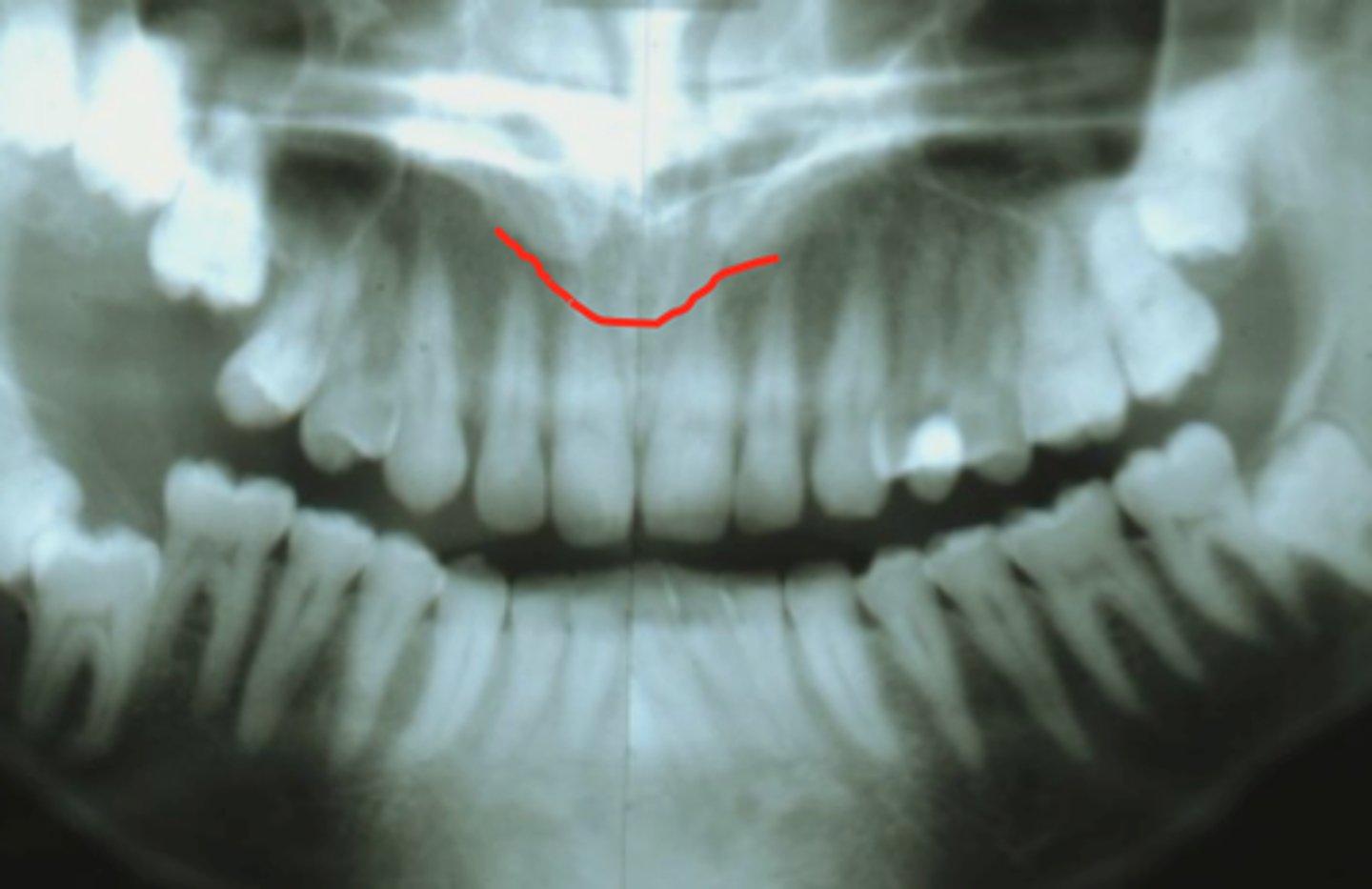
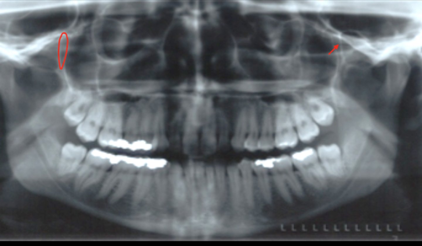
Nasal Fossa
The NASAL SEPTUM separates them.
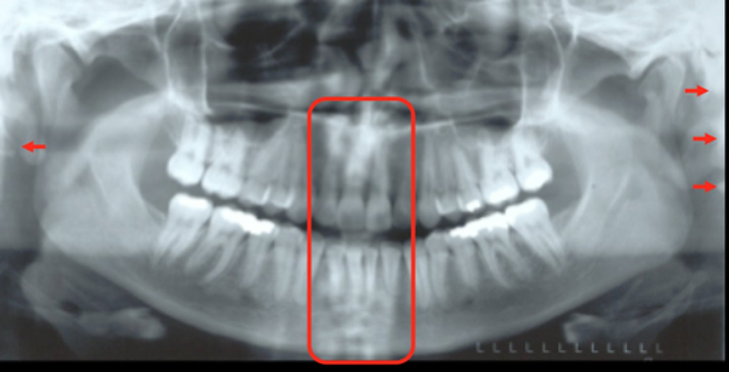
Name the structures that the red arrows points too?
What separates them?
L: Condyle
M: Coronoid Process
R: Inf. Alveolar Canal
Label from the left side of mouth to Right.
Mental Foramen
Lateral Walls of Nasal Fossa
Inferior Nasal Concha
Soft Tissue of Nose
radiopaque. on roots of maxillary anterior teeth
Shorter arrow: Inferior Meatus
Appears radiolucent (air filled spaces)
Longer arrow: Middle meatus
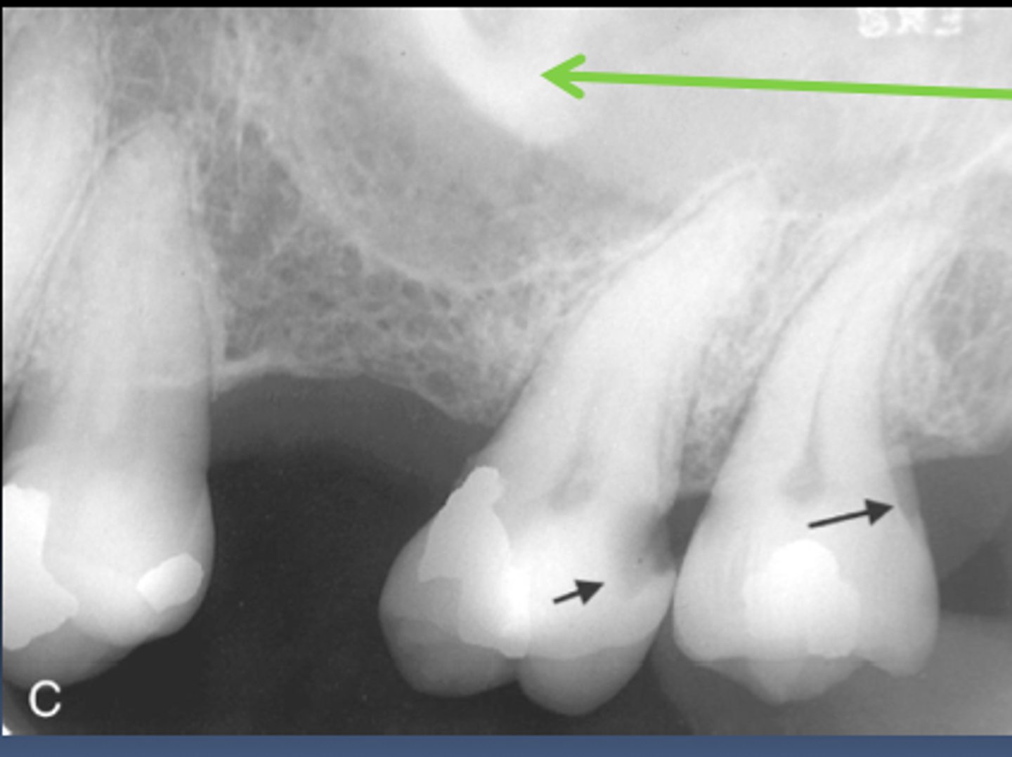
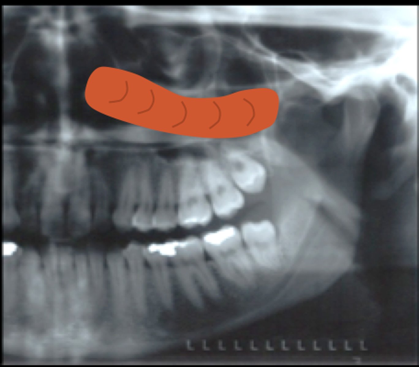
Walls of Maxillary Sinus
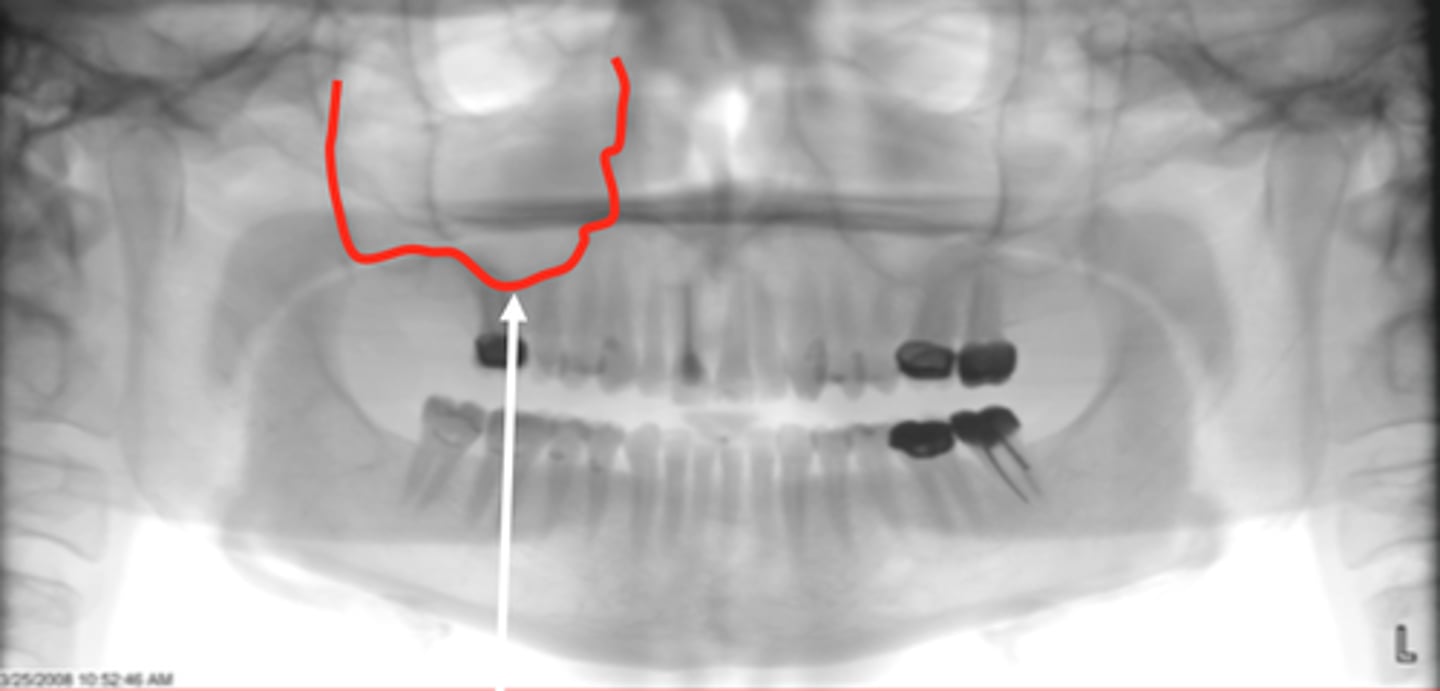
Zygomatic Process of Maxilla
seen as U, V, or J shaped entity. THICK walls compared to sinus. Located in maxillary 2nd molar area
Zygomatic Bone / Arch
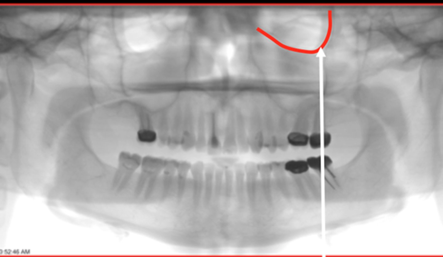
Pterygomaxillary Fissure
Upside down tear drop shape behind maxillary sinuses.
Pterygopalatine Fossa
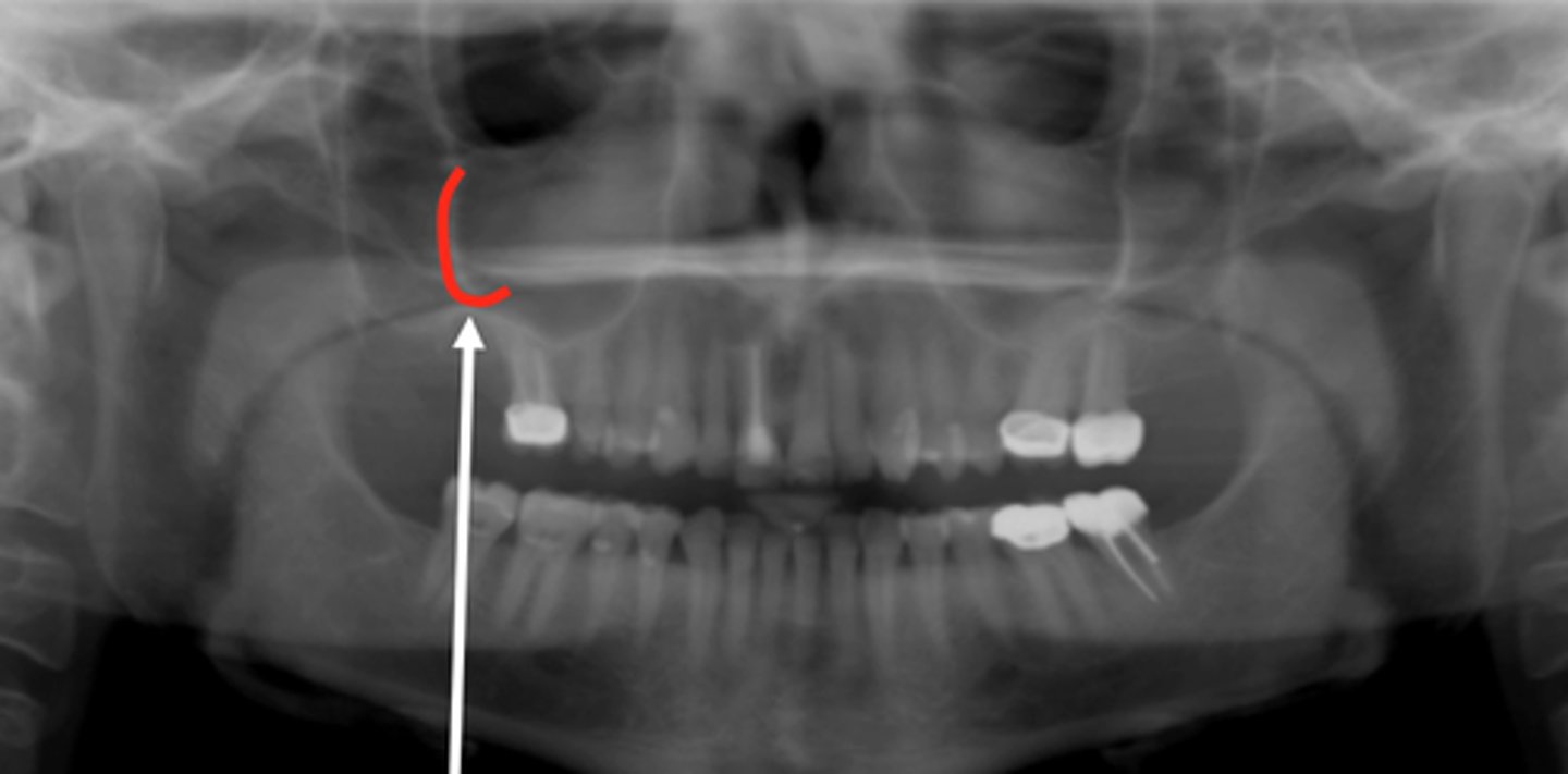
Orbital Rim
Curved radiopaque line superior to maxillary sinus
Floor of Nose = Hard Palate
Hard Palate on the Left
Soft Palate on the Right
Soft Tissue of the Tongue
Radiolucent line bw the palates and the tongue can be best seen curving down over max molar.
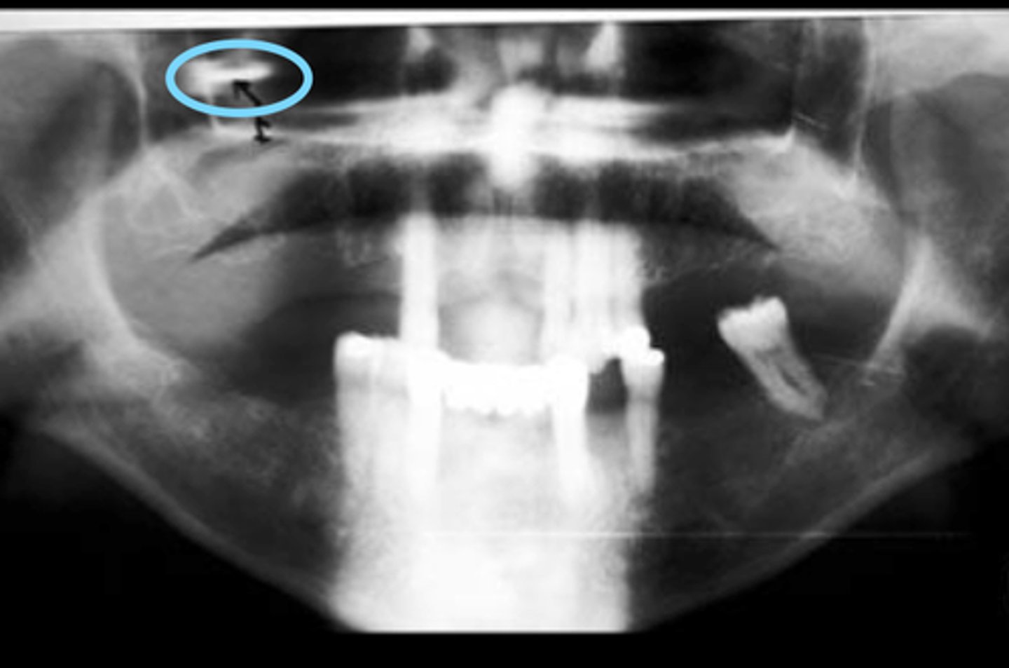
External Auditory Meatus
Radiolucent entities located posterior to condyle.
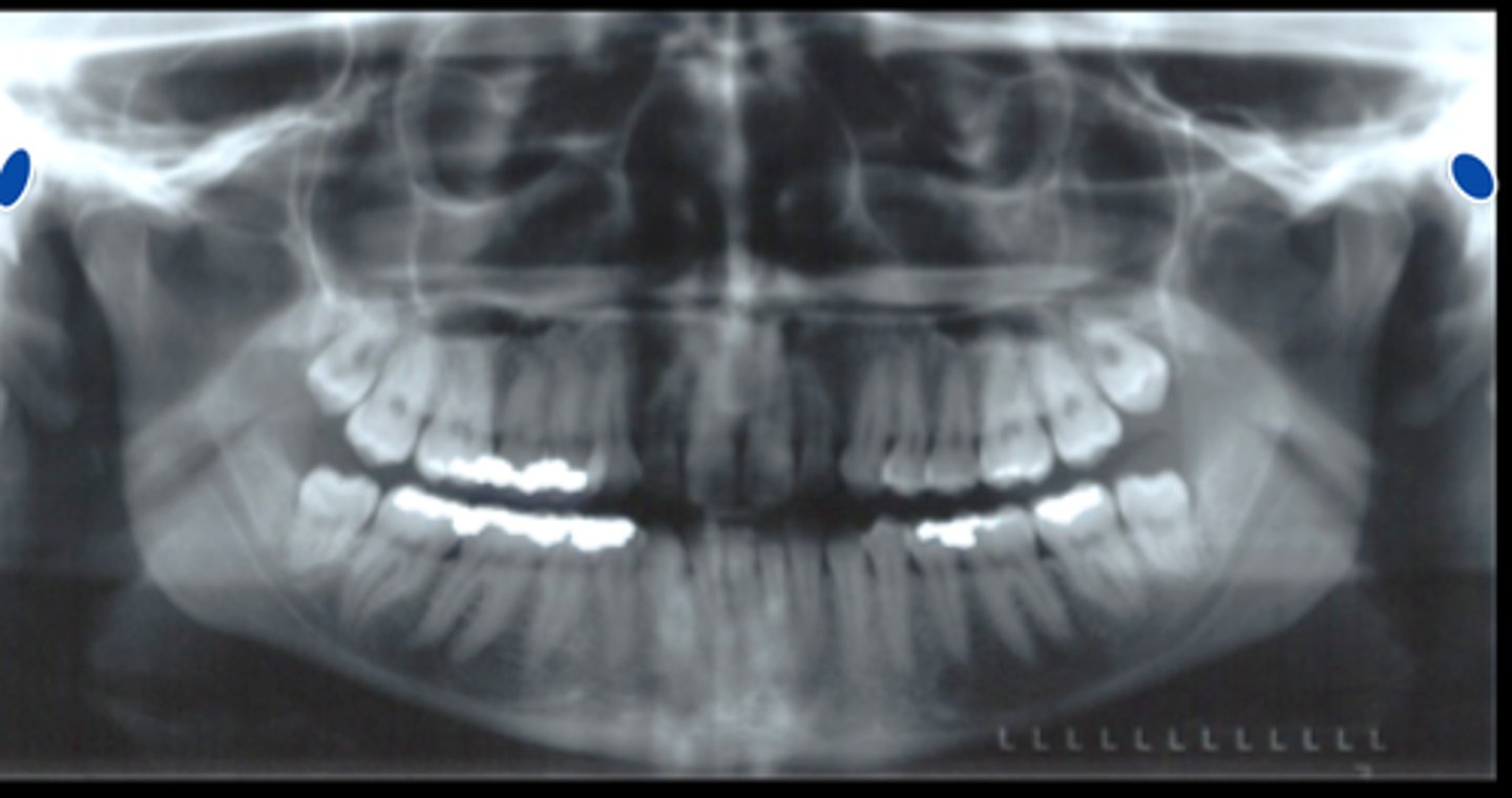
Soft Tissue of the Ear
Posterior to the mandibular condyle and inferior to the ext auditory meatus.
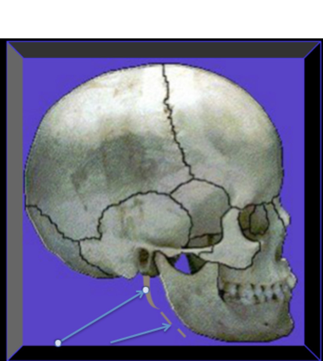
Stylohyoid Ligament
Hyoid Bone
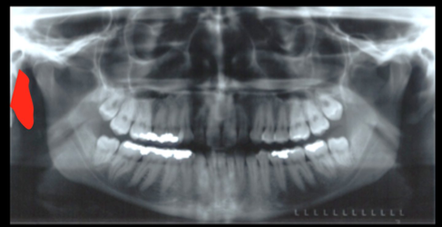
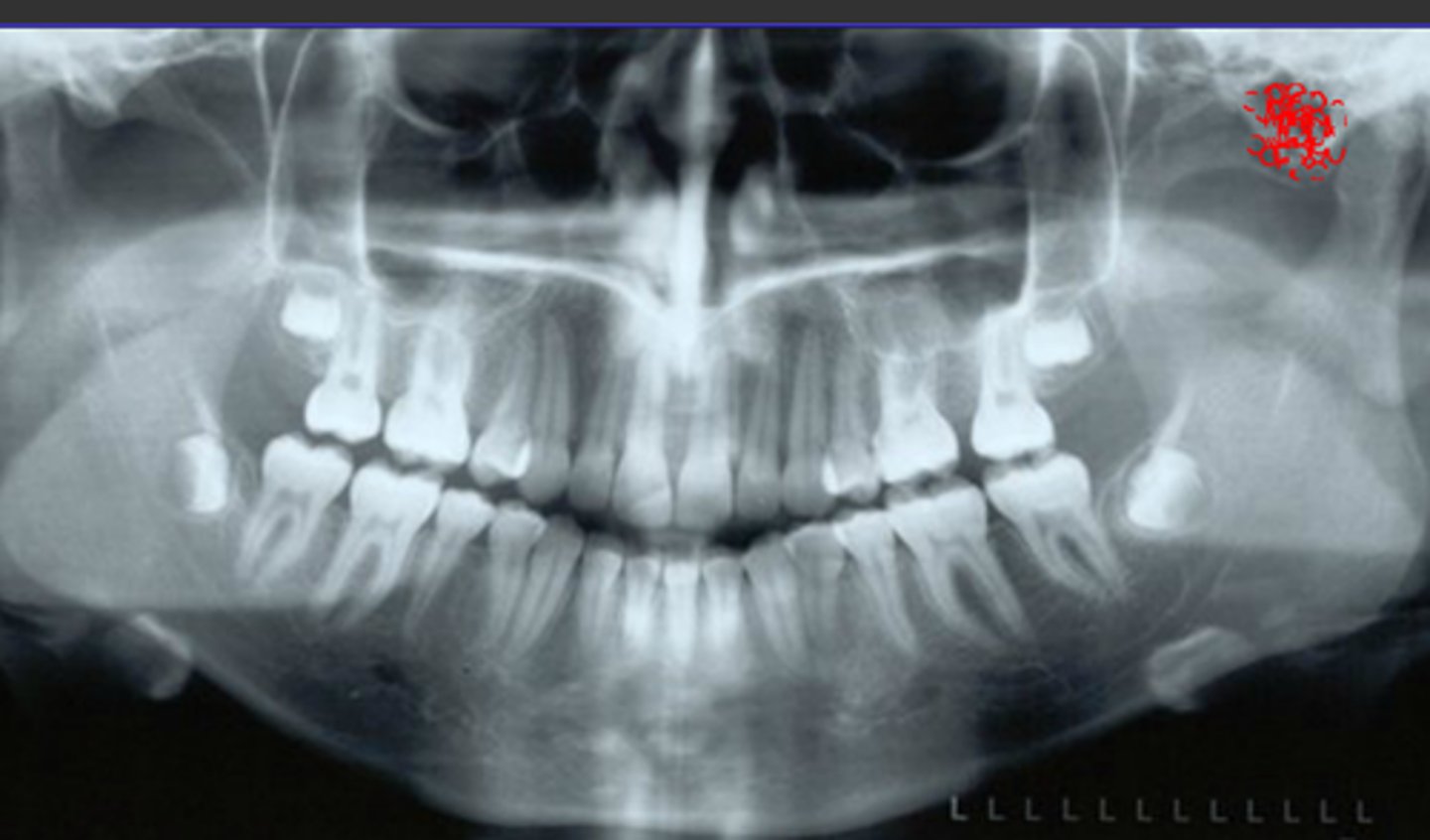
Identify the radiopaque object seen below the mandible on these images
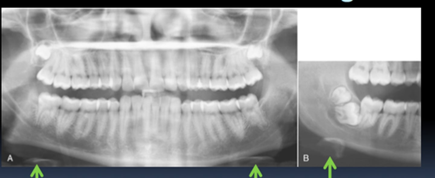
Vertebral Column
May appear both laterally and in the ant part of the image.
Ghost Images
(NOT ON LIST)
Mastoid Air Cells
Superimposed in condylar region (ant/post to condyles)
Small radiolucent soap bubble air filled cavities
Top arrow: Styloid Process of Temporal Bone
Bottom Arrow: Stylohyoid Ligament
1) Localize the abnormality
2) Assess the periphery and shape
3) Analyze the internal structures
4) Analyze the effect of the lesion on the surrounding structures
Systematic Radiographic Examination Steps
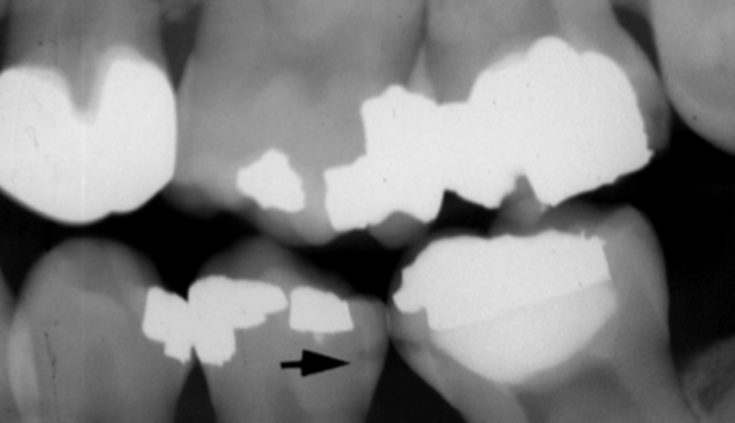
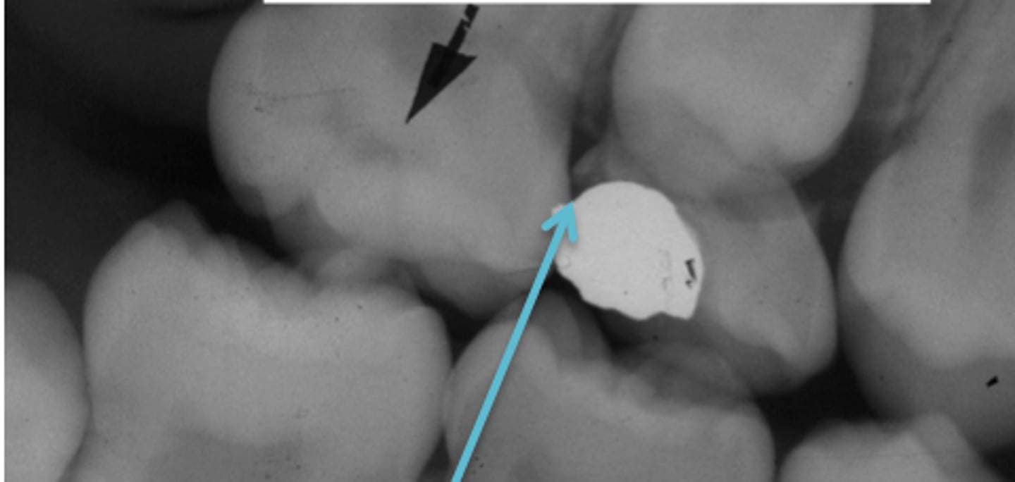
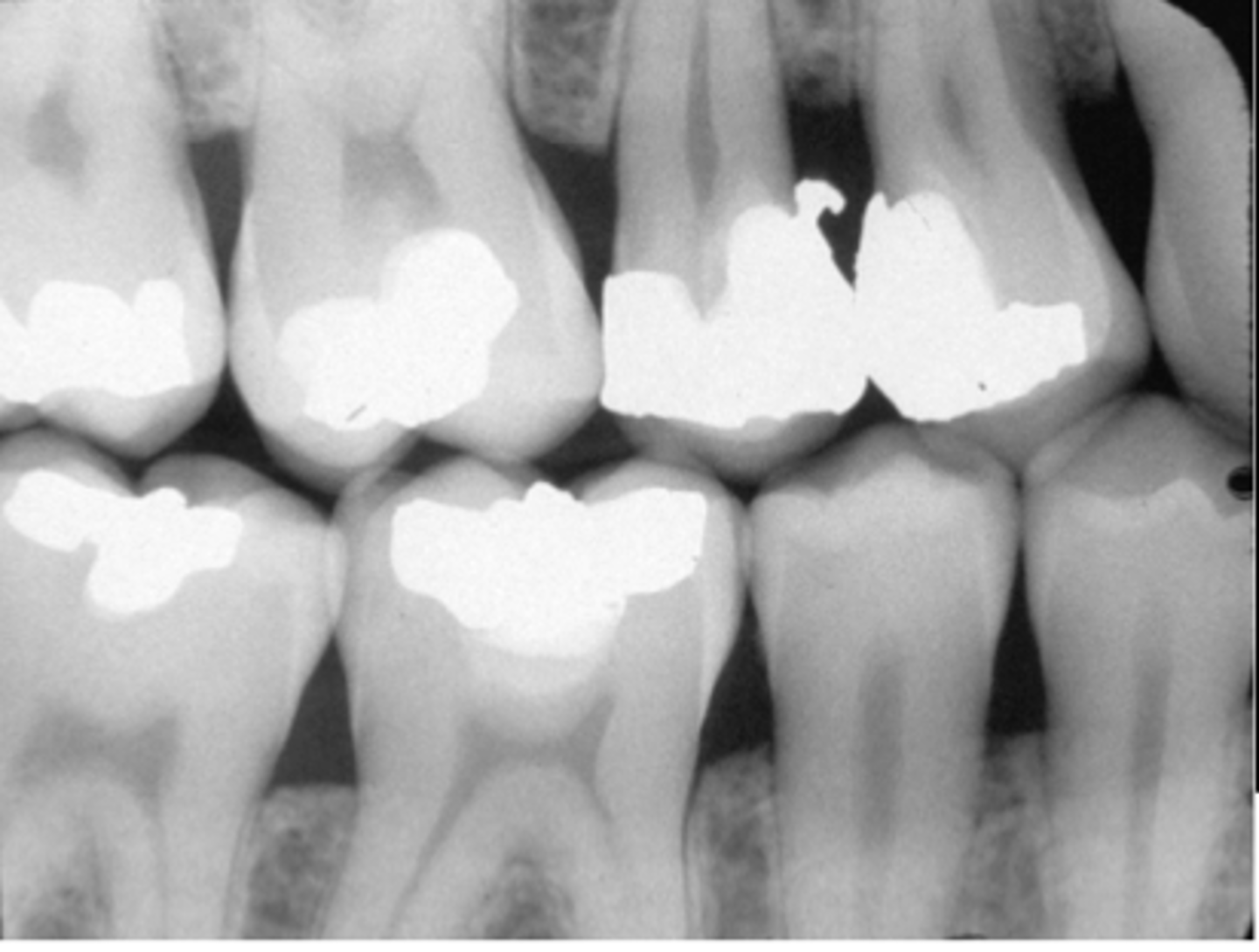
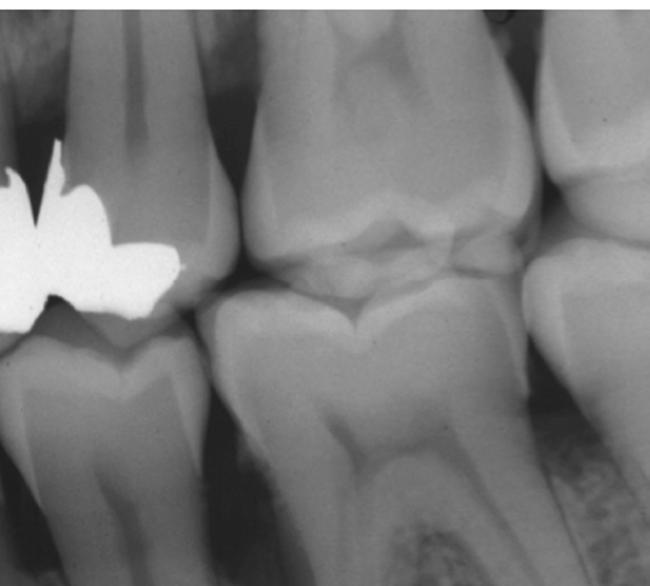
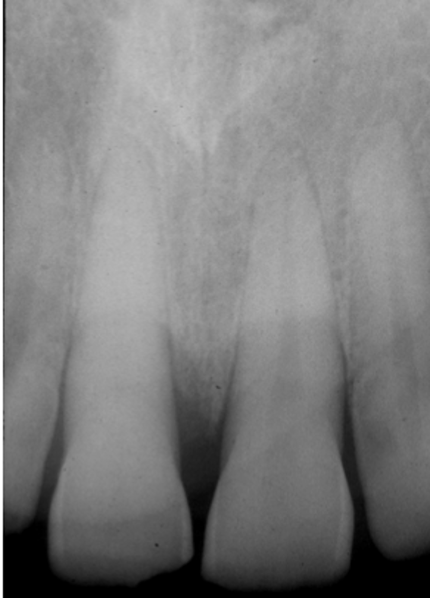
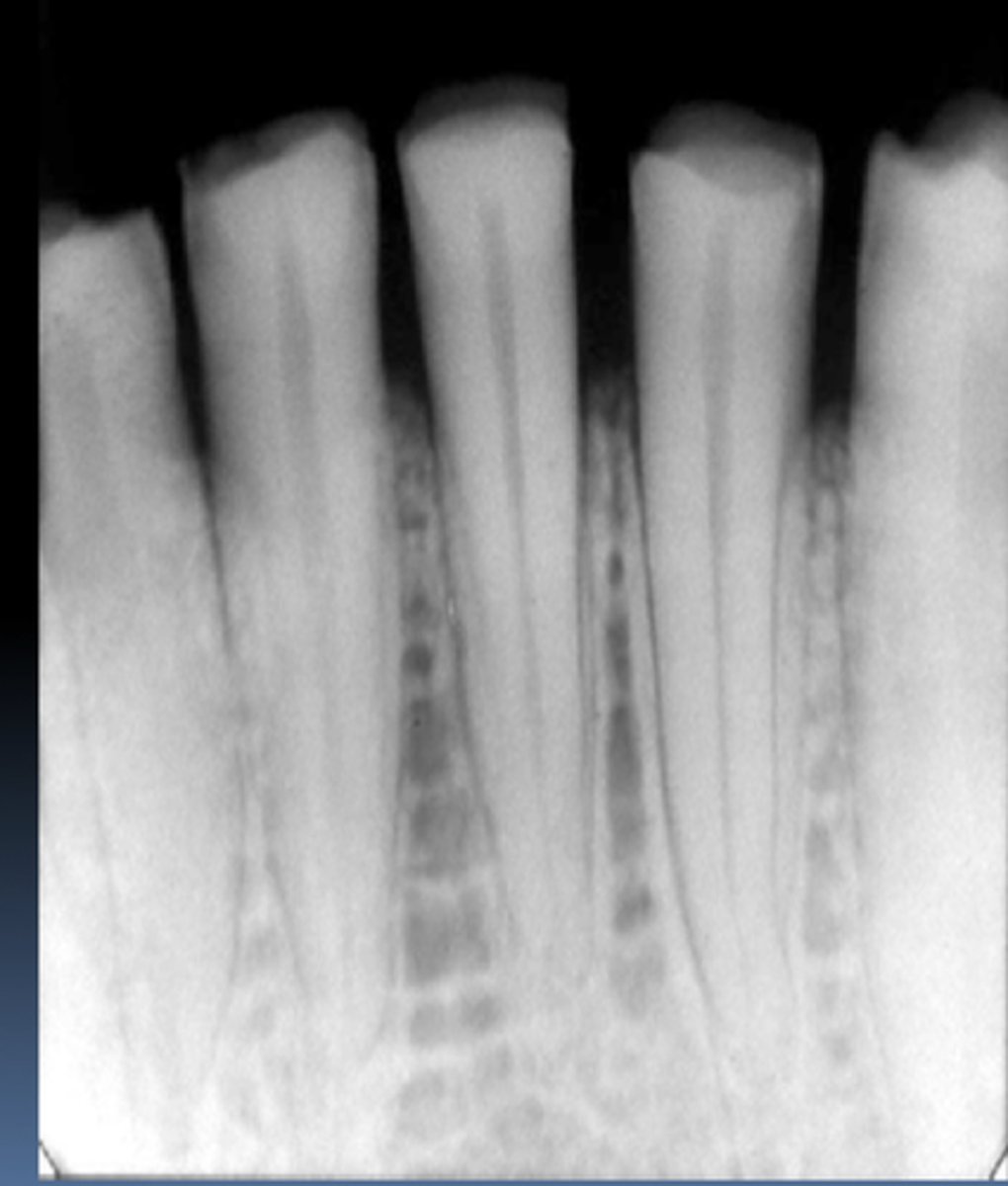
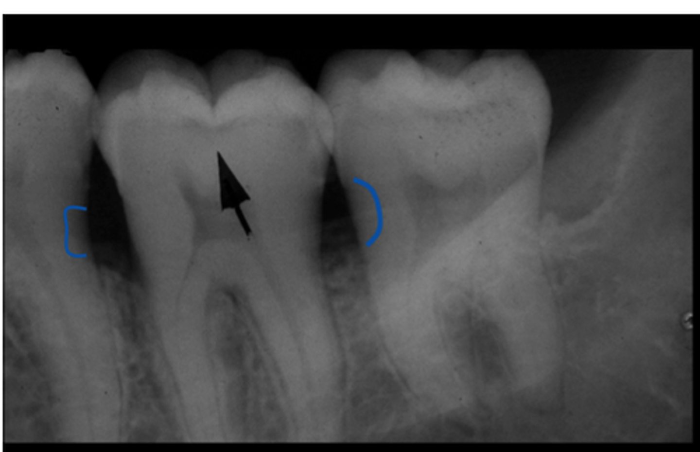
Proximal Caries Lesions
Occlusal Caries Lesions
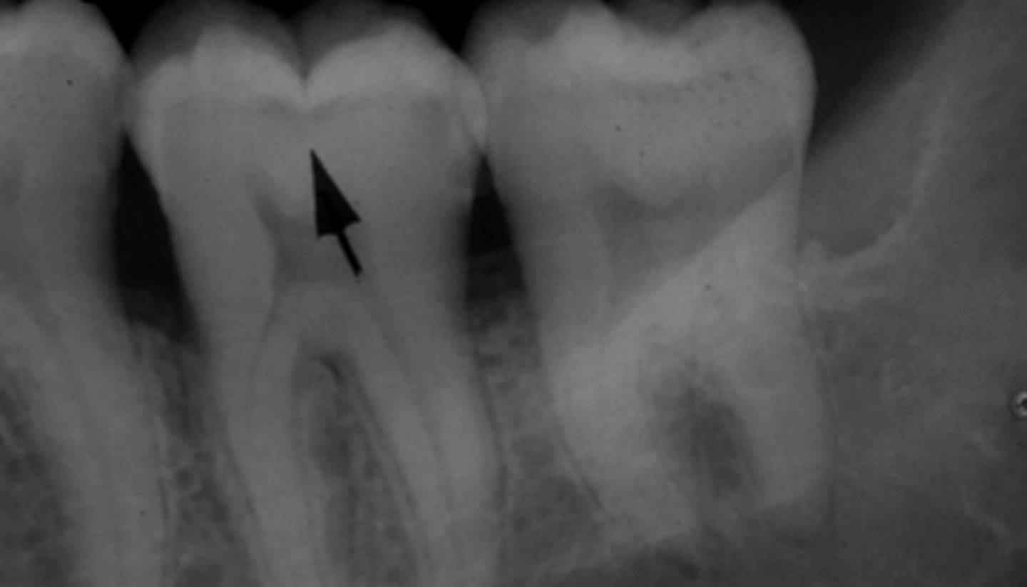
Mach Band
Sharp defined density difference such as between enamel and dentin.
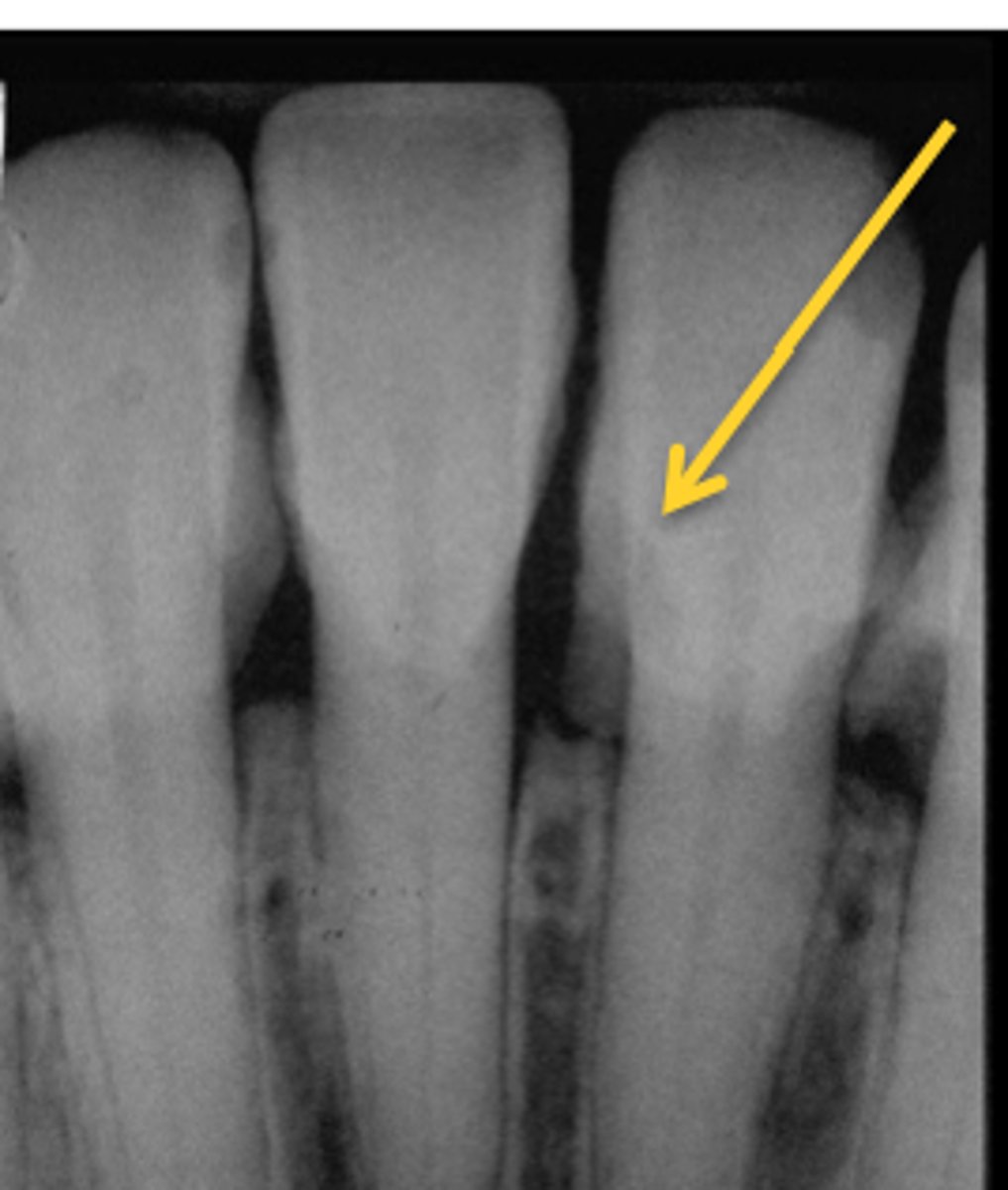
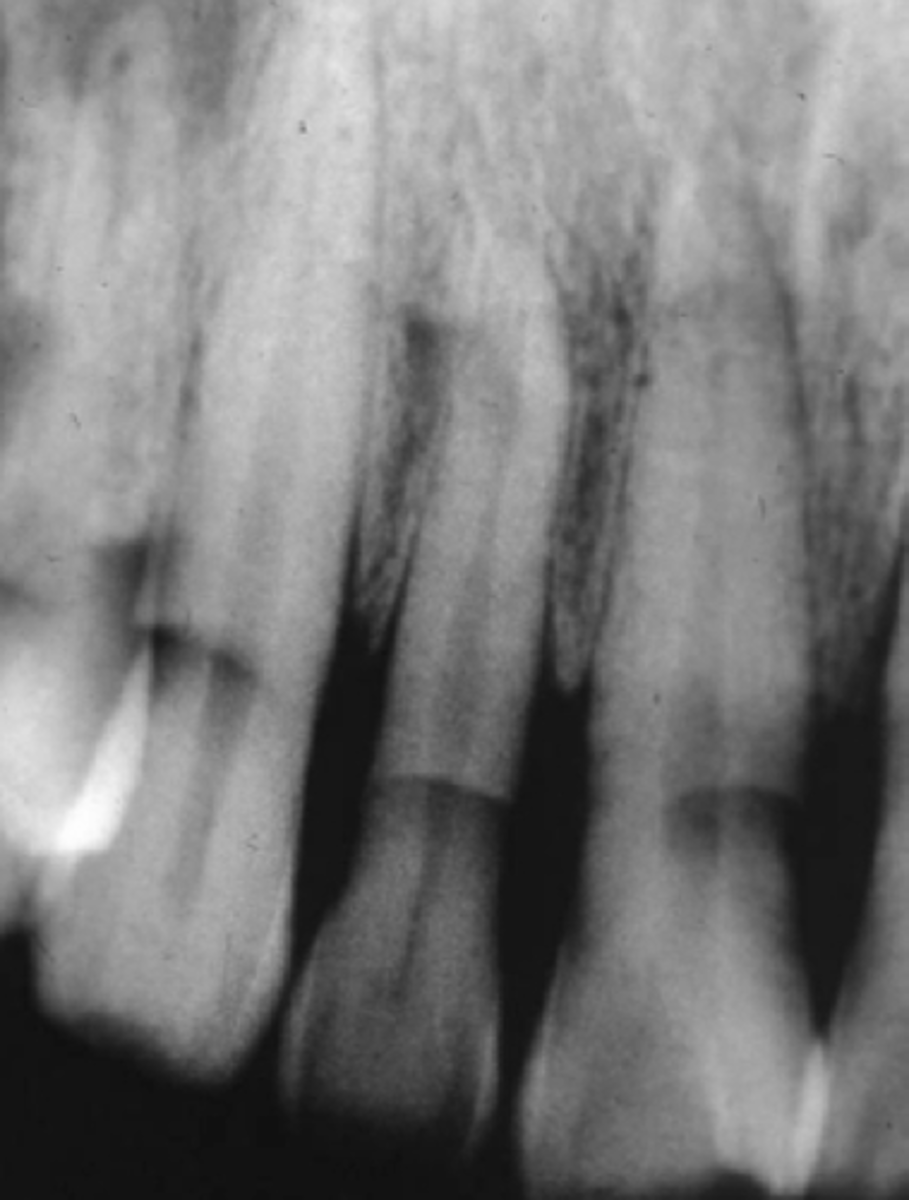
Root Caries Lesion
Restoration (D and M of 9) vs Lesion (D of 10)
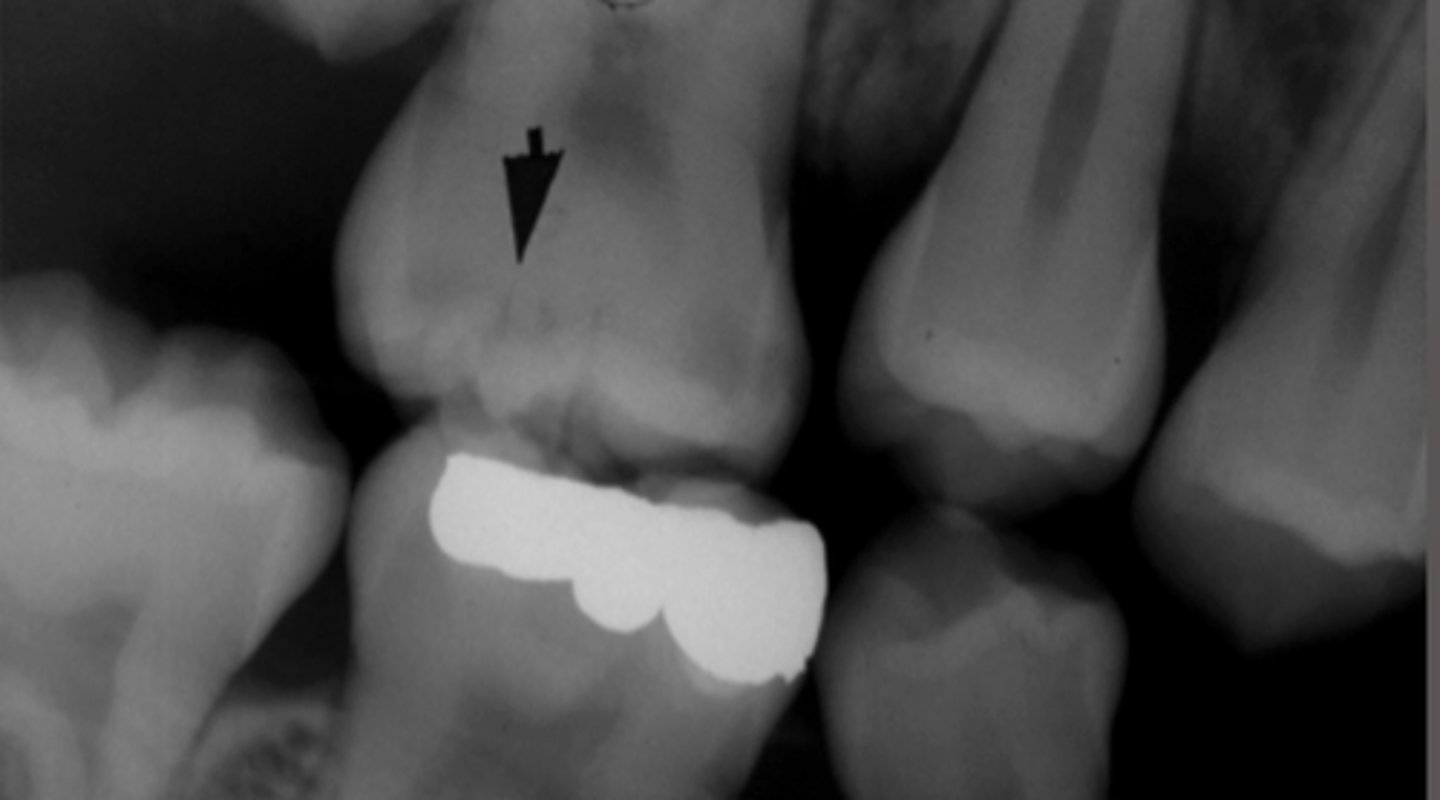
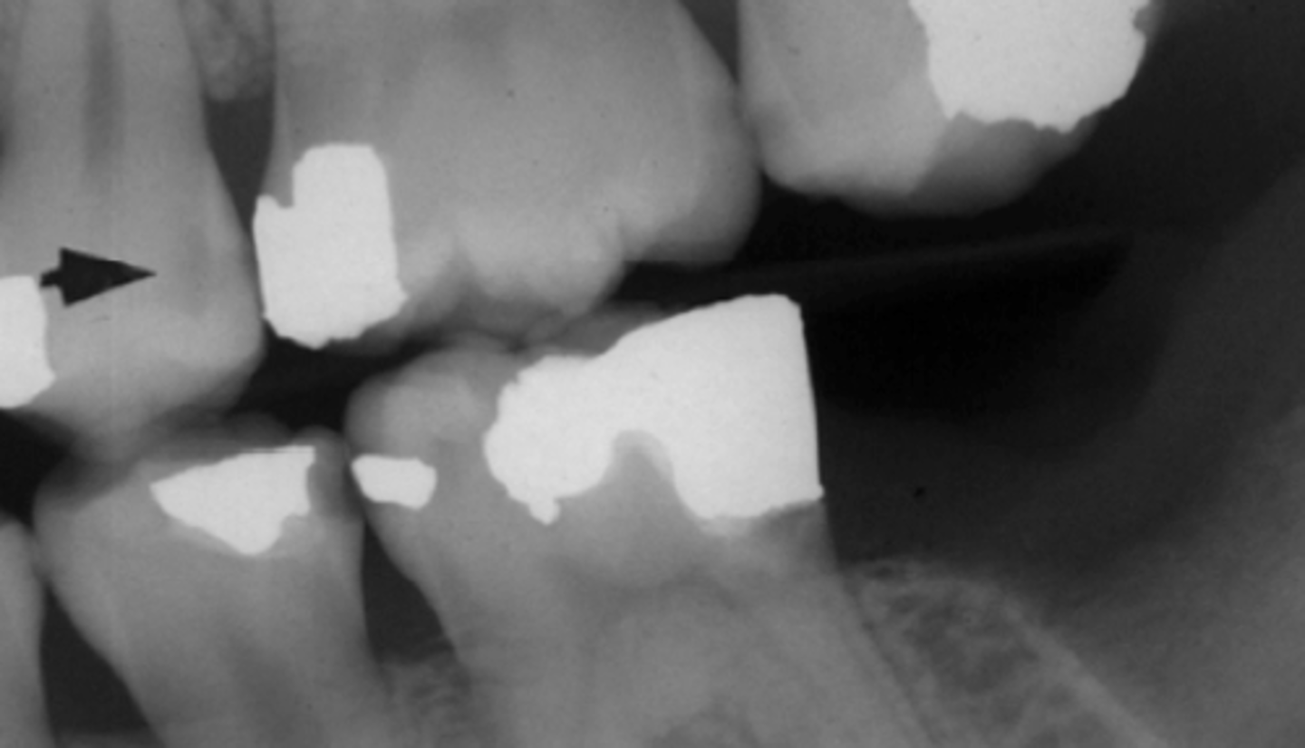
Recurrent Caries
Blue Arrow!?
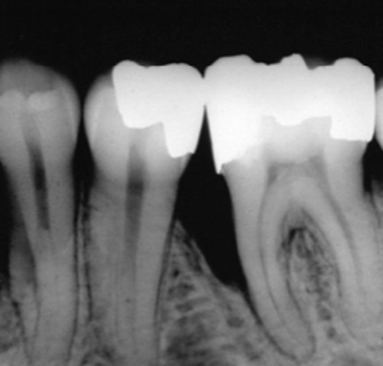
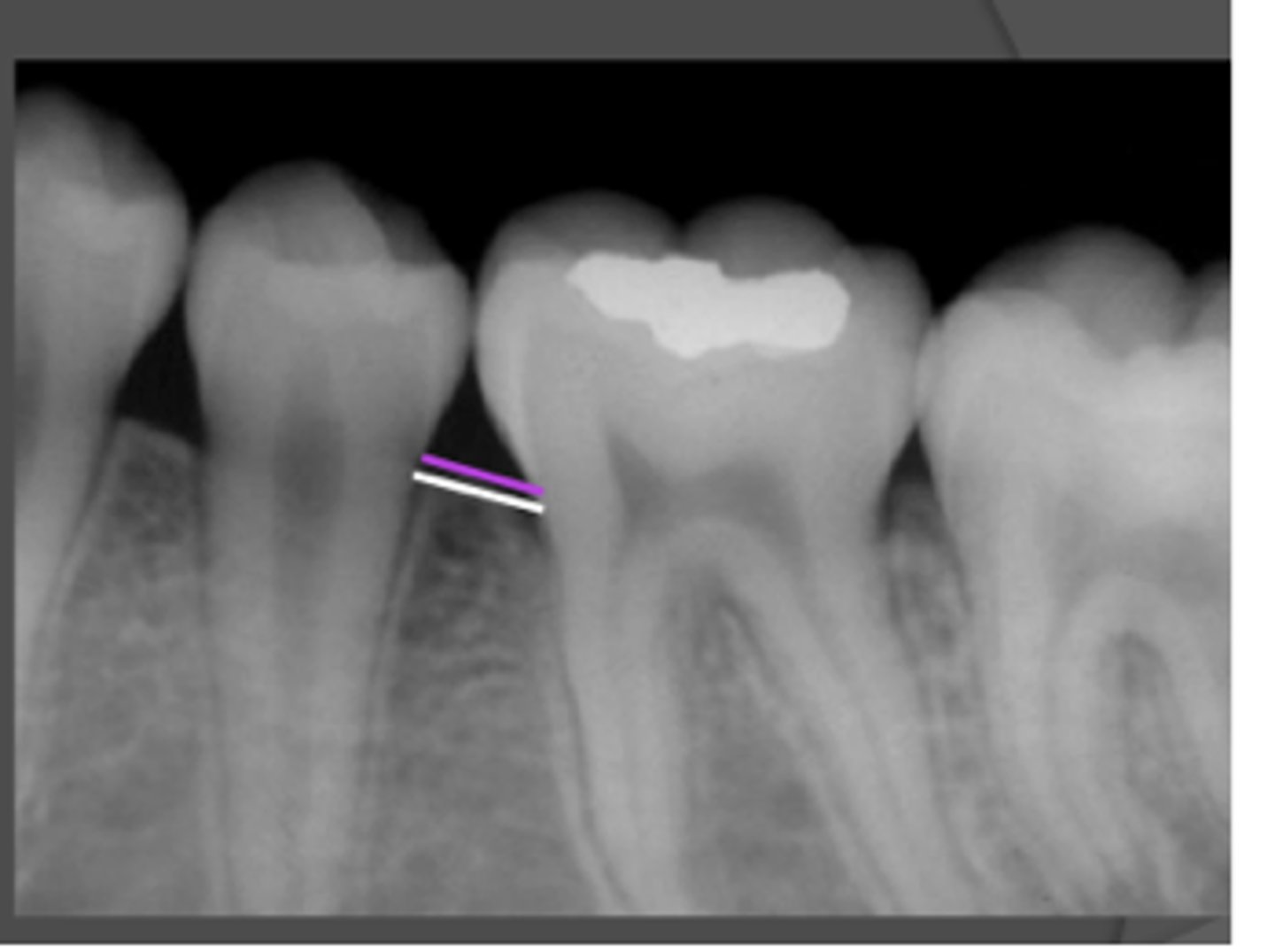
Horizontal Bone Loss
Vertical Bone Loss
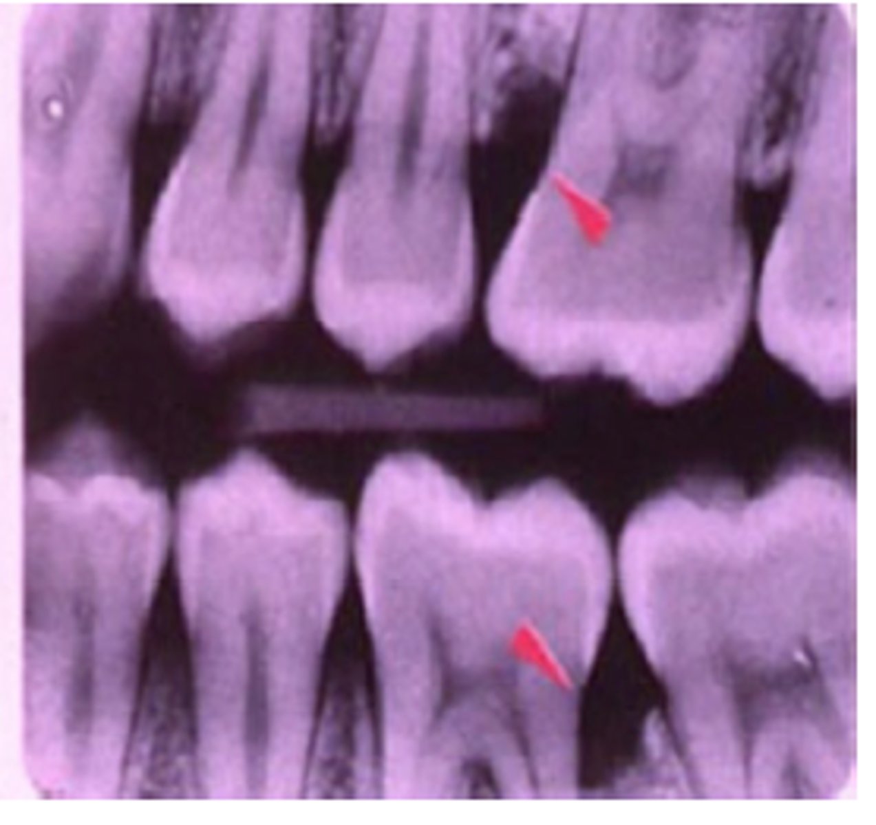
Calculus
Furcation Involvement
Crown To Root Ratio
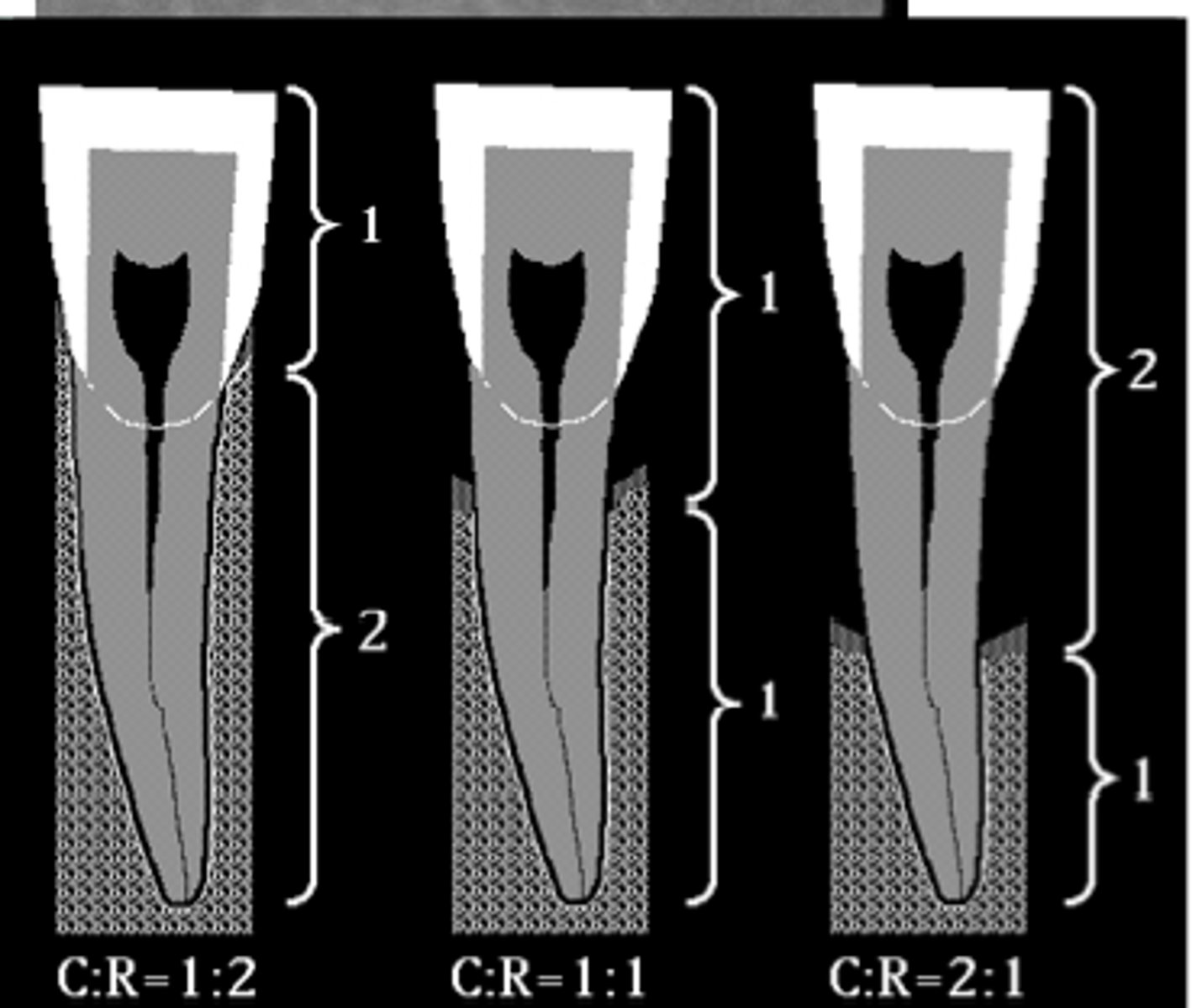
Normal Periodontal Bone Level
Aggressive Periodontitis
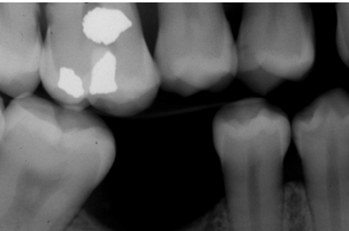
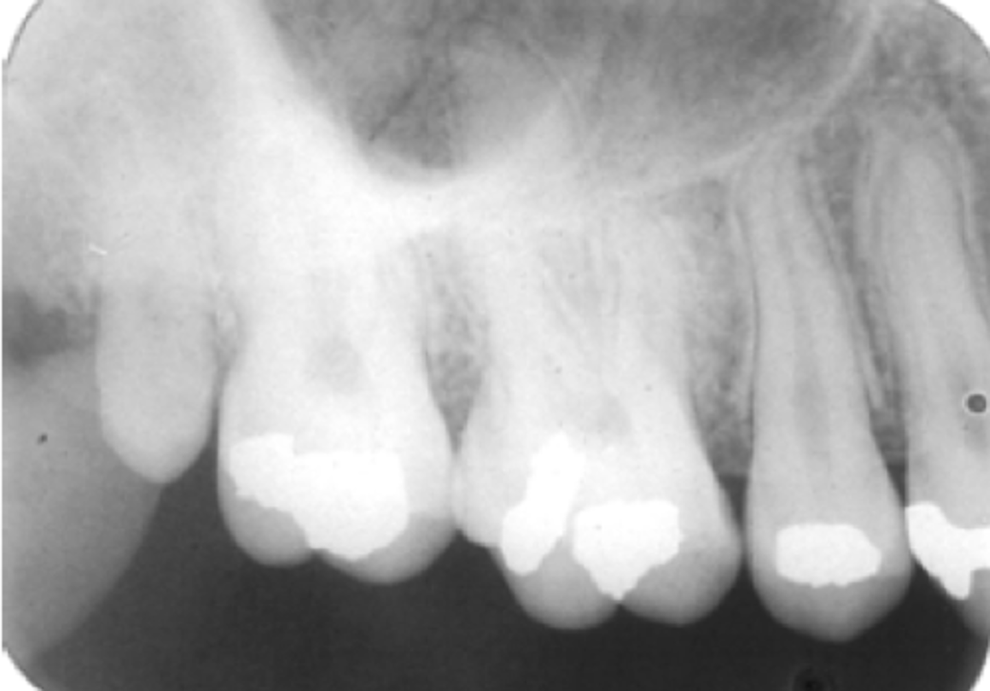
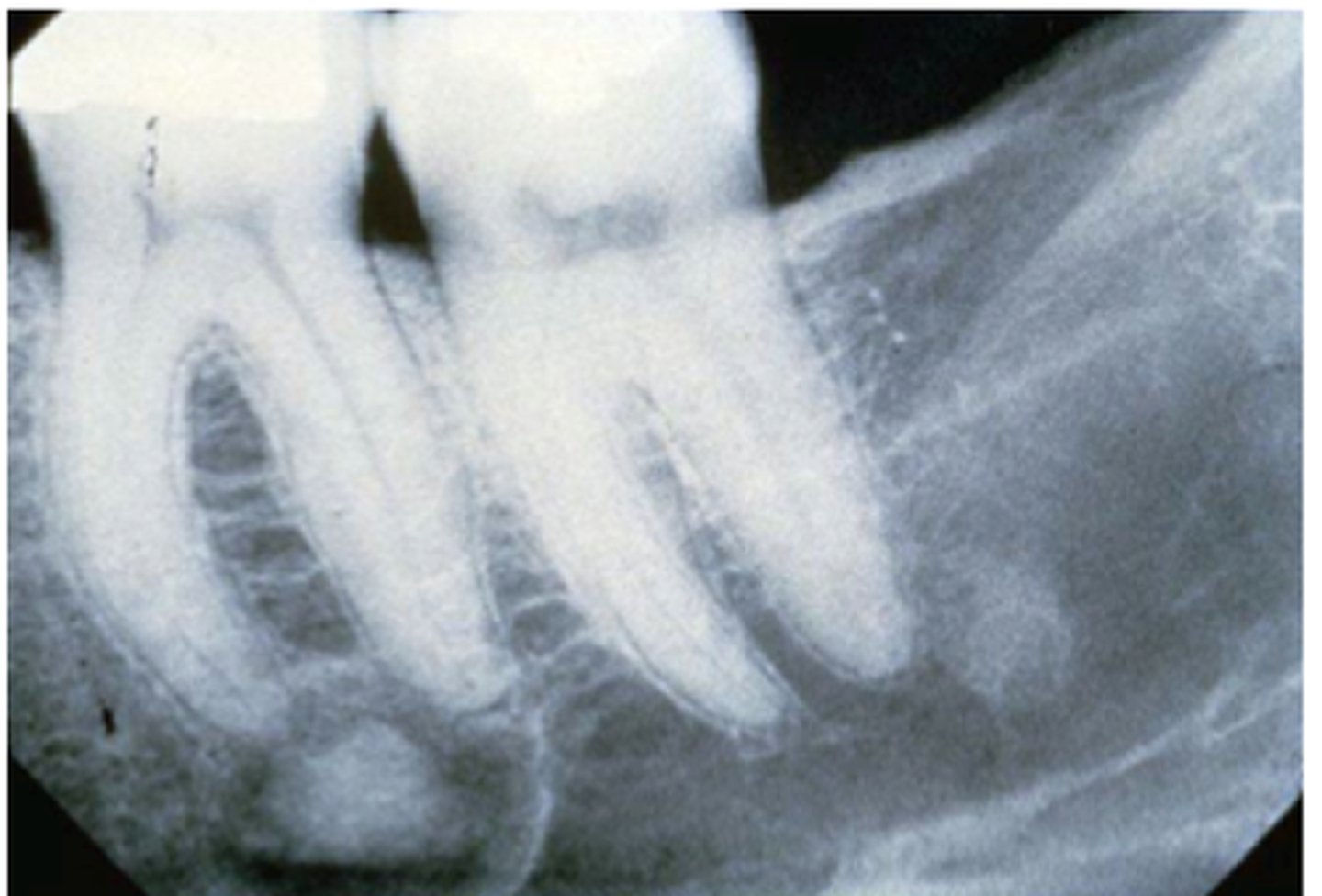
Overhanging Margins
Whats wrong w/ restoration?
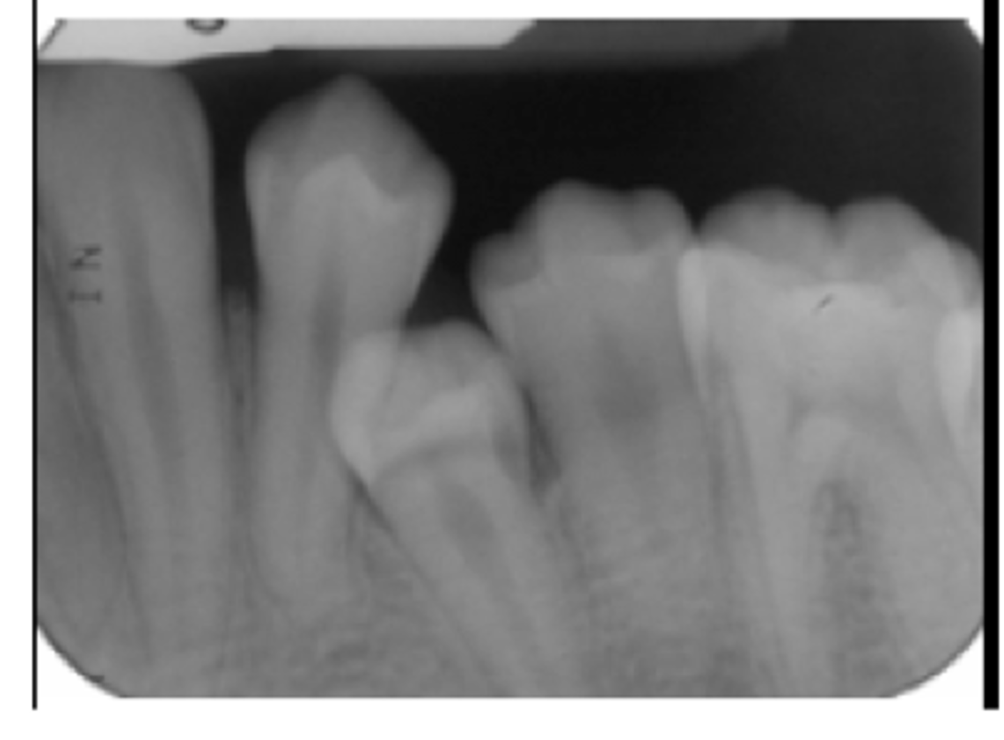
Distal Drifting
Only seen in premolars
Mesial Tipping
Tooth tips medially into edentulous space
Note: OPEN CONTACT
Open Contacts
If area not cleaned, should be monitored for periodontal disease.
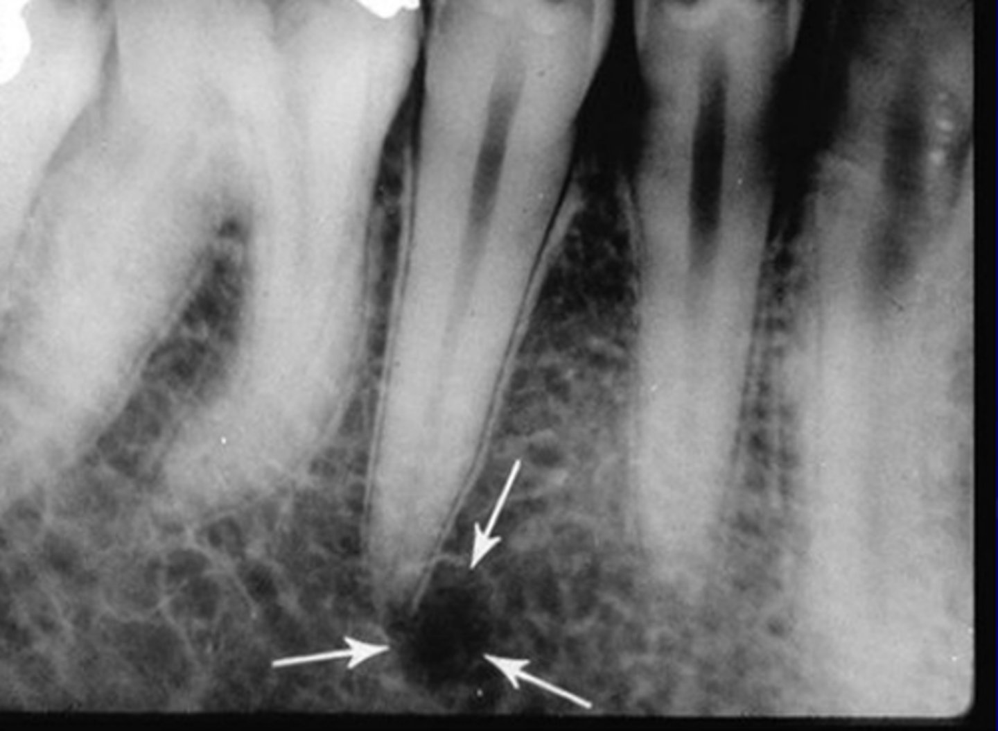
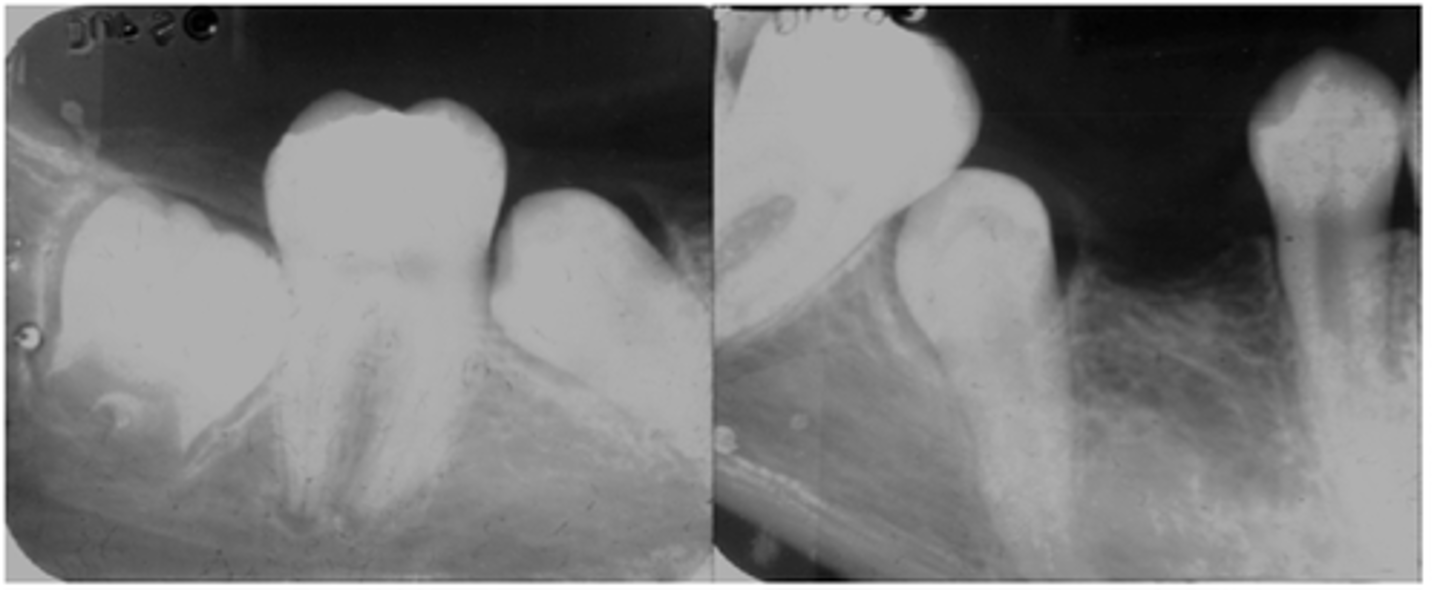
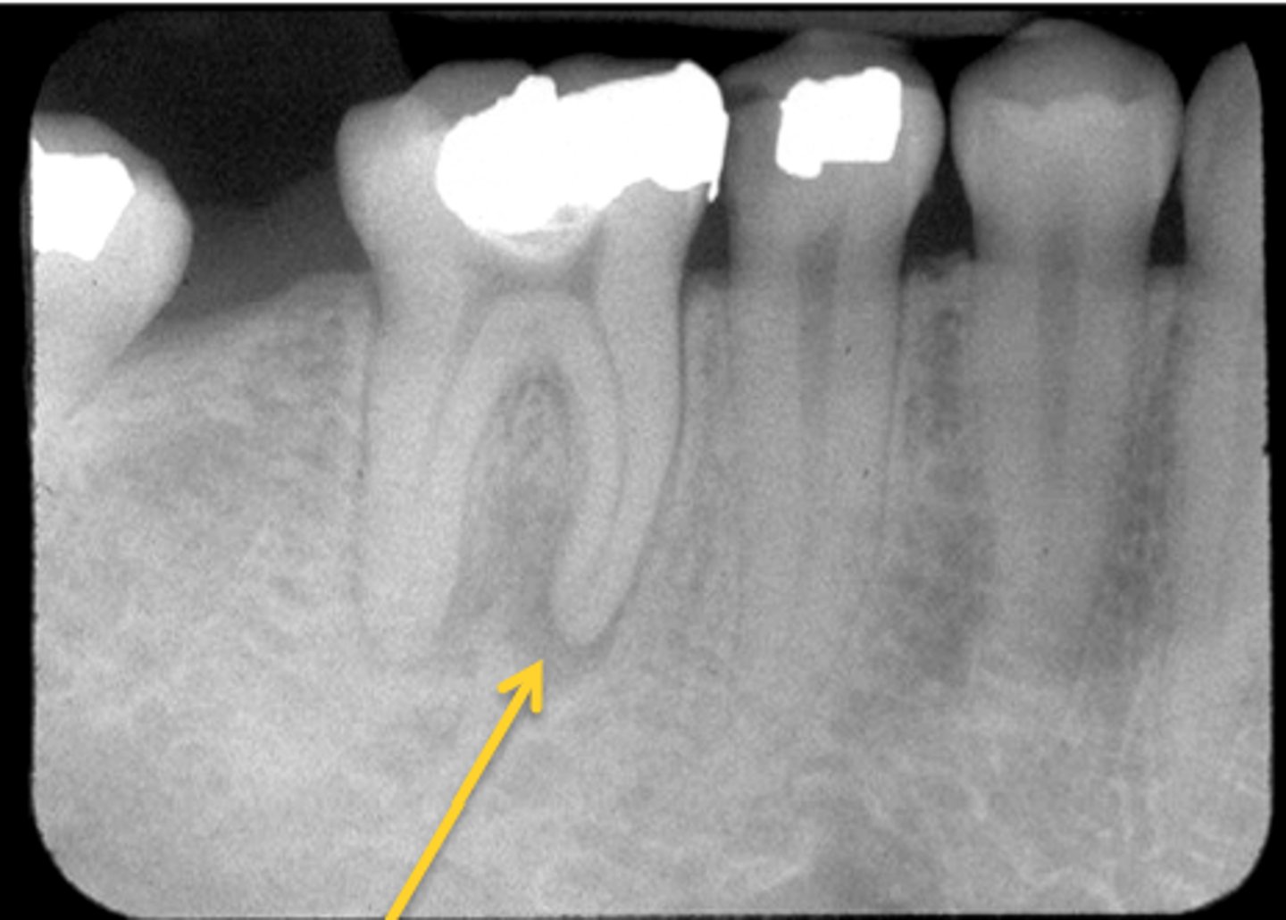
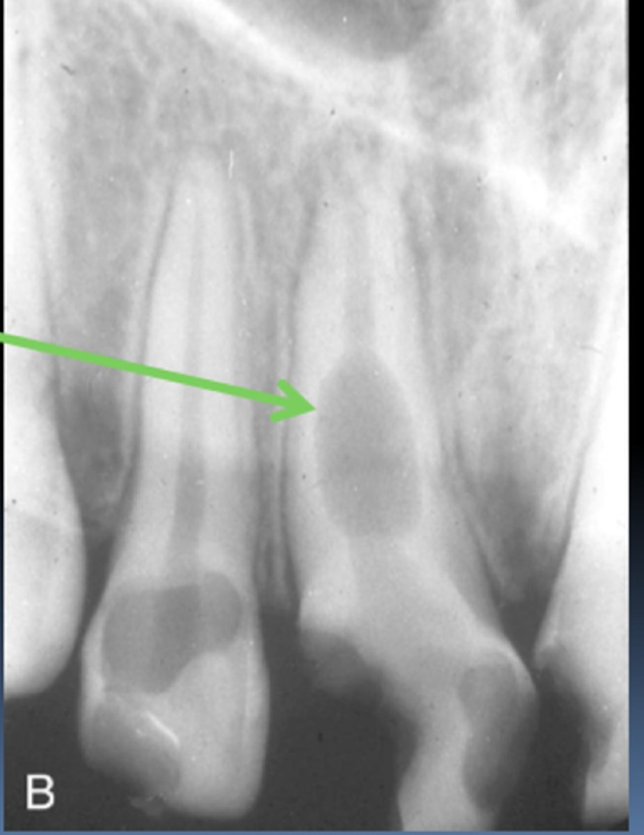
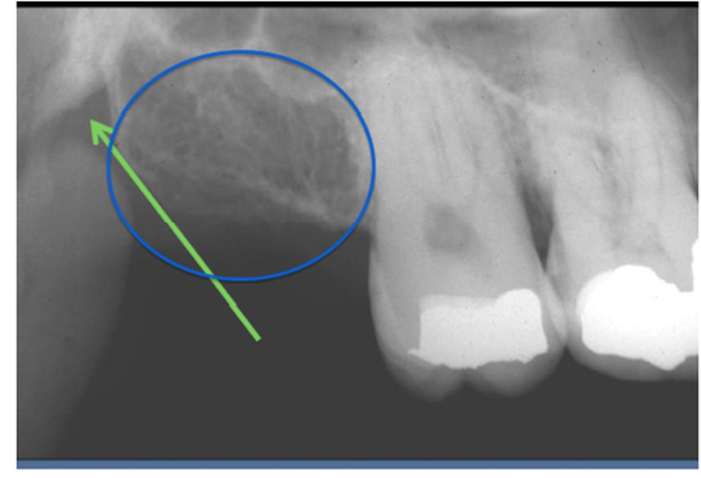
Rarefying Osteitis
radiolucent bone inflammation
irregular borders
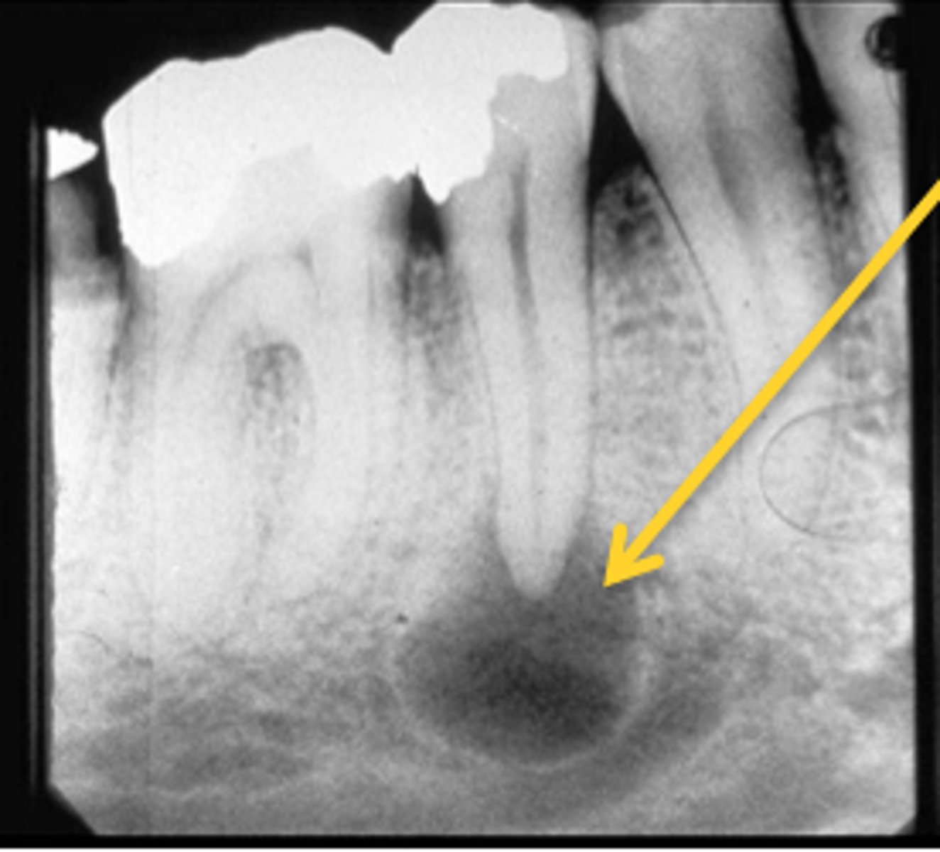
Radicular Cyst
Defined borders.
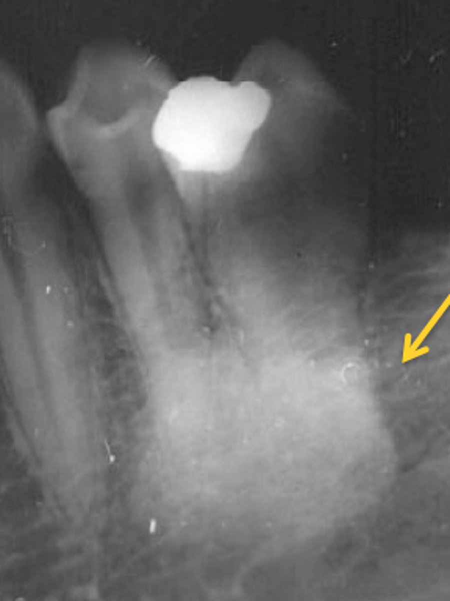
Sclerosing Osteoitis
radiopaque bone inflammation.
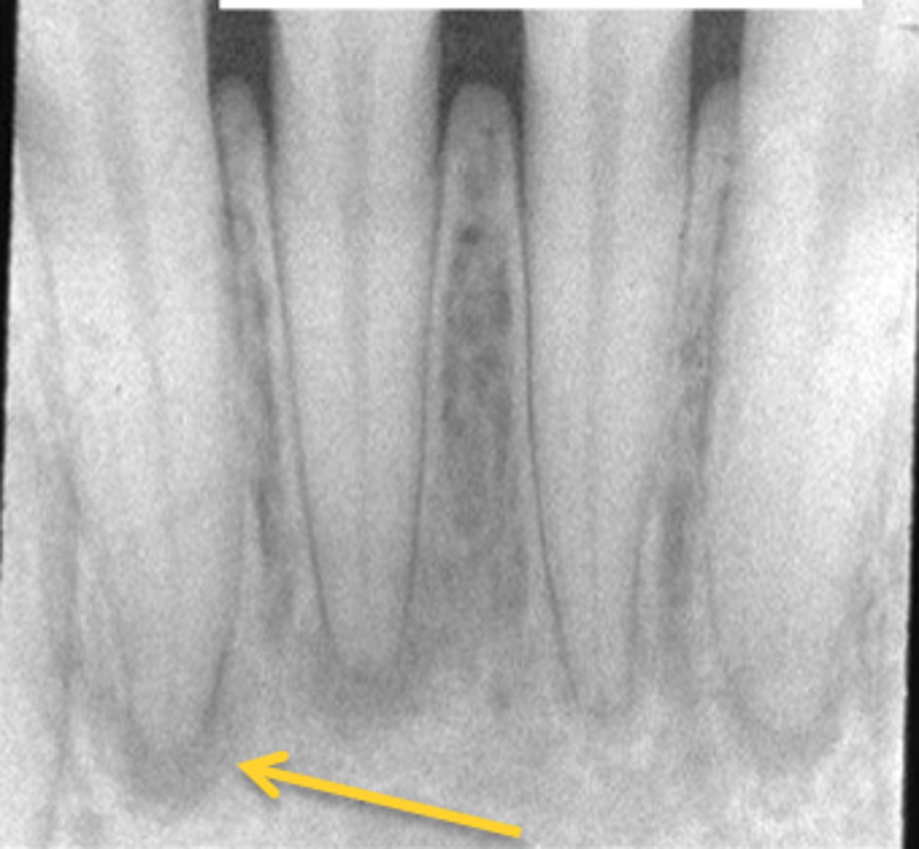
Periapical Cemento-Osseous Dysplasia
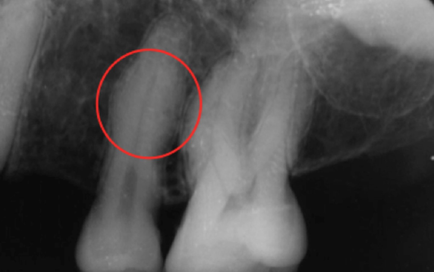
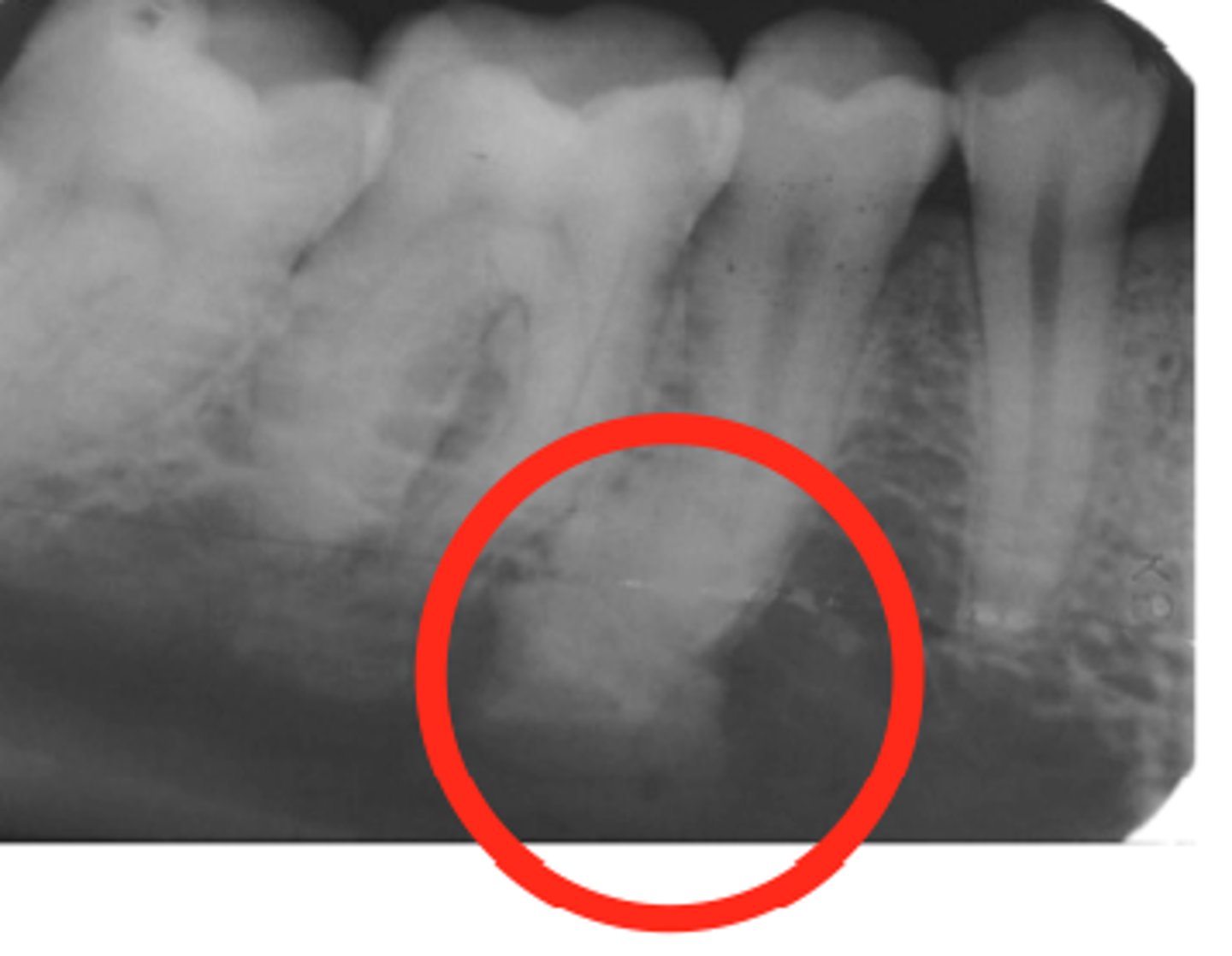
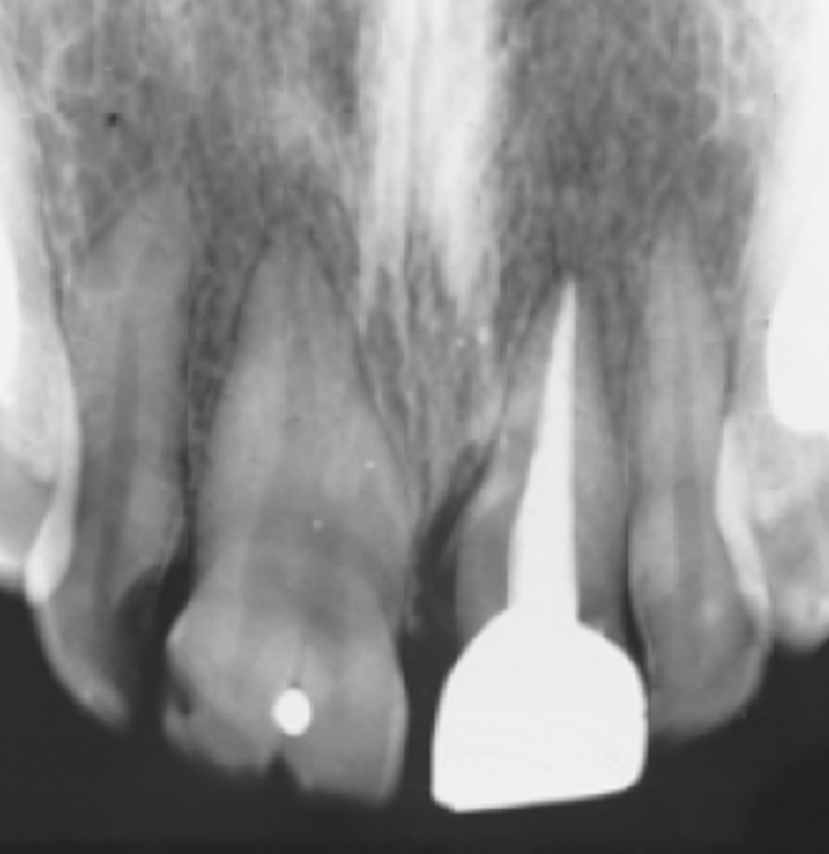
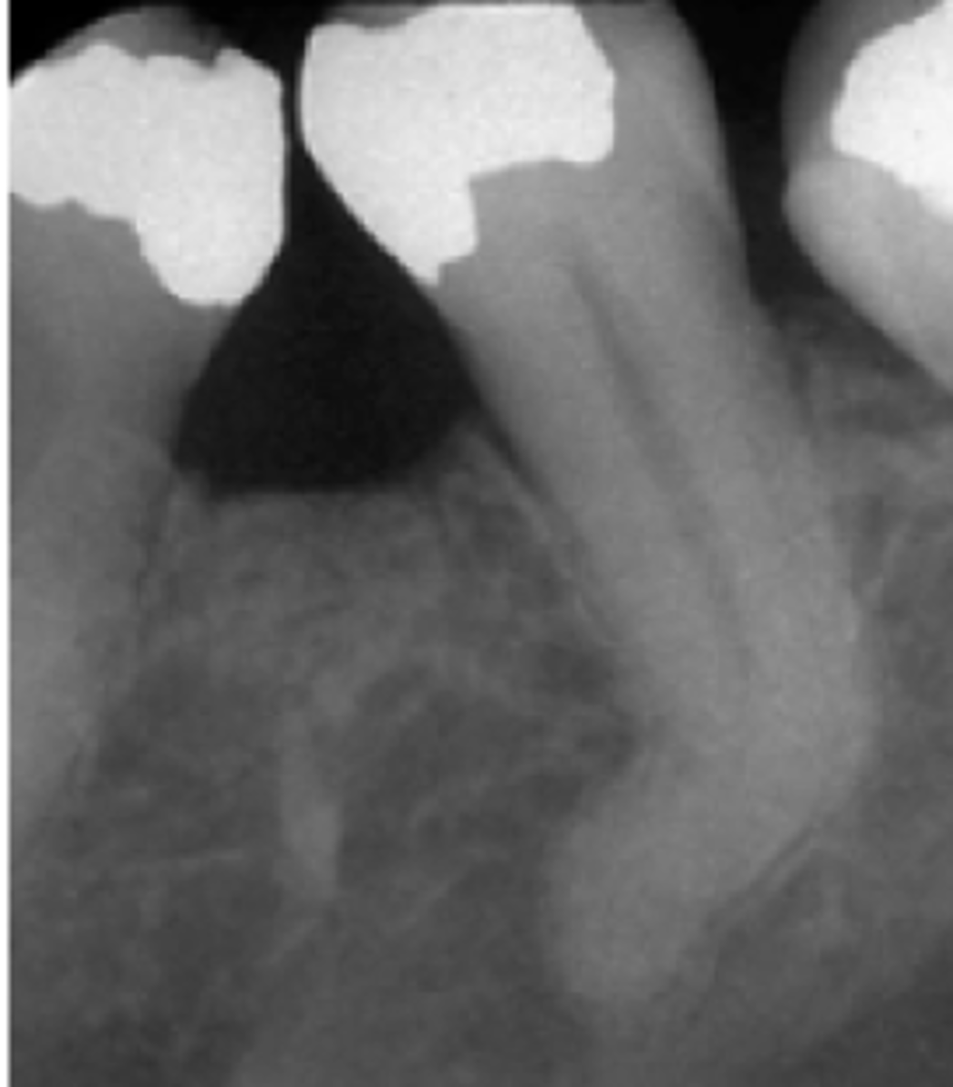
Hypercementosis
Wider root in apical and middle 3rd.
Enostosis
Island of radiopaque bone that is not associated w/ a tooth.
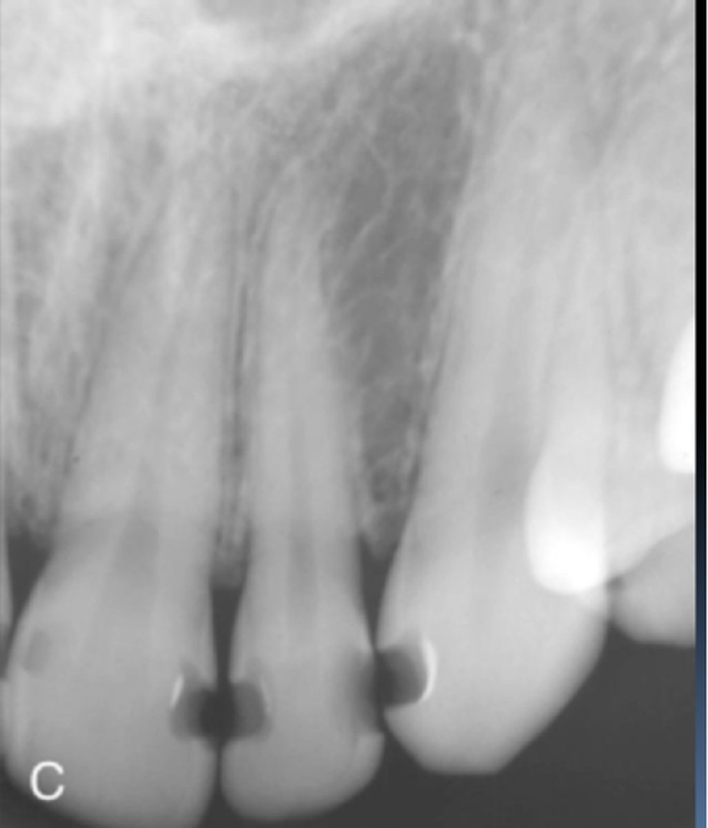
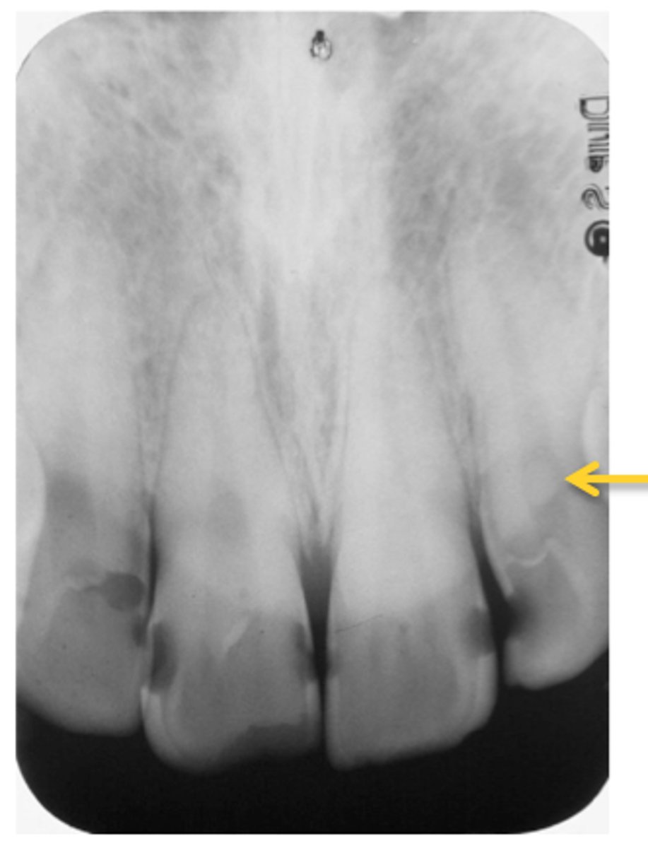
External Resorption
Osteosclerosis
radiopacity
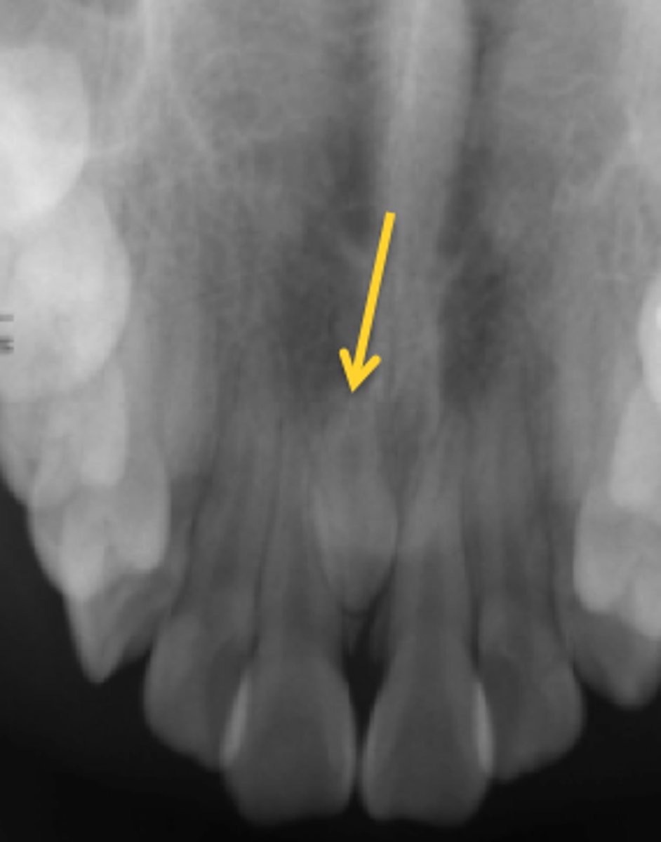
Internal Resorption
Calcification
Pulp Stones
Mesiodens
Supernumerary Teeth
Distodens
Supernumerary Teeth
Peridens
Supernumerary Teeth
Microdont
Macrodont
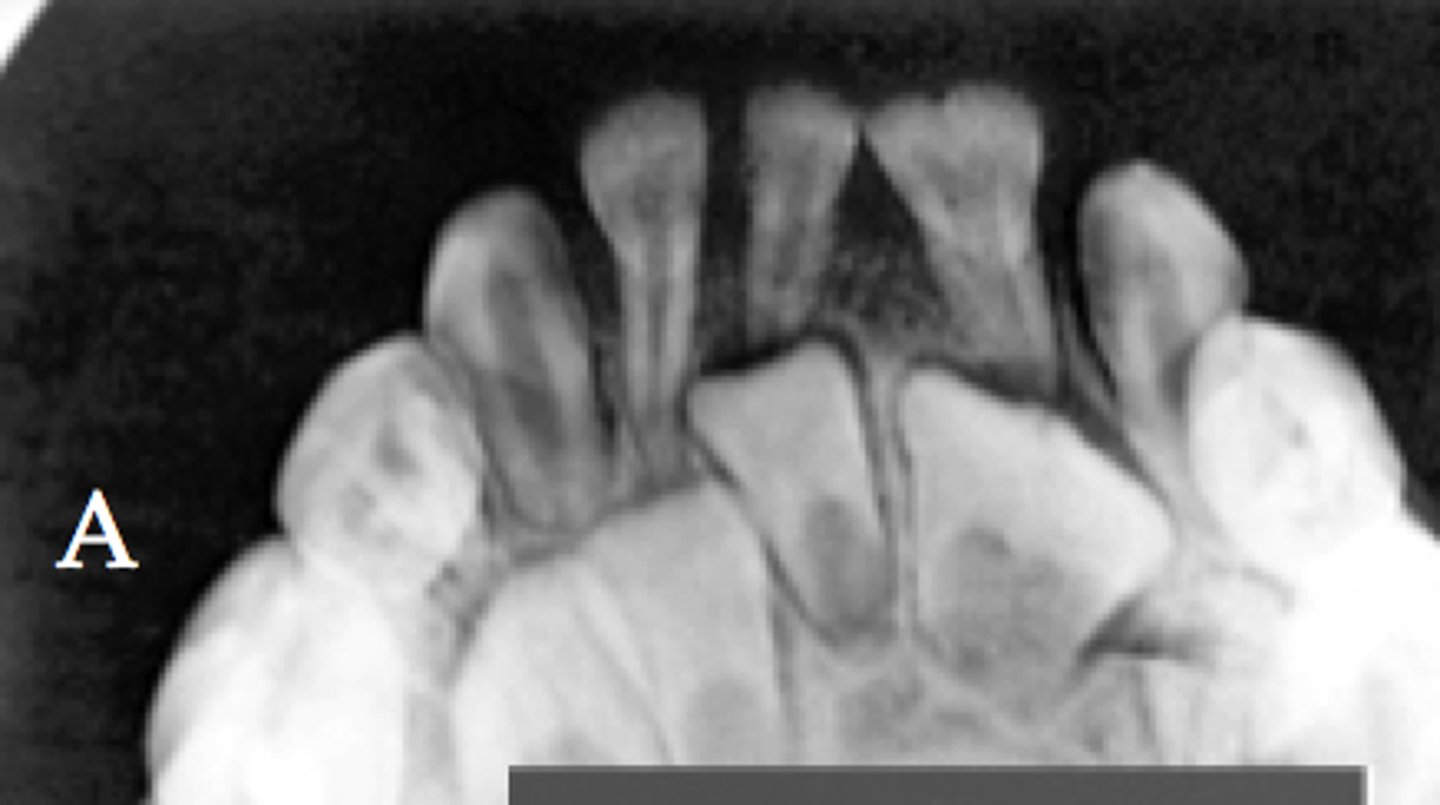
Fusion
Union of adjacent tooth germs of developing teeth.
Concrescence
Fusion at the root through cementum only.
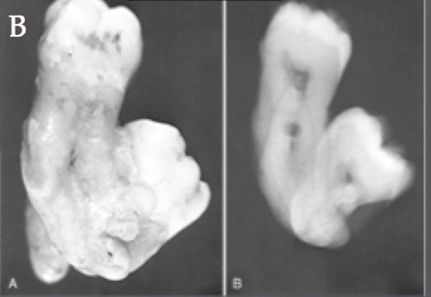
Gemination
"Twinning"
Single tooth bud attempt to divide
Taurodontism
Elongation of pulp chamber and surrounding tooth structure
Dilaceration
Dens In Dente when in Incisal Edge
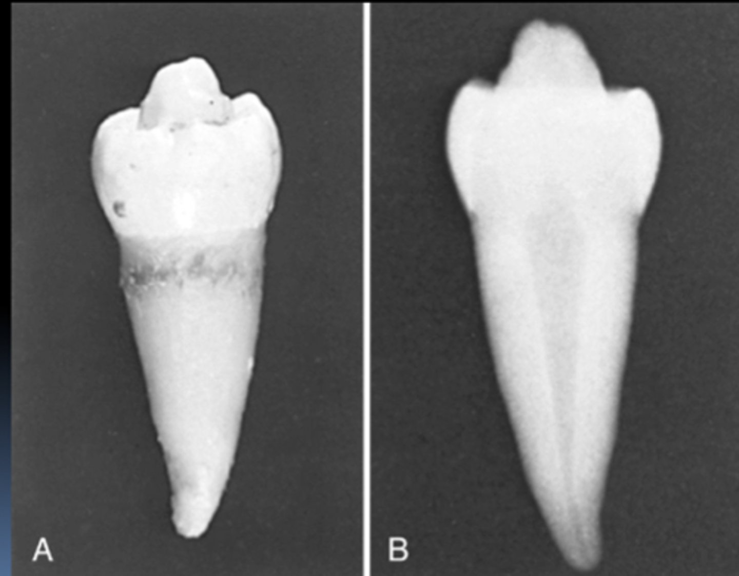
Dens Evaginatus
Outpouching of enamel organ
This tooth presents an example of
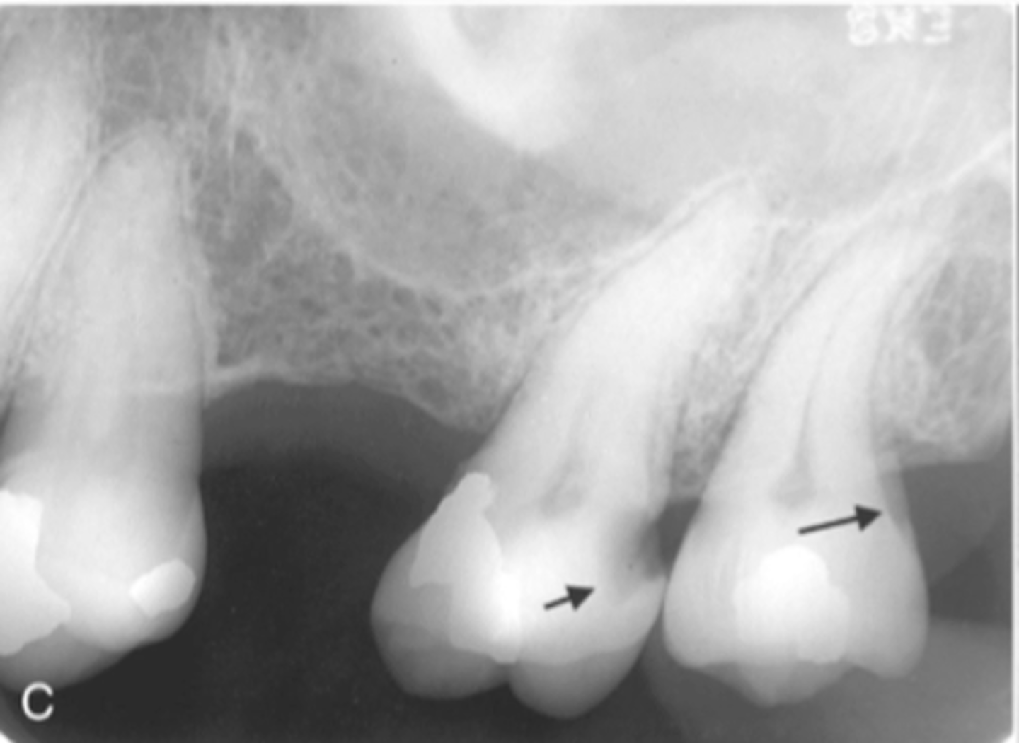
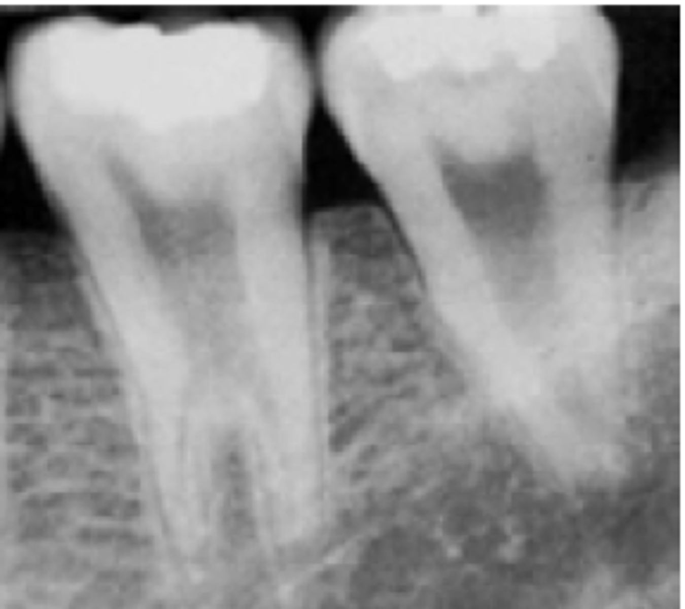
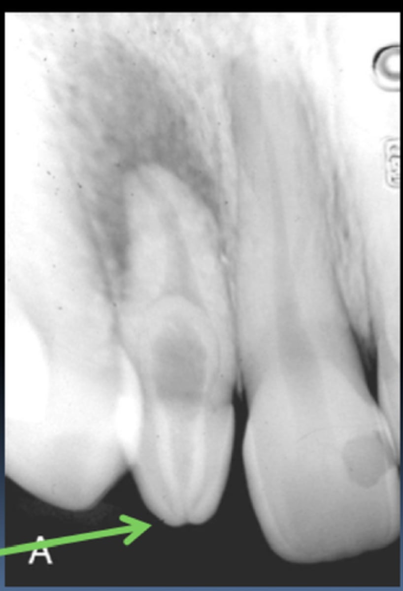
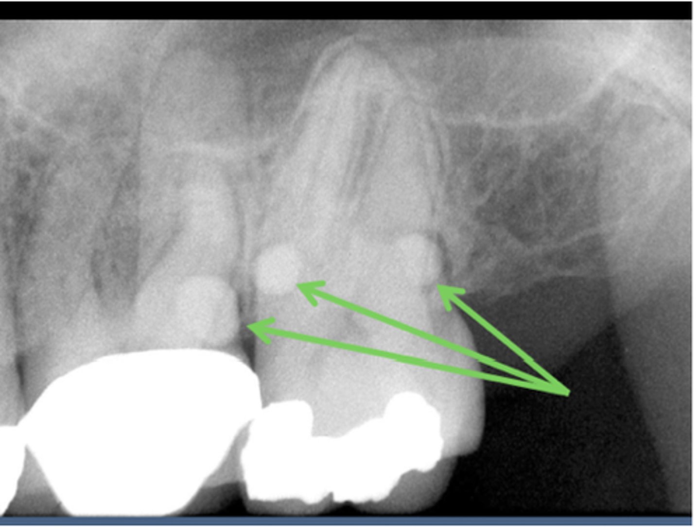
Enamel Pears
Enamel globules of 1-3mm in diameter on root of molars.
Attrition
Abrasion
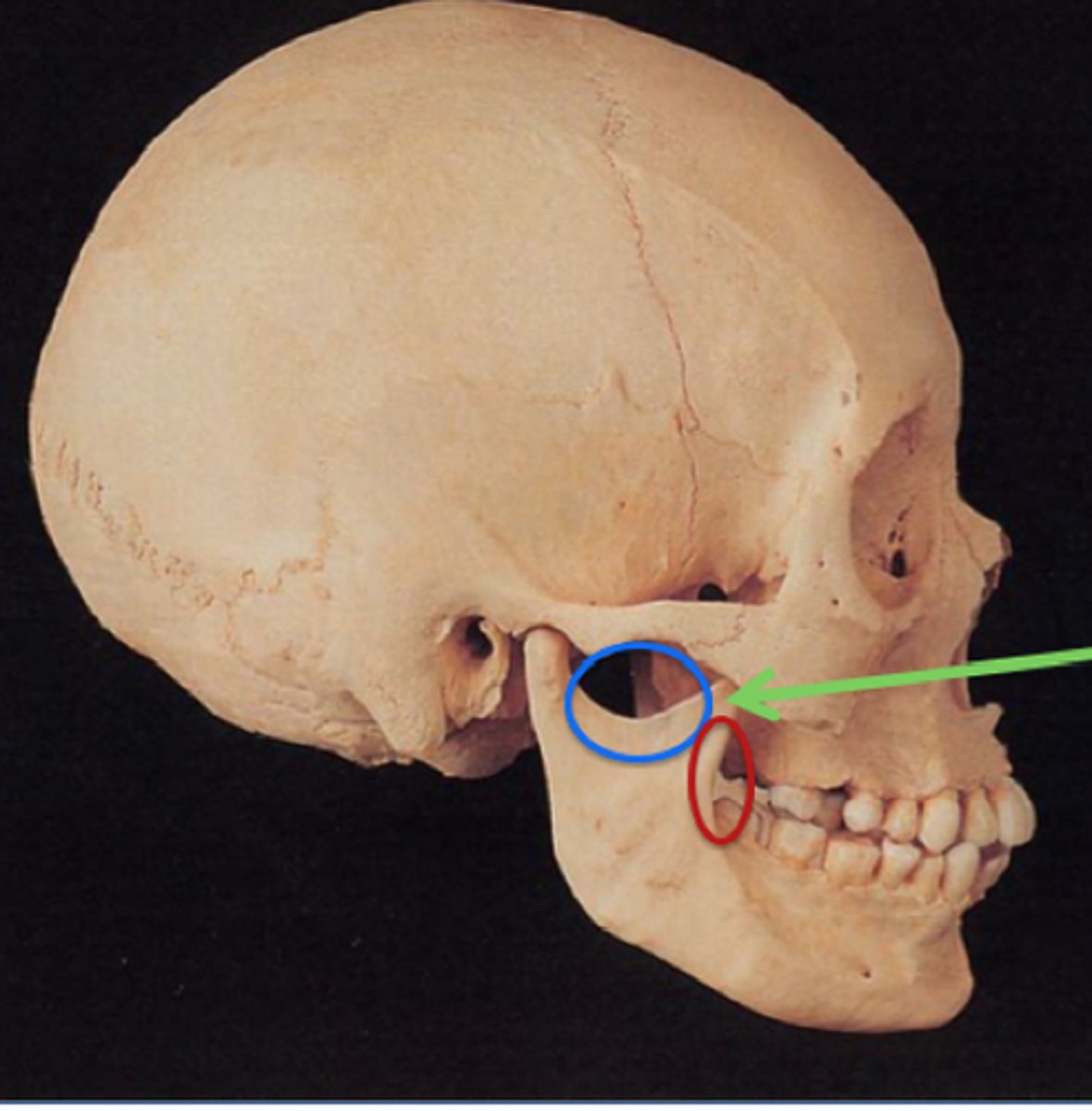
Blue - Mandibular Notch
Green - Coronoid Process
Red - Coronoid Notch
Label blue red green
Nasal Aperature (Anterior Nasal Rim)
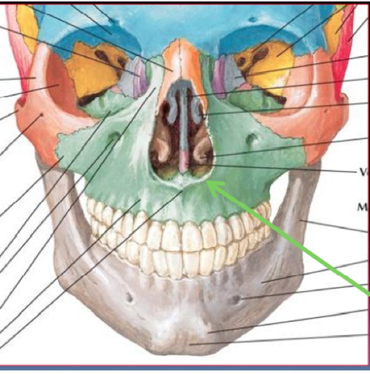
Ethmoid Bone
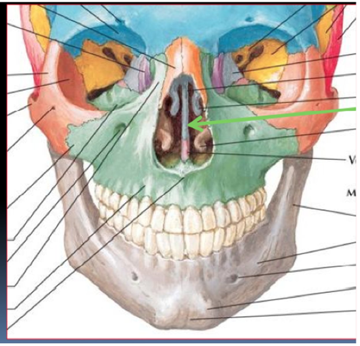
green - Alveolar Process
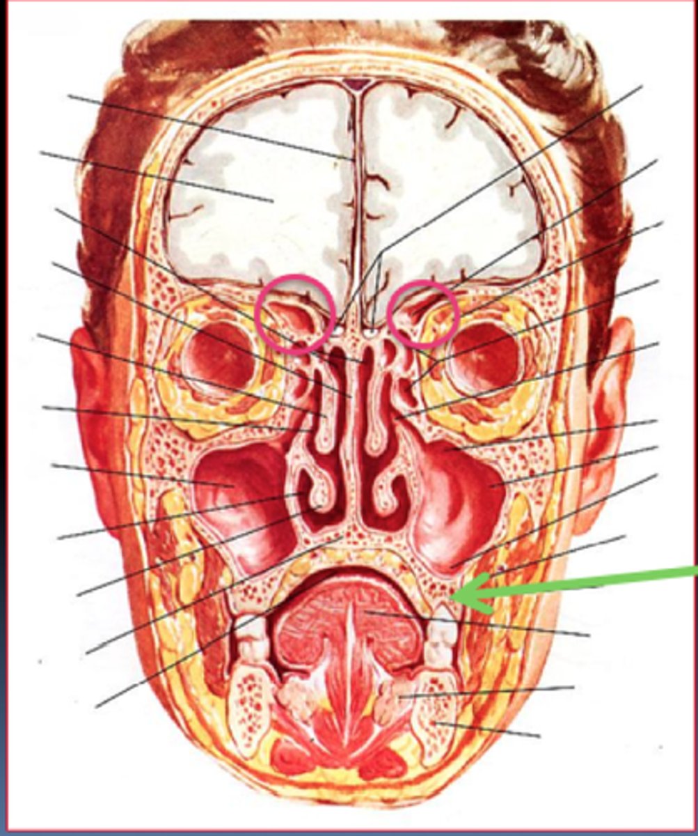
pink - frontal paranasal sinuses
Pink Circles?
Green arrow?
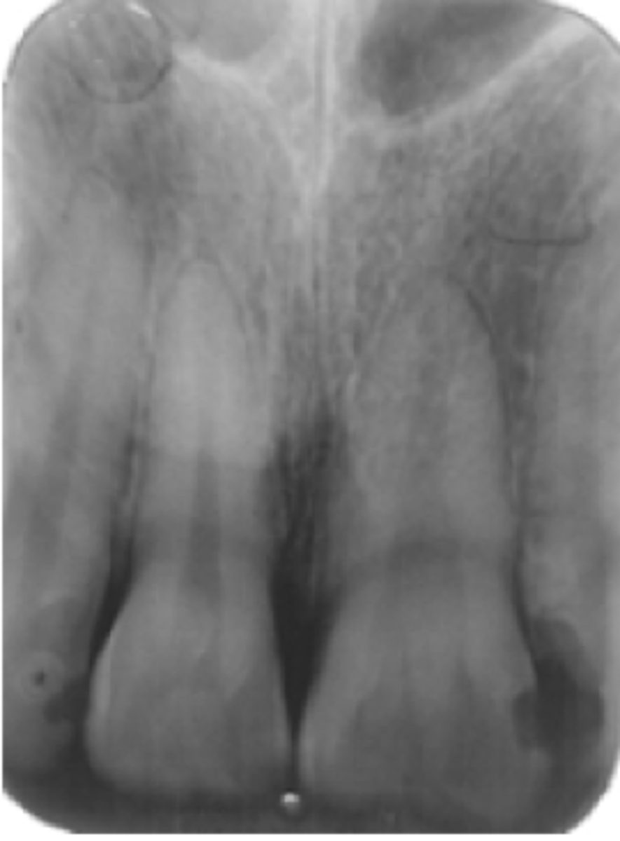
Anterior Nasal Spine
Anterior Nasal Spine
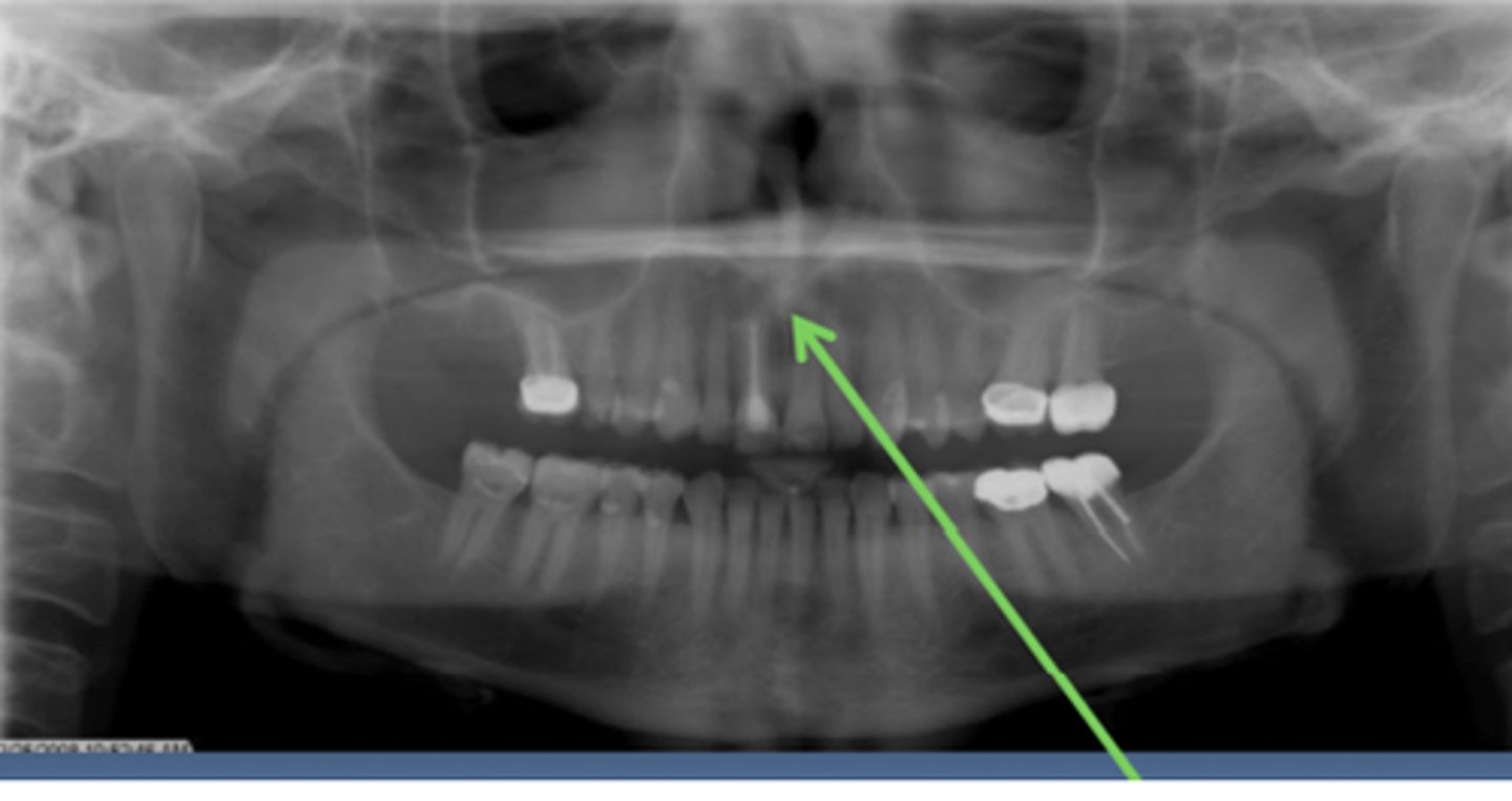
Hamular Notch (Green)
Maxillary Tuberosity (Blue)
Identify the triangular space the green arrow is pointing too.
Identify what is circled in blue
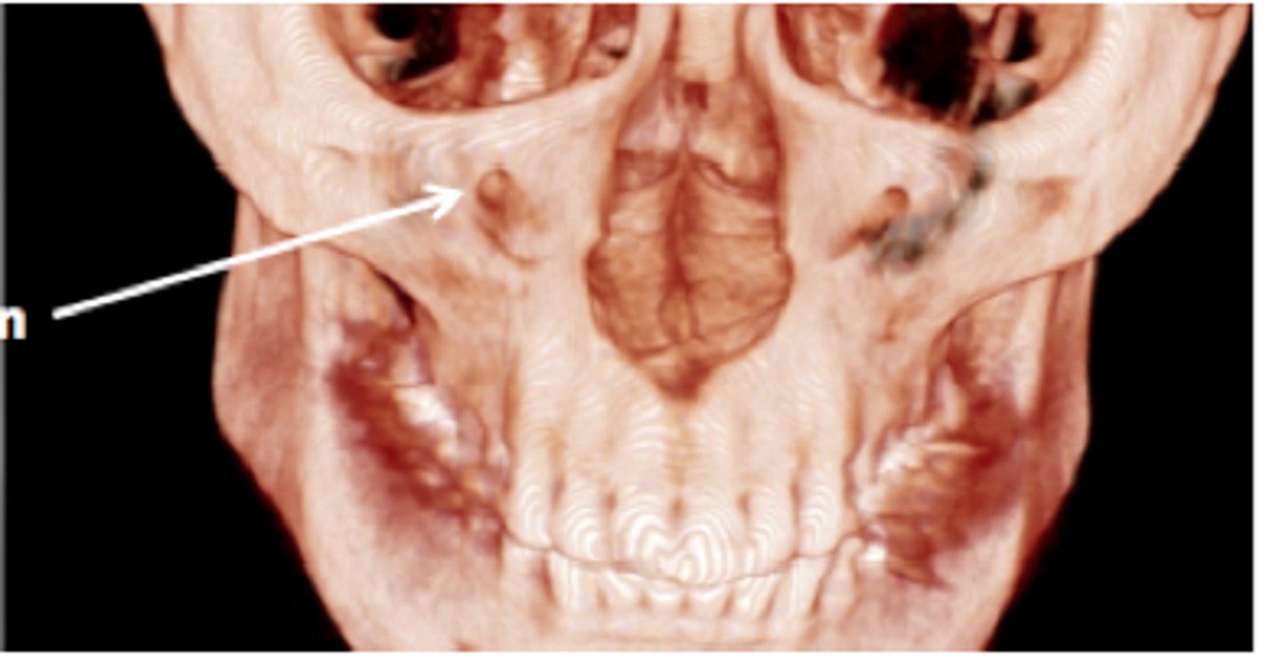
Infraorbital Foramen
Pterygoid Hamulus
Recurrent Caries Lesion
On M and D of 19
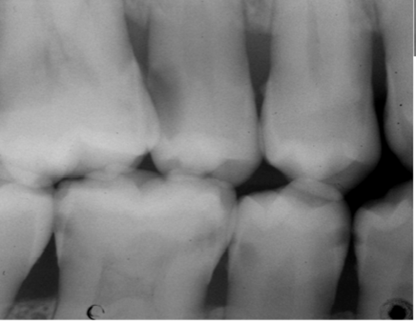
Cervical Burn Out (Blue)
Arrow points to Occlusal Caries Lesions
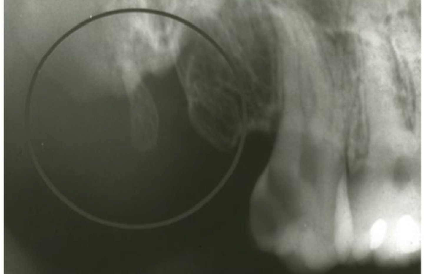
Focal Cemento-Osseous Dysplasia
Lesser Wing of Sphenoid Bone