Practical exam #2
1/62
There's no tags or description
Looks like no tags are added yet.
Name | Mastery | Learn | Test | Matching | Spaced | Call with Kai | Chat |
|---|
No analytics yet
Send a link to your students to track their progress
63 Terms
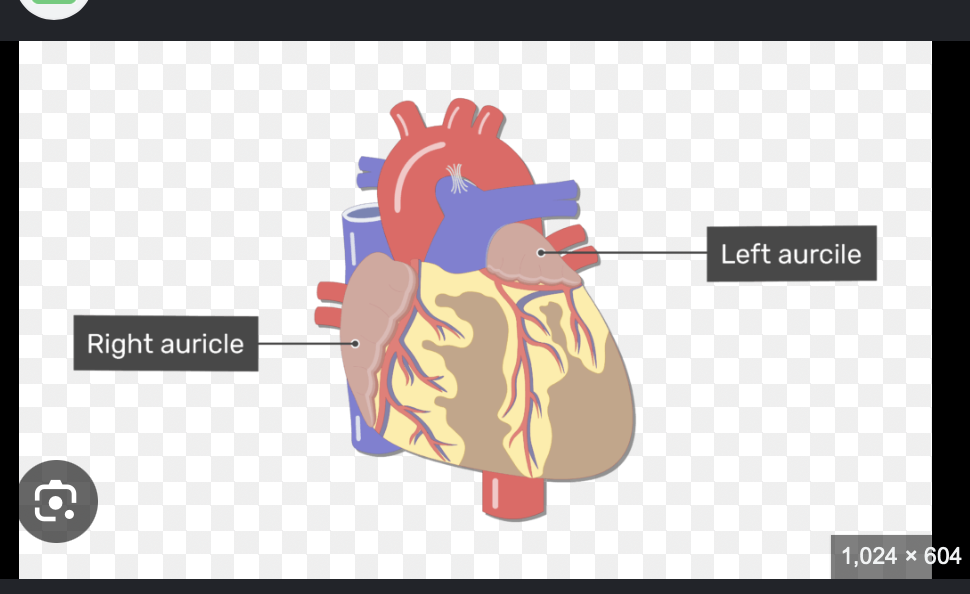
Auricles of the heart
They are flaps on top of the atria that extend when the atria fills up a lot with blood and needs extra space
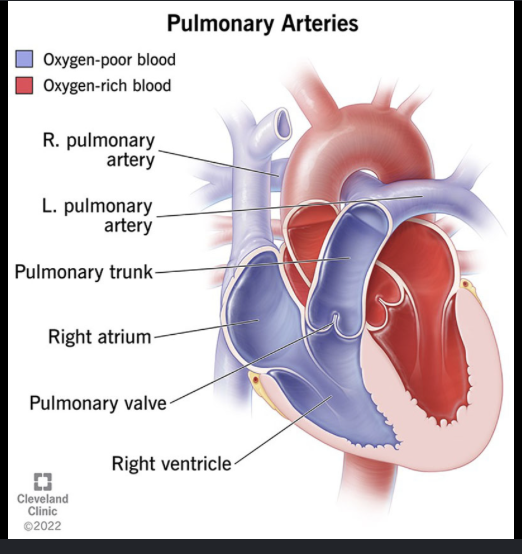
Pulmonary arteries
Pulmonary veins
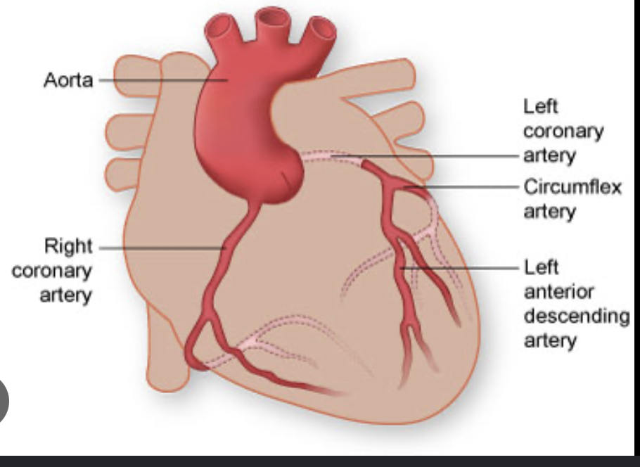
Coronary arteries
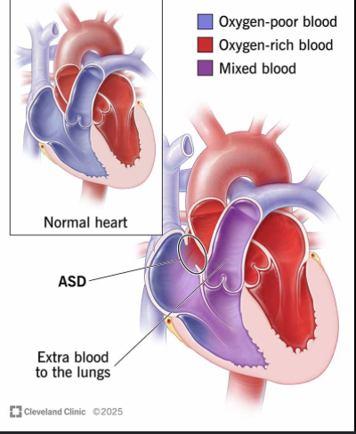
Atrial septum
CABG stands for?
coroanry artery bypass grafting
When plaque builds up in an artery they take a smaller blood vessel (vein or artery) and stitch it onto the healthy part of the artery to bypass onto another healthy part
Main diferences between fetal heat and blood flow
It’s 3 shunts
Foramen Ovale (door between the atrias) where blood skips ventricles and jsut goes from atrium to atrium through this hole
Ductus arterious (vessel connecting right atrium to the pulmonary artery), this is incase the blood slips from aorta into the ventricle so that blood goes back to circulation instead of lungs
Ductus venousus: (connects umbilical vein to the inferior vena cava) allows for fresh oxygen from palcenta to bypass liver to heart and brain
Foramen ovale
door between the atriums where blood skips ventricles and jsut goes from atrium to atrium through this hole
Ductus arterious
(vessel connecting right atrium to the pulmonary artery), this is incase the blood slips from aorta into the ventricle so that blood goes back to circulation instead of lungs
Ductus venousus
(connects umbilical vein to the inferior vena cava) allows for fresh oxygen from palcenta to bypass liver to heart and brain
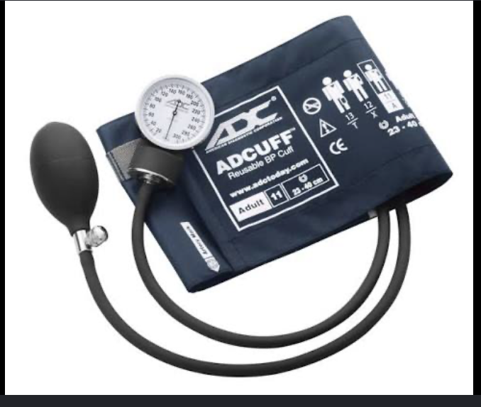
Sphygmomanometer
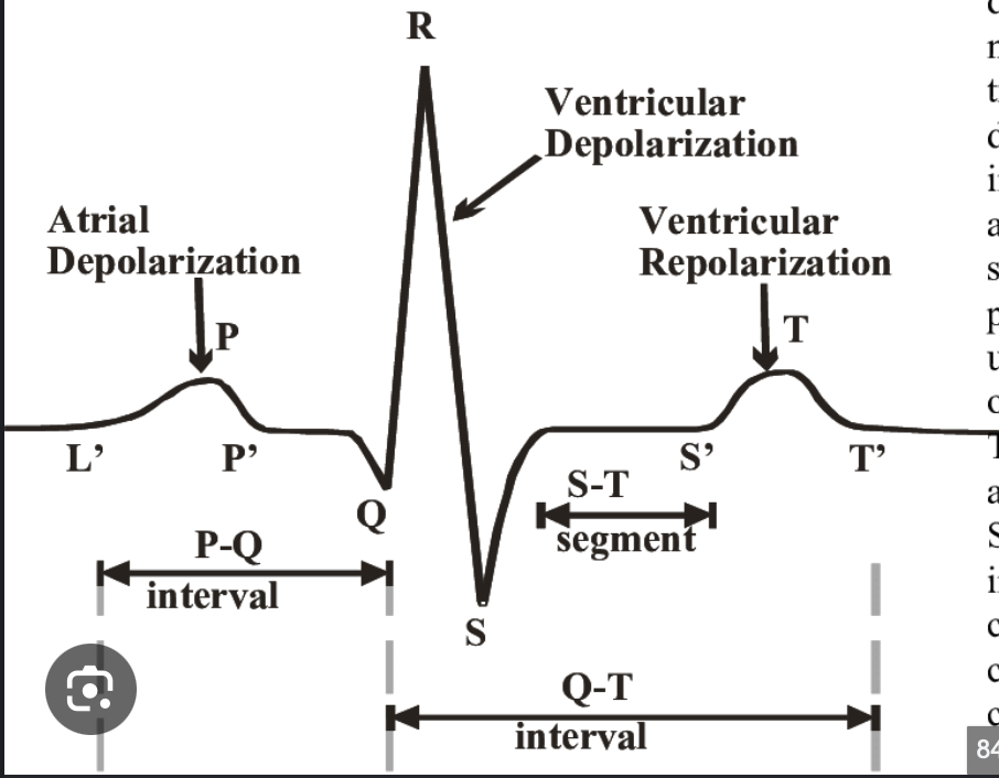
ECG trace
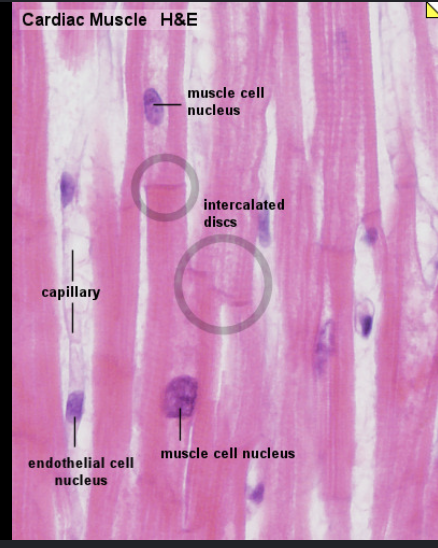
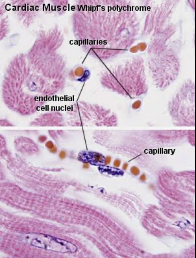
Heart muscle/thin cardiac muscle
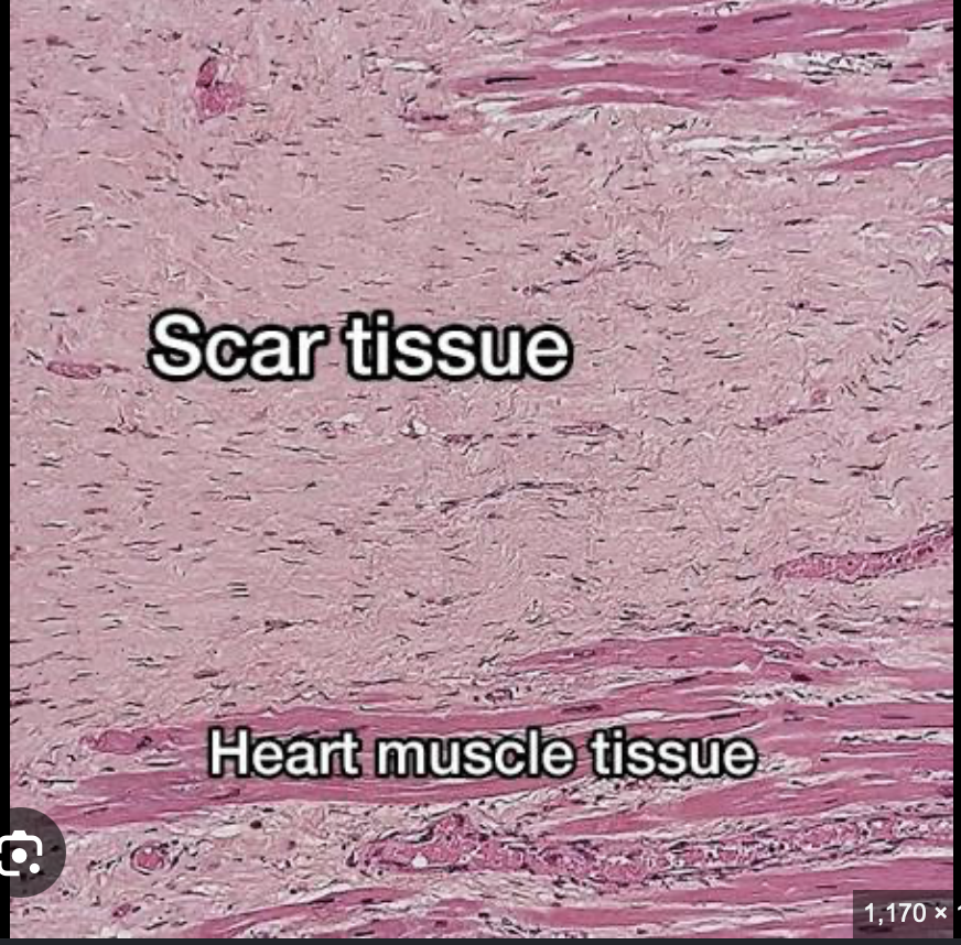
Myocardial infraction
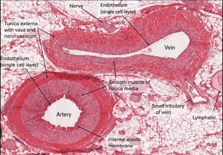
Vein vs Artery
Capilalries (3 main types)
Continous: cells packed with/ tiny cracks (only allows small molecules like water & ions through)
Fenestrated: tiny pores to allow bigger molcules to pass found in kidney & small intestine for absorbtion
Sinusoidal: massive large cells that allow for whole cells & proteins to pass (liver, bone marrow & spleen)
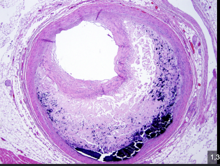
atherscerosis
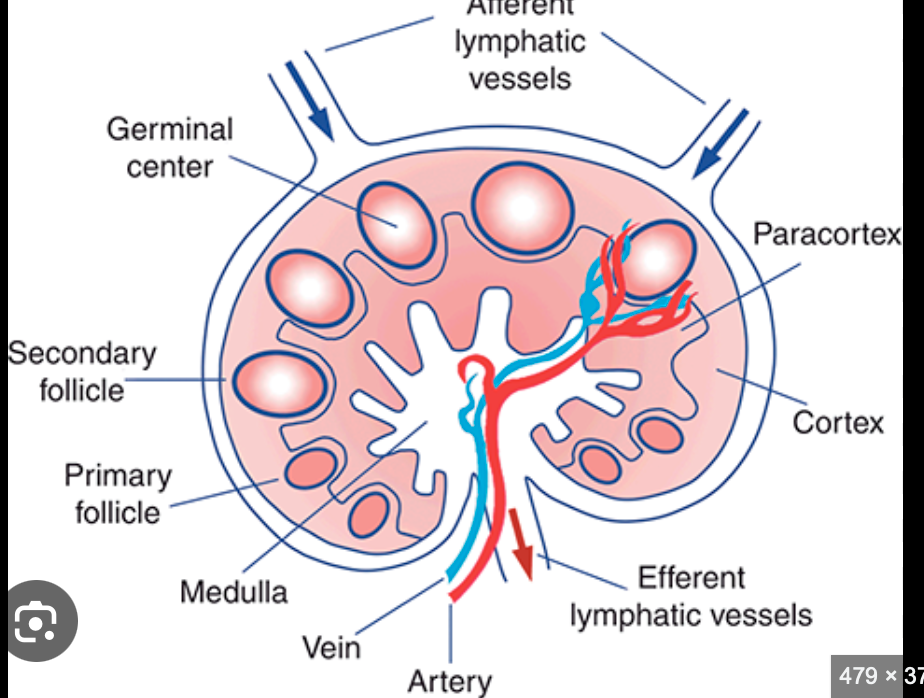
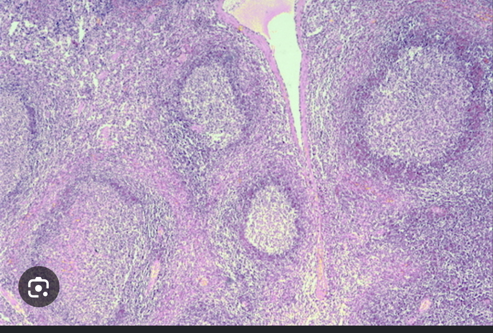
lymph node
capsule
deep cortex
medulla
efferent afferetn vessles
germinal center
Where are T cells created?
in the bone marrow but mature in the thymus
Stored in paracortex
Where are B cells created?
created and mature in bone marrow
Stored in germinal centers/ cortex of lymph node
Flow of lymph in lymph node
Afferent lymphatic vessels
subscapular sinus
trabecular (cortical) sinuses
medullary sinuses
efferent lymphatic vessel
Lymph node
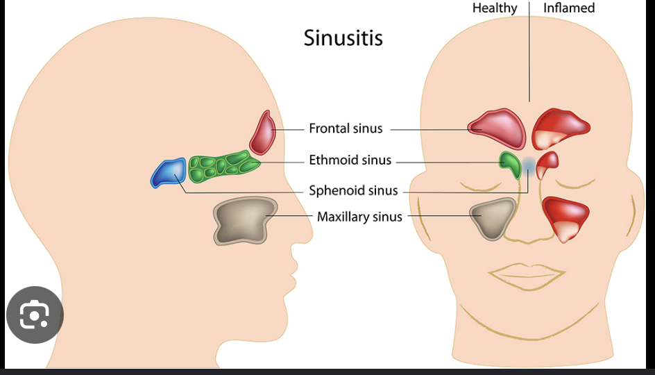
Sinuses
Pharynx
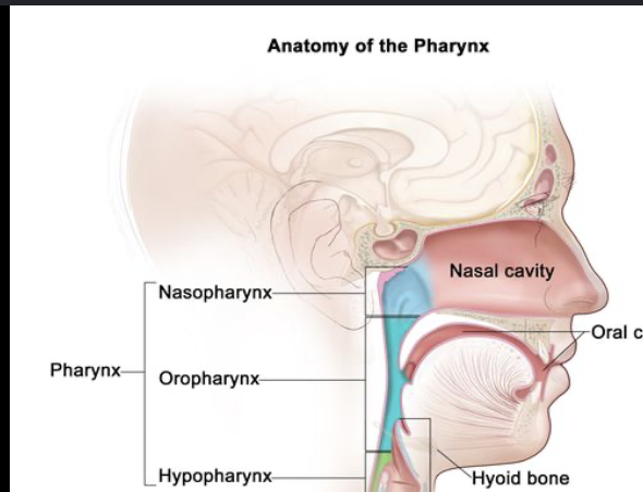
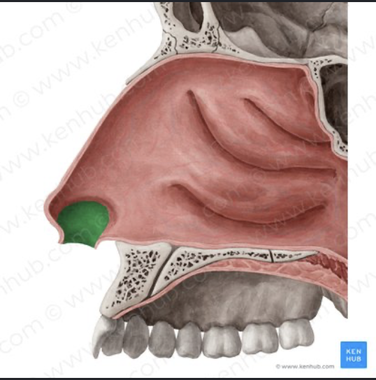
Nasal vestibule
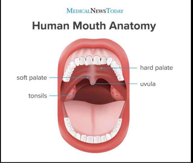
Hard palate vs soft palate
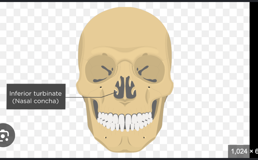
Nasal conchae
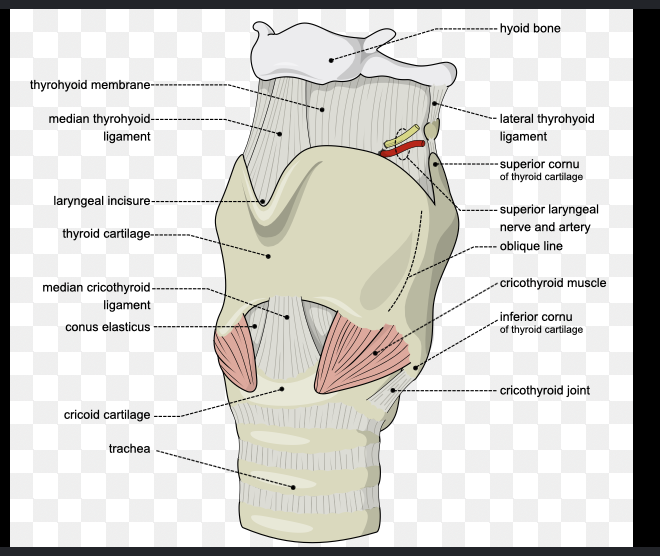
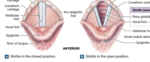
Larynx
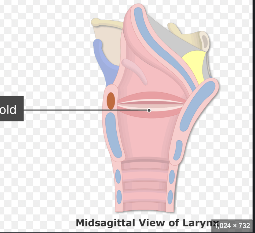
Vestibular folds/vocal folds/glottis
Epiglottis
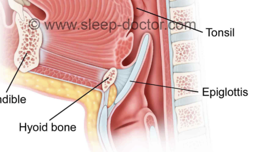
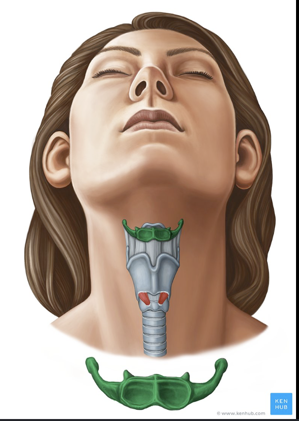
Hyoid bone
Seondary and tertiary bronchi plus bronchioles
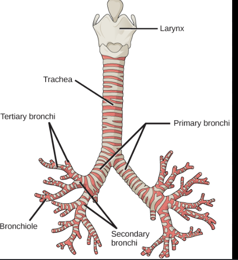
What bronchi seerates into lobes?
The secondary bronchi, they seperate giving the air to the 2 lobes on the left and the 3 lobes on the right
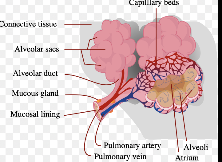
Alveoli sacs, individual alveolus and capillaries
steps in involuntary reflex
once bolud reaches pharynx ts inovulntary
soft palate rises to close nasal cavity
Larynx (voice box) moves up & forward
Epiglottis folds over larynx to prevent food from going into trachea
Upper esophageal sphincter relaxes letting food into esophagus
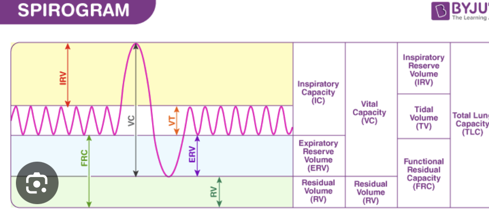
How does a wet spirometer measure each of the volumes?
CANNOT measure total lung capacity bc we cant egt the unmeasurable residual volume
Inspiratory reserve volume: bell drops lower than normal breathing
Expiratory reserve volume: bell goes higher than normal breathing
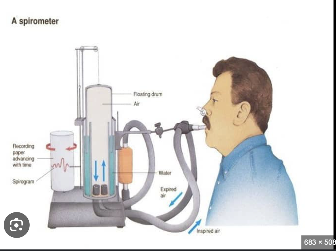
What does a wet spriometer help diagnose?
Restrictive and obstructive lung disease
Asthma
COPD
Cystic fiboriss
Pulmonary fibrosis (restrictive)
Scoloiosis (restrictive)
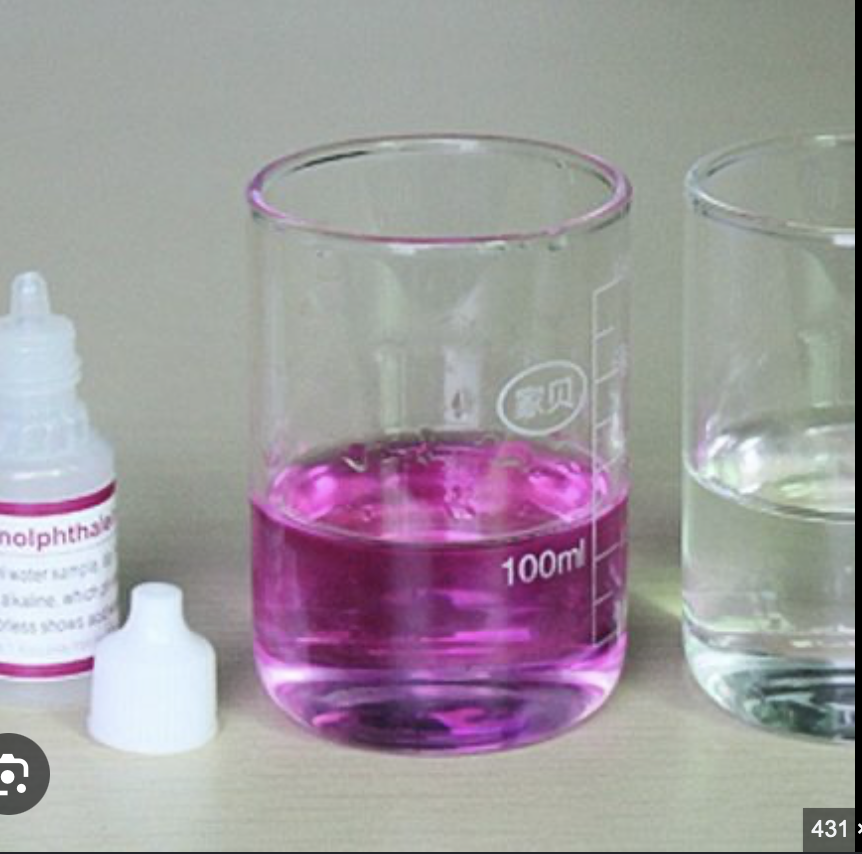
phenolphthalein
measures acidity
pink when basic, colorless with onyl CO2 exposure NO O2
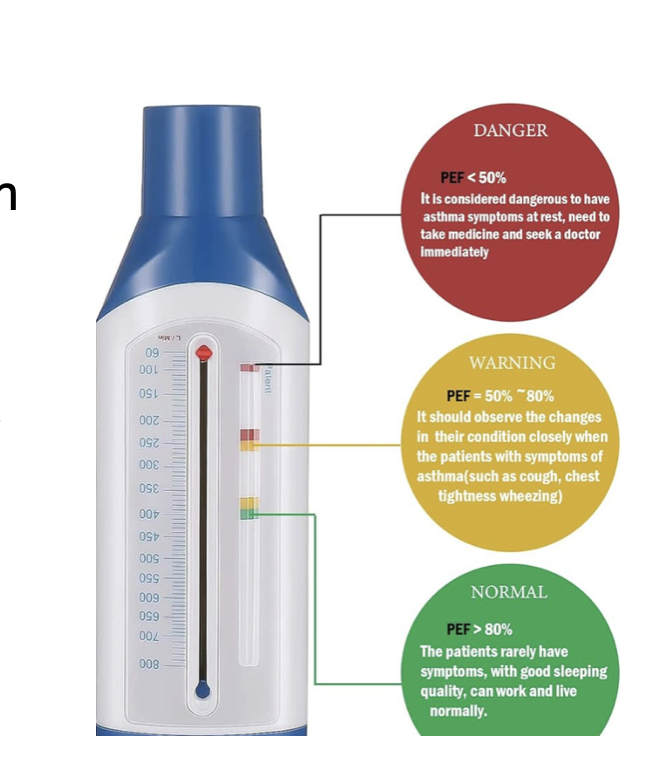
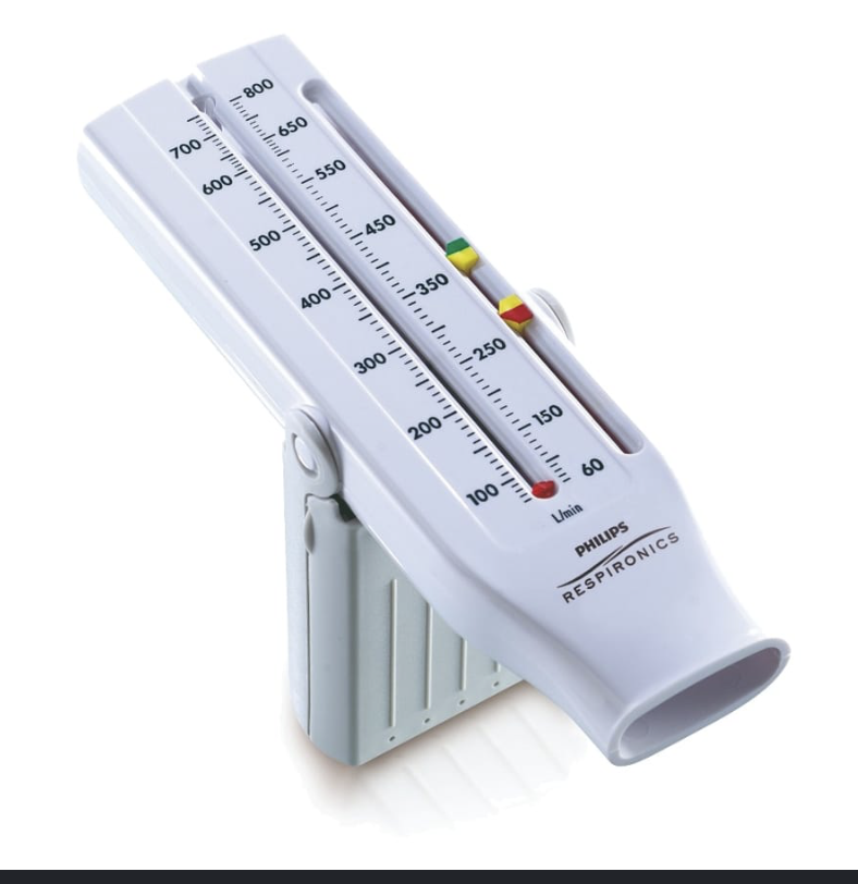
Peak flow meter
Measures how fast you can push air out of your lungs can predict asthma attacks before they happen
Oximeter
finger thing that measure how much of your blood is carrying oxygen
Hypoxemia or COPD can be diagnosed
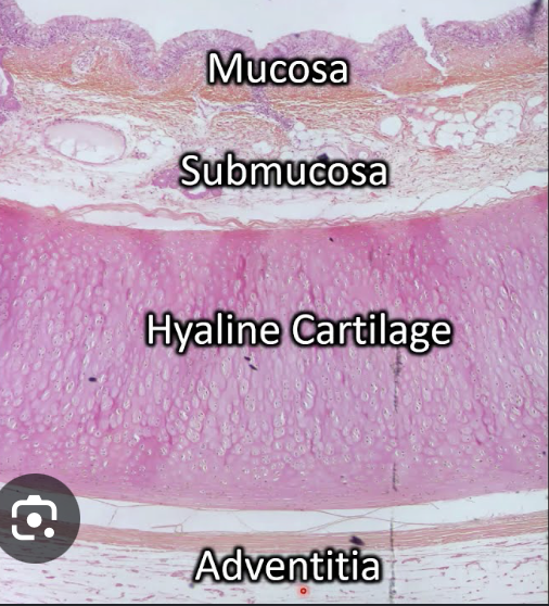
Trachea histology tissue types
Mucosa: pseudostartfiied columnar epithelium, loose conenctive laminia propia tissue
Submucosa: Connective tissue
Hyaline cartilage: cartilage plus smooth muscle
Adventitia: areolar connective tissue
Terminal bronchiole
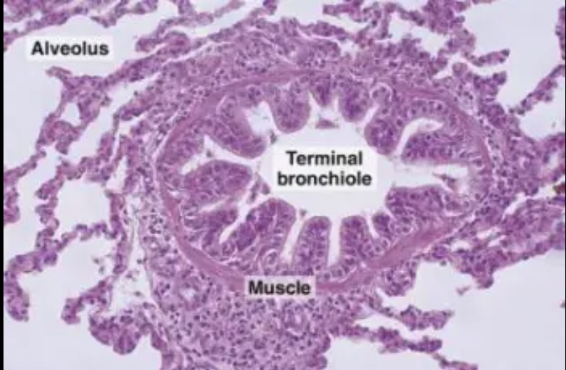
Alveolus
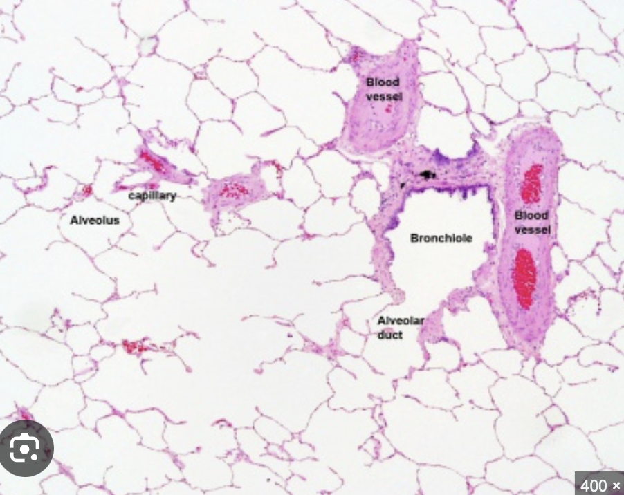
Alveolar sac
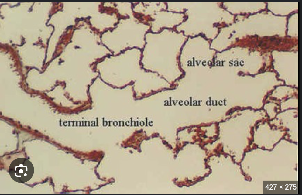
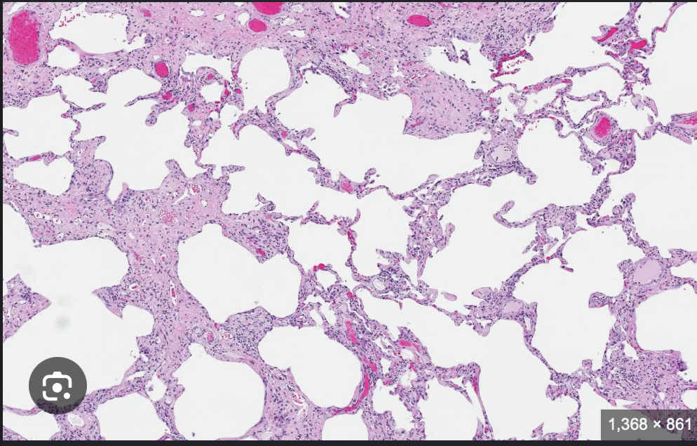
Smokers lung notice how you can hardly norice difference between alveolus & terminla bronchiole bc vronchiole lack thickening around them and too wide
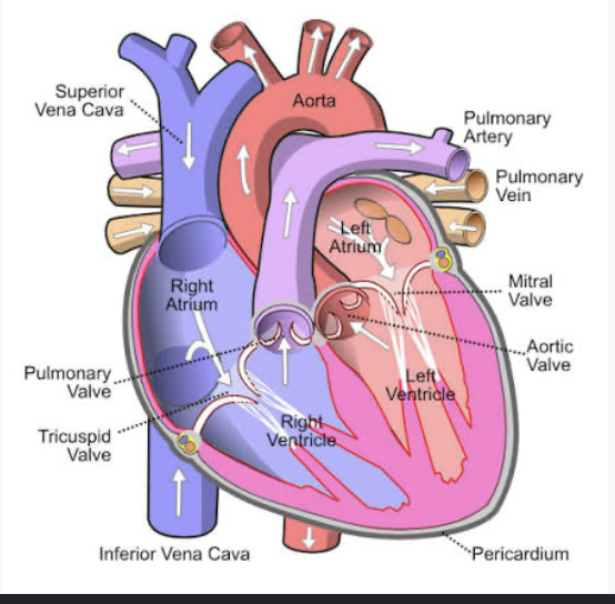
“Lub” sound
Atria valves closing (tricuspid & mitral)
"dub” sound
saortic and pulmonic valves closing
What valves open at the same time?
Aortic, pulmonary, mitral & tricuspid valves
The heart has its own personal blood supply, what supplies it?
coronary arteries and veins
Humans have an inferior vena cava while sheep have:
a posterior vena cava
Heart wall from outer to deepest
Pericardium: outer
Myocardium: middle
Endocardium: deepest (areolar tissue)
Nodal cells
specialized cardiac muscle cells that produce and conduct electrical currents to myocardium for beats
SA controls the atria, what does the AV node do?
AV node delays electrical signals so atria can fully contract and empty into ventricles
What do the bundle of HIS and purjinke fibers do?
just make sure the ventricles act right
Spleen
largest lymphatic organ in the body
red pulp of spleen
Blood filters here
phagocytes remove abnoraml RBCs and other antigens from blood
lymphocytes becomes acustomed ot the blood anitgens and makes antibodies
White pulp of spleen
Contains lymphocyte nuclei (stains purple)
Glottis is the full structure? epiglottis is the smile like fold on bottom. vocal fold= split in middle
Pulmonary Ventilation
breathing, moving air in & out of lungs
External Respiration
exchange of gases between gas and blood
Internal Respiration
Exchange of gases between lung and tissues
spirogram
Peak flow meter levels of danger
400=normal good PEF= 80%