Sectional Sonography Exam 3: Heart and Hemodynamics
 Call Kai
 Learn
Learn Practice Test
 Spaced Repetition
 Match
Match Flashcards
 Knowt Play
1/100

There's no tags or description
Looks like no tags are added yet.
Name | Mastery | Learn | Test | Matching | Spaced | Call with Kai | Chat |
|---|
No analytics yet
Send a link to your students to track their progress
101 Terms
What does the circulatory system include ?
heart blood vessels and lymphatics
left vetricle > aorta
small arteries
arterioles
capillaries
venules
small veins IVC and SVC > right atrium
What is the purpose of the circulatory system ?
transports O2, gases, nutrients, and other essential substances to tisssues and working muscles. Carries away waste products of cells to be excreted.

Thorax
upper part of the body, greater part is occupied by the lungs, which are enclosed by the pleural sac
separates the addominal cavity from the thorax
diaphragm
list the medial and lateral portions of the thorax
medial: mediastinum
lateral: pleurae and lungs
what does the sternum consist of anteriorly ?
manubrium, sterni body, xyphoid process

what does the thymus gland get replaced with with age ?
fat
Heart
hollow muscular organ
250-300 gm

What is the heart considered ?
anteposterior structure with its right sided chambers located more anterior than its left-sided chambers
What is the heart protected by ?
the sternum and rib cage anteriorly and the vertebral column and rib cage posteriorly
Heart Surfaces (3)
1. Sternocostal is anterior
2. Diaphragmatic (inferior and apex) Apex is inferior and forms left ventricle.
3. Base is posterior
APEX
formed by the left ventricle. The apex points downward, anteriorly and to the left
BASE
formed by the left atrium into which the pulmonary veins enter from the lungs. Broad superior portion of the heart that is opposite the apex. Projects superiorly and posteriorly to the right. Sometimes called the posterior surface
Pericardium
A fibroserous sac that surrounds the heart and proximal portions of the great vessels.
Serous pericardium
a thin, transparent serous membrane which has two layers.
Parietal pericardium
(outer layer) forms smooth moist lining for the fibrous pericardium.
Visceral pericardium
(inner layer) or epicardium covers and forms the outer layer of the heart wall
Pericardial Cavity
area in between visceral and parietal layers is a potential space
contains small amounts of serous fluid to prevent friction and allow for easy gliding during heart contractions
pericardial effusion
Excess fluid (fluid accumulation)
3 layers of heart wall
Epicardium: visceral layer- outer most layer.
Myocardium: heart or cardiac muscle - middle layer
Endocardium: a thin layer of endothelium, inner most layer. *Also covers the heart valves and is continuous with the endothelial lining of the blood vessels.
Atrioventricular valves
allows inflow of blood. Blood from atria’s to ventricles. Low pressure.
Right AV valve =
tricuspid valve
left AV valve =
bicuspid/mitral valve
Semilunar valve
allows outflow or exits of the ventricles. High pressure
Pulmonary semi lunar valve
outflow of blood from RV into pulmonary trunk
Aortic semi lunar valve
outflow of blood from LV into aorta
Rt atrium
Receives blood from the coronary and systemic circulation. Receives blood from the IVC & SVC. Coronary sinus receives blood that supplies the heart itself
Rt. Ventricle
anterior surface of the heart. Receives blood from RA through the Tricuspid Valve and ejects blood into the pulmonary artery/trunk for 02 exchange from lungs.
Left atrium
most posterior, forms most of the base. Four pulmonary veins return oxygenated blood to the heart form the lungs to the LA.
Left ventricle
receives blood through the mitral valve or bicuspid valve
has much thicker wall because it has to pump blood throughout the body
Blood exits into aorta through the aortic semi lunar valve
INTRAVENTRICULAR SEPTUM
Thick and muscular partition between the right and left ventricles. Has a muscular interventricular septum and a membranous interventricular septum.
5 Sections of Aorta
1. Root of Ao (which comes from LV) 2. ascending 3. descending 4. abdominal 5. bifurcation into iliacs

Aortic Arch
a continuation of the ascending aorta, Lies behind the manubrium sterni and runs upward, backward and to the left anterior to the trachea then passes down to the left where is continues as the descending aorta.
• Brachiocephalic artery arises on the right and divides into the right CCA and Subclavian arteries.
• The left CCA and subclavian arteries comes directly off the aortic arch.

3 layers of arterial wall
Tunica Intima: most inner layer consists of endothelial cells, which allows for smooth blood flow
Tunica Media: middle layer consists of smooth muscle fibers with elastic and collagenous tissues.
Tunica Adventitia: outer layer of loose CT with bundles of smooth muscle fibers and elastic tissue. Thicker in arteries than veins.
ARTERIES ARE…..
• Vessels that carry away from the heart
• Hollow Elastic Tubes– the elasticity of larger arteries helps maintain steady flow.
• Pulsatile (from heart contractions)
• Faster flowing than veins
• Enclosed within a sheath that include an accompanying vein and nerve.
where is plaque found?
arteries
Where are thrombus/clots found?
veins
Veins appear and can collapse because
they have less elastic tissue than arteries
3 layers of vein wall
Inner single endothelial layer ***Extensions of this layer called semi lunar valves.
semi lunar valves
prevent blood reflux
3 layers of vein wall
Tunica Intima: most inner layer consists of endothelial cells, which allows for smooth blood flow
Tunica Media: Middle layer consists of smooth muscle & elastic fibers. Much thinner than arteries.
Tunica Adventitia: Outer layer consists of strong CT. Smaller than arteries.
VEINS ARE……...
• Vessels that carry blood back to the heart
• Hollow compressible tubes
• Have valves that allow blood flow only towards the heart (EXCEPT: IVC, SVC & COMMON ILIACS)
VALSALVA MANEUVER
Flow pattern with respiration
Inspiration
Vein lumen narrows
Expiration:
Vein lumen widens
CAPILLARIES
branches of arterioles
• Site of exchange of gases, nutrients, wastes etc. between organs, working muscles and other tissues.
• Single layer of endothelial cells..allows for permeability and/or diffusion.
VENULES
the smallest veins
Vasa Vasorum
Comprise the tiny arteries and veins that supply the walls of the blood vessels.
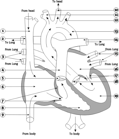
1
superior vena cava

2
Right pulmonary arteries
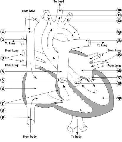
3
pulmonary veins

20
atrial septum

7
tricuspid valve

9
inferior vena cava
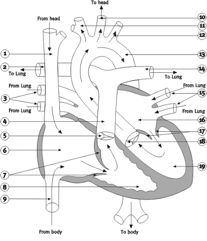
6
right atrium

8
right ventricle

5
pulmonary valve

4
pulmonary artery

11
brachiocephalic

10
left CCA

12
left subclavian

14
left pulomonary arteries
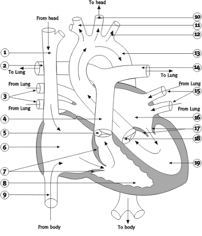
15
left pulmonary veins

17
mitral valve

18
aortic valve

21
ventricular septum

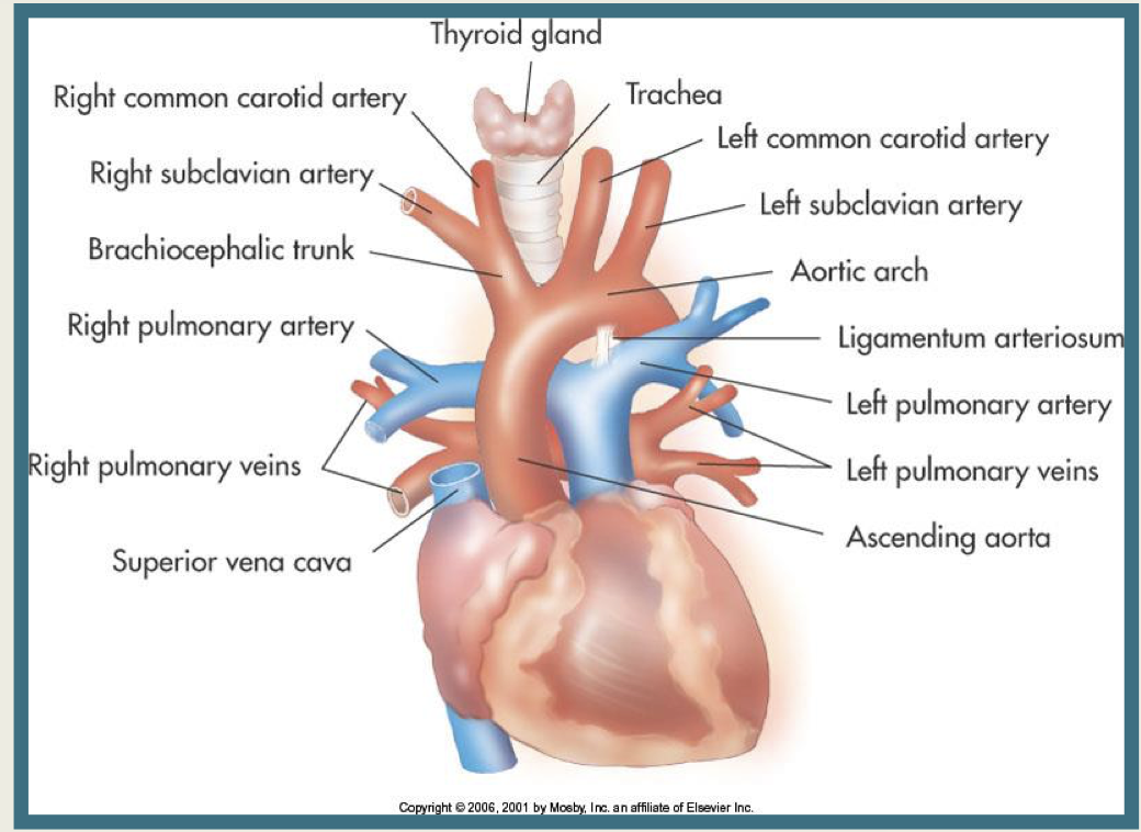
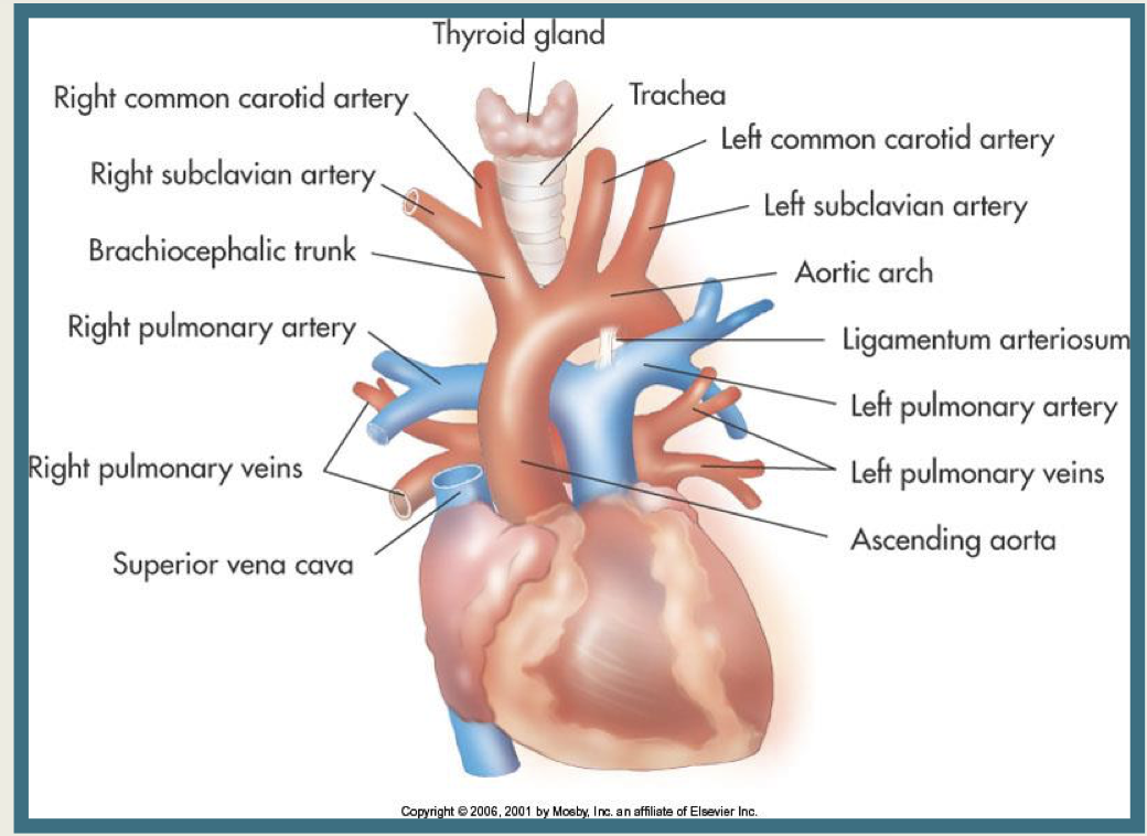
1
Right common carotid artery

2
right subclavian artery

3
brachiocephalic trunk

4
right pulonary artery

5
right pulmonary veins

6
superior vena cava

7
left common carotid artery

8
left subclavian artery
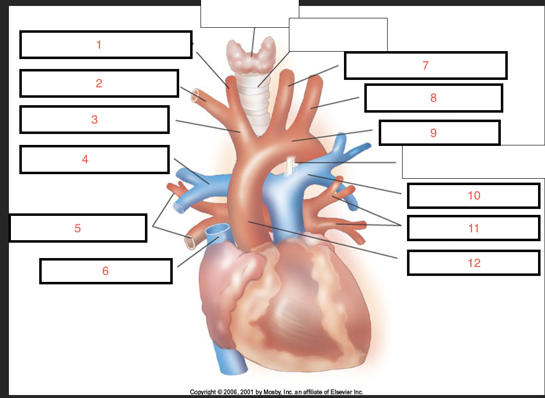
9
aortic arch
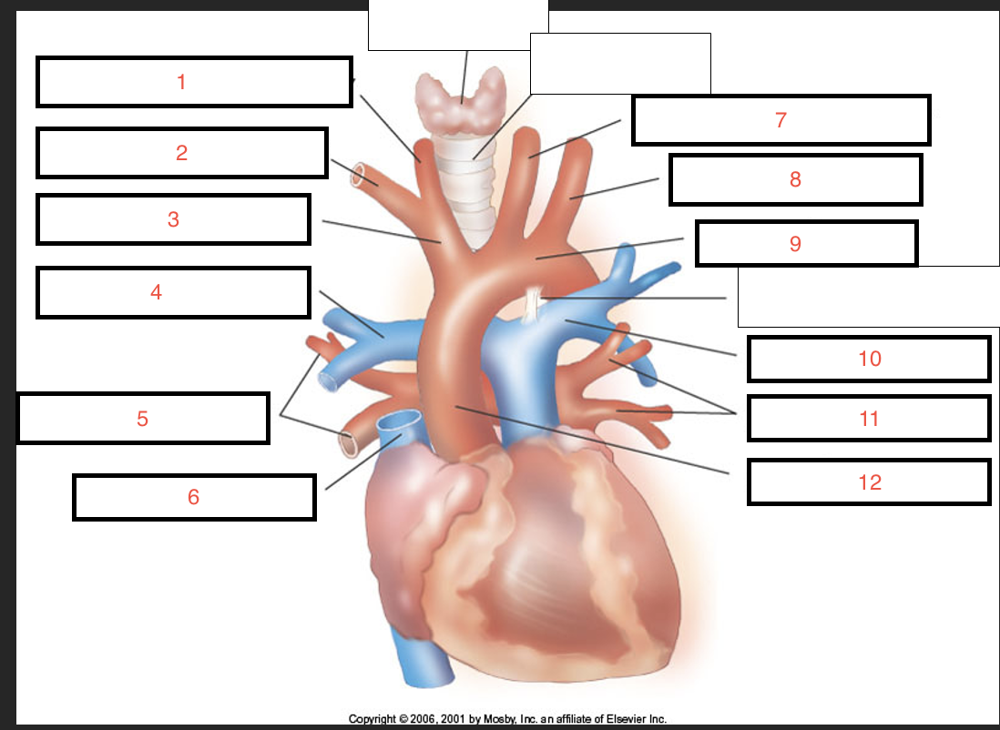
10
left pulmonary artery

11
left pulmonary veins

12
ascending aorta
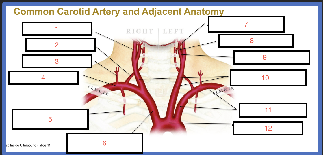
1
internal carotid

2
vertebral

3
thyrocervical trunk

4
subclavian
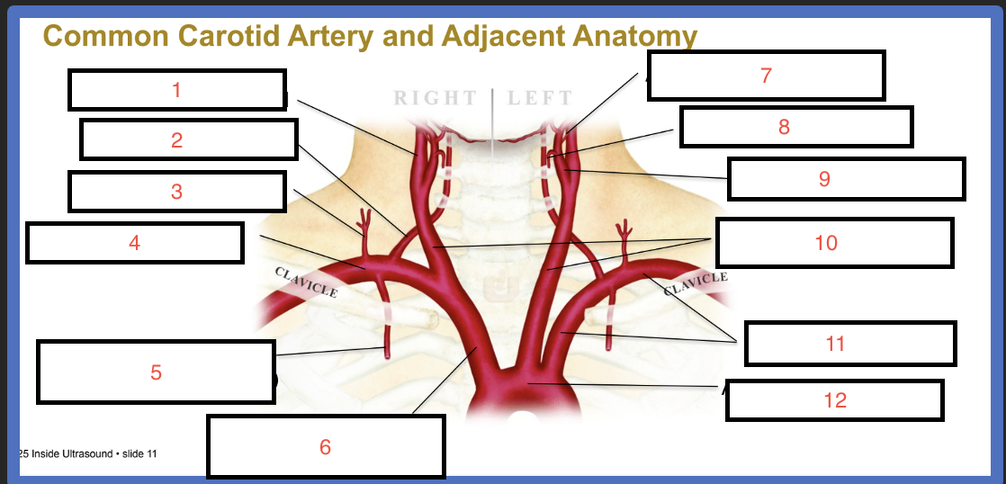
5
internal thoracic (internal mammary)

6
brachiocephalic (innominate)

7
ascending pharyngeal

8
superior thyroid
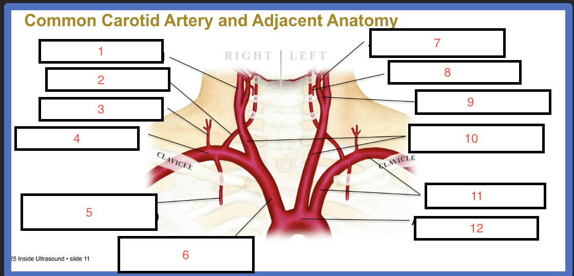
9
external carotid

10
common carotid

11
subclavian
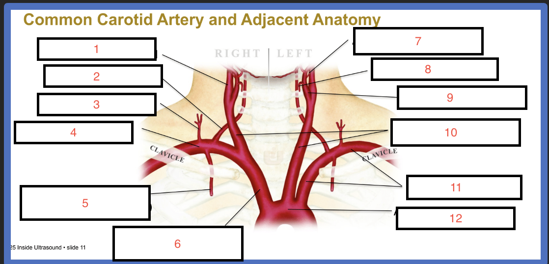
12
aortic arch
step 1 of blood flow in arteries
Heart: The heart is potential/pressure energy and is the source of energy. Pressure is greatest at the heart. Blood flowing away from the heart has a decreased pressure and is considered a pressure or energy wave.
step 2 of blood flow in arteries
Energy gradient must exist in order to move blood around.
step 3 of blood flow in arteries
Velocity: Velocity is greatest during systole or the acceleration phase. Pressure is evenly distributed in all directions. PEAK SYSTOLE is the greatest amount of energy. There is a decrease during diastole, the deceleration of the the pulse phase and when there is change in the direction of flow.
step 4 of blood flow in arteries
Compliance: Blood vessels will stretch with increased pressure unless there is plaque build up.
step 5 of blood flow in arteries
Resistance: High resistance vascular beds get a quick burst of blood during systole. Low resistance vascular beds get a constant blood supply.
Increase in vessel length = increase resistance
Increase in vessel diameter = decrease in resistance
step 6 of blood flow in arteries
Viscosity: Blood thickness/viscosity is the main resistance to flow. Increase in hematocrit = an increase in RBS and the thicker the blood. The interaction of the RBC to each other slows the blood down.
Fourth step of blood flow in veins:
4) Gravity: When erect, venous blood flow must overcome the effects of gravity, also known as hydrostatic pressure, in order to return the blood to the heart. Hydrostatic pressure results in venous distention and pooling of blood in the lower leg.
Second step of blood flow in veins:
Calf/muscle pump: Calf muscles surround the intramuscular and deep veins of the leg, propelling blood toward the heart. The effectiveness depends on the strength of the contraction and the existence of properly functioning valves, which prevent retrograde flow.
First step of blood flow in veins:
1) 1) Pressure/energy gradient: Left ventricle contracts at a pressure of 100 mmHg which drops to 20mmHg by the time the blood flow has reached the venules. Therefore a pressure gradient of 20mmHg must exist in order to get blood to flow from the venules to the RT ATRIUM
Third step of blood flow in veins:
Respiration: During inspiration the intrabdominal pressure increases and intrathoracic pressure decreases, resulting in the collapse of the IVC which will decrease or stop flow coming from lower extremities. During expiration the intrabdominal pressure decreases and intrathoracic pressure increases and flow to the heart is increased from lower extremities. VALSALVA