MRI Brain Pathology
 Call Kai
 Learn
Learn Practice Test
 Spaced Repetition
 Match
Match Flashcards
 Knowt Play
1/47

Earn XP
Description and Tags
vocabulary flashcards covering brain pathologies and related MRI appearance including neuro-oncology, vascular lesions, inner ear/orbit conditions, infectious/inflammatory diseases, and congenital brain malformations
Name | Mastery | Learn | Test | Matching | Spaced | Call with Kai | Chat |
|---|
No analytics yet
Send a link to your students to track their progress
48 Terms
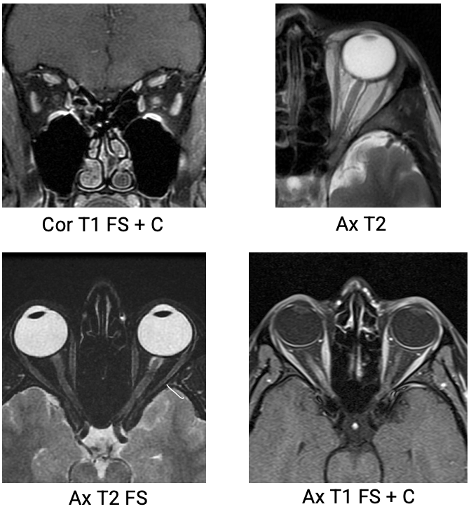
Optic Neuritis (Devic’s Disease)
Inflammation of the optic nerve
SYMPTOMS: blurred vision, loss of vision, eye pain, photopsia (flashes of light)
MR APPEARANCE:
Unilateral nerve swelling
T2: hyper
Flair: hyper
T1 + Gad: hyper enhancing region
Optic Nerve Glioma
low-grade, slow-growing tumor of the optic nerve
common in peds
frequently associated with Neurofibromatosis type 1 (NF1).

Acoustic Neuroma (Vestibular Schwannoma)
Benign tumor on 8th cranial nerve within the vestibule
SYMPTOMS: balance issues, sensorineural hearing loss, tinnitus, facial numbness when large
more common in adults in their 30’s
MR APPEARANCE:
T1: hypo to brain (63%), iso to brain (37%)
T2:
Heterogenously hyper to brain
may have associated peritumoral arachnoid cysts
T1 + GAD: intense enhancement
SWI: some show blooming due to presence of intratumoral microhemorrhages

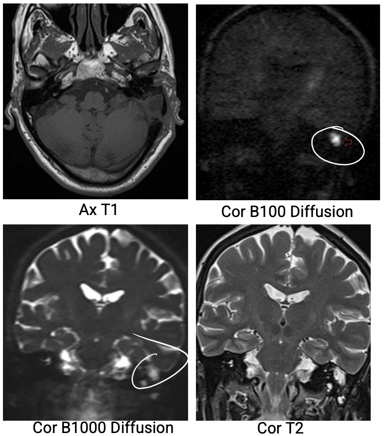
Cholesteatoma
Abnormal collection of skin cells deep within the ear
benign but can cause mass-like effects
hearing loss, dizziness, bleeding
MR APPEARANCE:
T1: hypo
T2: hyper ( > CSF)
T1+ GAD: no enhancement
may see thin, linear enhancement of margins
DWI: restricted diffusion
Meniere's Disease (Endolymphatic Hydrops)
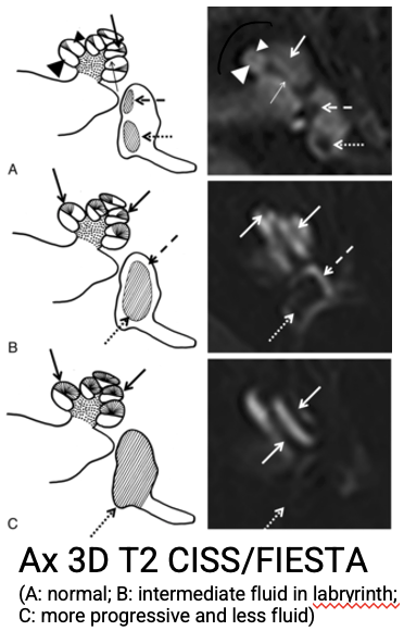
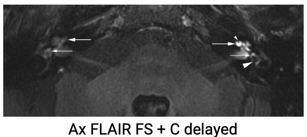
Inner ear disorder characterized by episodic bouts of vertigo, fluctuating sensorineural hearing loss, tinnitus, and ear fullness.
MR APPEARANCE:
FLAIR + GAD: 4 or 24hr delay shows IAC enhancement
T2 3D: eval of saccule, endolymphatic duct, choclear aqueduct
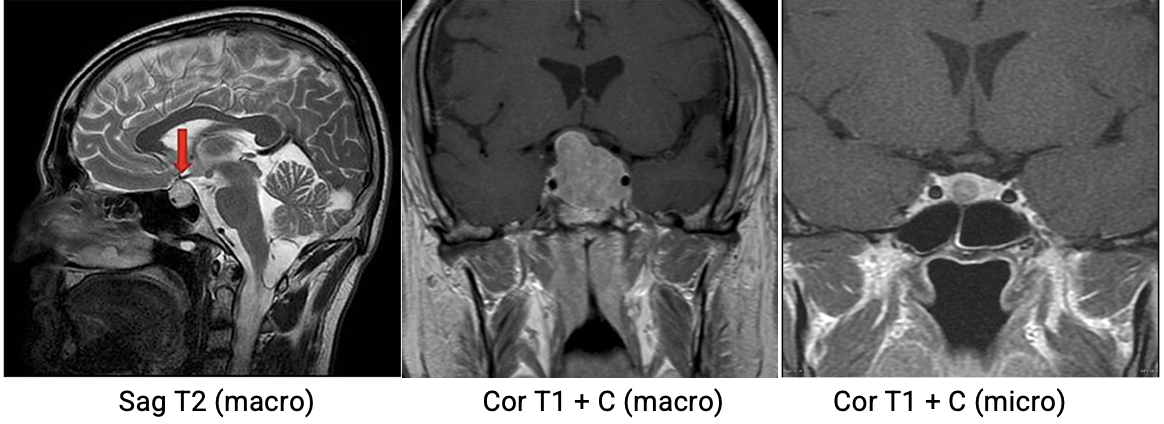
Pituitary Adenoma
Benign pituitary gland tumor
Microadenoma:
10mm or less
Macroadenoma:
10mm+
2x more common than micro
MR APPEARANCE:
T1: iso
T2: hyper
T1 + GAD: hypo to iso
Dynamic sequences beneficial for micro studies (mass will have delayed (90-120 sec) enhancement vs. normal pituitary tissue (60-80 sec))
Empty Sella
pituitary gland is shrunken or absent from the sella → filled with CSF
commonly correlated with idiopathic intracranial hypertension
Craniopharyngioma
Grade I tumor arising from squamous epithelium cells in the sella region
Adamantinomatous
most common
predominantly children
Papillary
predominantly adults
SYMPTOMS:
Headache, increased ICPs, visual disturbance, hormonal issues
MR APPEARANCE:
SOLID:
T1: iso - hypo to brain
T1 + GAD: vivid enhancement
T2: variable
CYSTS:
T1: iso - hyper to grey matter
T2: variable
~80% hyper

Pilocytic (juvenile) Astrocytoma
Grade I, slow-growing brain tumor
most common primary brain tumor in children
LOCATION: cerebellum and optic pathways
SYMPTOMS: increased ICP, hydrocephalus, headache, vision changes
MR APPEARANCE:
T1:
SOLID: iso - hypo to brain
CYSTIC: hypo unless hemorrhagic
T1 + GAD: vivid enhancement
cyst wall enhances in ~50% of cases
T2:
SOLID: hyper to brain
CYSTIC: hyper
T2*/GRE/SWI: signal loss if calcification or hemorrhage present

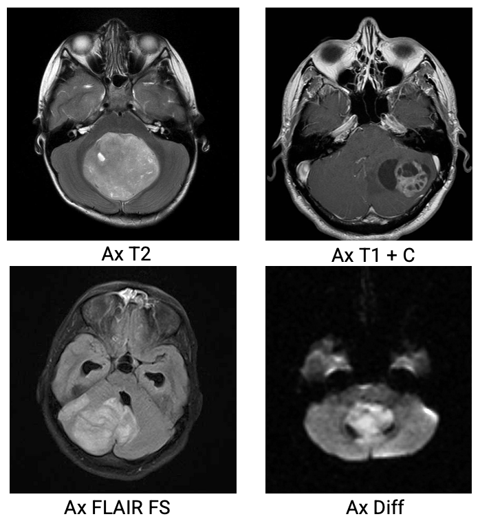
Ependymoma
Broad group of tumors that involve the lining of the ventricles
~10% of ped tumors
1/3 of tumors in kids 3 or younger
Grade II or III (Anaplastic Ependymoma)
Most located in posterior fossa
poor prognosis
MR APPEARANCE:
T1: solid parts iso to hypo to white matter
T2: hyper to white matter
SWI: foci of blooming from hemorrhage or calcification
T1 + GAD: heterogenous enhancement
DWI: solid parts may show restricted diffusion (esp. in anaplastic tumor)
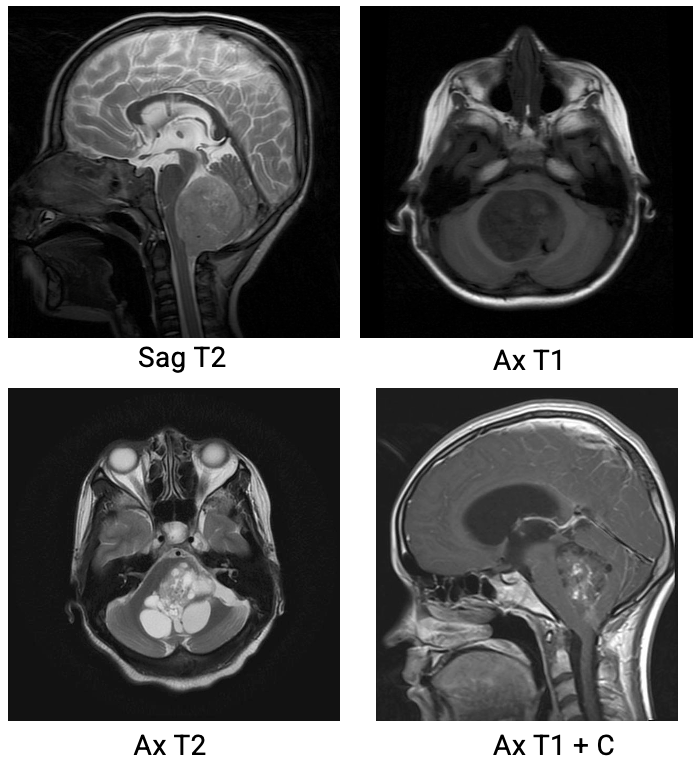
Medulloblastoma
Aggressive Grade IV tumor originating in posterior fossa
30-40% of peds post fossa tumors
Very fast-growing
Obstructive hydrocephalus common
Associated with Li-Fraumeni Syndrome
MR APPEARANCE:
T1: hypo to grey matter
T1 + GAD: 90% of tumors show heterogenous enhancement
T2/FLAIR:
iso - hyper to grey matter
hetero due to calcification, necrosis, and cyst formation
surrounding edema common
DWI: restricted diffusion due to hypercellularity
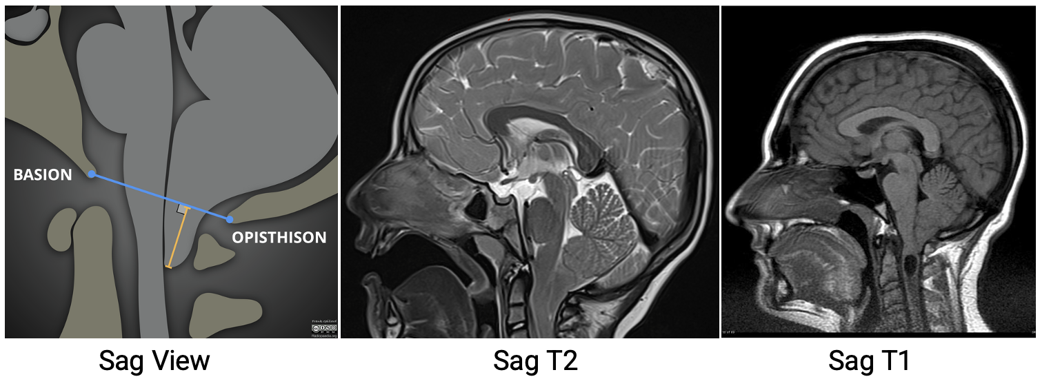
Chiari Malformation (Arnold Chiar)
Cerebellum and/or brainstem migration out of the cranial cavity
Cerebellar Tonsils most inferior portion of cerebellum
6 Variations
describe different degrees of brain tissue displacement or underdevelopment at the base of the skull
Type 1 most comon
Can be associated with Syrinx in cord due to increased ICP
Best demonstrated on SAG image
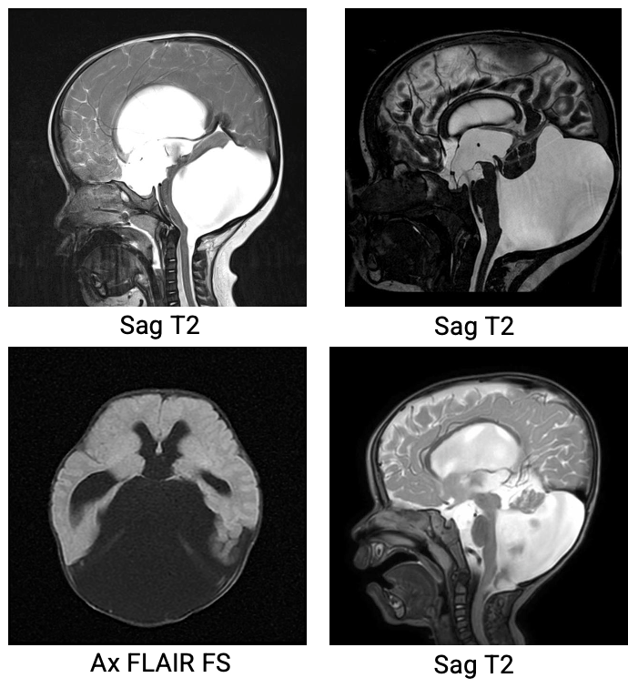
Dandy-Walker Malformation
Rare congenital brain defect that affects the cerebellum and the fluid-filled spaces around it.
Most common posterior fossa malformation
Key features:
hypoplasia of the cerebellar vermis (missing/underdeveloped)
cystic dilatation of the fourth ventricle
enlarged posterior fossa (pushes brain structures upward)
Hydrocephalus (excess CSF → macrocephaly)
Symptoms:
motor delays, balance/coordination issues, increased ICP → headache, vomiting, irritability, seizures
Meningocele
Skull or spinal defect resulting in the protrusion of the meninges filled with CSF
congenital, iatrogenic, or spontaneous.

Ischemic Stroke
rapid-onset neuro deficit from reduced/blocked blood supply to brain tissue
caused by thrombus, embolization, or systemic hypoperfusion.
2nd leading cause of death worldwide (MI is #1)
Symptoms of POSTERIOR CIRCULATION Infarct
Dizziness/vertigo, dysphagia, unilat limb weakness, ataxia, diplopia
Symptoms of ANTERIOR/MIDDLE CIRCULATION Infarct
unilateral weakness of face, arm, and/or leg, visual disturbances, dysphagia
Hemorrhagic Stroke
Bleeding directly into-
brain tissue (intracerebral) or
subarachnoid space around the brain tissue
caused by weakened vessels, aneurysms, AVMs, tumors
neurological emergency; 20% of all strokes (less common than ischemic)
Symptoms of HEMORRHAGIC Stroke
sudden neuro deficits and/or decreased level or consciousness, headache, possible seizure
Stroke Risk Factors
Atherosclerosis, age, family hx, smoking, HTN, high cholesterol, diabetes
DWI for Stroke Eval
ACUTE:
DWI signal - high
ADC appearance - low
EARLY/LATE SUBACUTE:
DWI Signal
Early - Low
Late - High
ADC appearance - low (both)
CHRONIC
DWI signal - Low
ADC Appearance - high

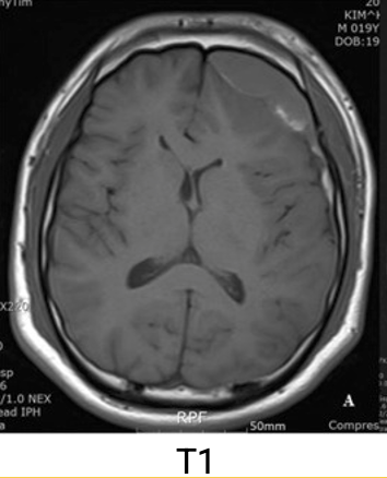
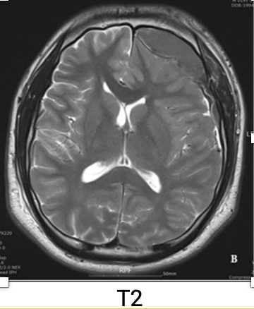
Subdural Hematoma
A/traumatic blood accumulation b/w Dura and Arachnoid space
MRI appearance depnds on acuity/stability
Subarachnoid Hemorrhage (SAH)
Bleeding inside the subarachnoid space
sudden, severe "thunderclap" headache
appearance: hyperintense on T2 FLAIR in the first 12 hours, SWI/GRE dark blood pooling post bleed

Epidural Hematoma
Traumatic arterial bleeding between the skull and the dura (endosteal layer)
can cause mass effect, midline shift
Most occur in temporal*, parietal, and frontal regions
MR appearance:
Displaced dura dark on T1/T2
T1:
Acute-iso
chronic-hyper
T2:
Acute-hypo
chronic-hyper

Aging Blood Mnemonic
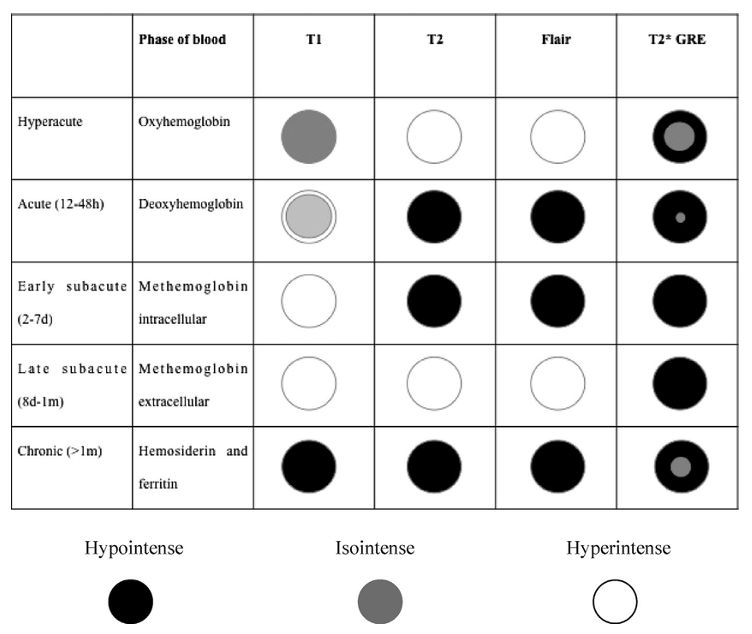

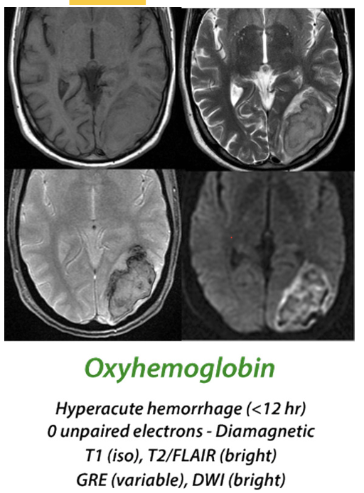
Oxyhemoglobin
Makes up most arterial blood (95%), venous 75%
weakly diamagnetic form of oxygen-bound hemoglobin
predominant in hyperacute hemorrhage (<12 hrs)
MR Appearance:
T1- iso
T2- hyper
Deoxyhemoglobin
No oxygen bound to hemoglobin
paramagnetic hemoglobin
predominant during acute phase (1-3 Days)
MR Appearance:
T1 iso
T2 dark
Methemoglobin
oxidized form of hemoglobin containing 5 unpaired electrons
paramagnetic
MR Appearance:
T1 Hyper during early and late subacute

Hemosiderin
insoluble iron-storage complex
superparamagnetic properties (103-106 unpaired electrons)
MR Appearance:
T1- iso
Flair- hyper
SWI- blooming (chronic)
DWI- hypo

Glial Cell Tumor: Diffuse Glioma
High grade glioma (III & IV)
very poor prognosis
Further broken into:
Diffuse Hemispheric Glioma
Pediatric type high grade Glioma
Diffuse Midline Glioma
MR APPEARANCE:
T1: hypo - iso
T2: hyper
T1 + GAD: non-enhancing
DWI: facilitated diffusion, some areas of restricted diffusion in higher grades
T2*/SWI: areas of marked hypo/blooming (intralesional hemorrhage)
DTI: disruption of white matter tracts indicates diffuse midline glioma > focal brain stem gliomas

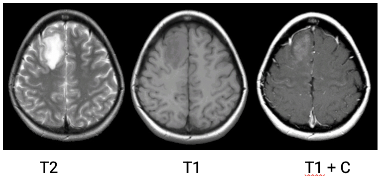
Glial Cell Tumor: Oligodendroglioma
Primary brain tumor originating from fat-producing oligodendrocytes
low-grade (Grade II or III)
Grade II: slow growing, better prognosis
Grade III:
Anaplastic, more aggressive
Treatment includes surgery, chemo, radiation
MRI Apperance:
T1: typically, hypointense
T2: typically, hyperintense
GRE/SWI: calcium can be seen as areas of "blooming"
T1 C+ (Gd): heterogenous enhancement common (~50%)
not a reliable indicator of tumor grade
DWI: usually no diffusion restriction
SWI: frequently contain calcs that bloom
Glial Cell Tumor: Astrocytoma
Primary brain tumor originating from star-shaped neuron support cells called astrocytes
Symptoms:
headache, dizziness, motor disfunction, cognitive decline, vision/speech changes
Prognosis depends on grade, size, location, and pt condition
GRADE I: Pilocytic Astrocytoma
GRADE II: Low Grade (diffuse) Astrocytoma
GRADE III: Anaplastic Astrocytoma
GRADE IV: Glioblastoma Multiforme (GBM)
Glial Cell Tumor: Glioblastoma Multiforme
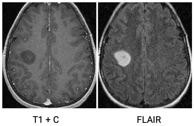
Most common and aggressive adult primary glioma tumor that typically develop from astrocytes
Grade IV astrocytoma
rapid expansion into adjacent tissues
associated w/ swelling around mass, nectrotic cavity
Symptoms:
headache, seizure, vision changes, LOC, slurred speech, cognitive decline, decreased motor function
Poor prognosis
Median survival 12-18 months
MRI Appearance:
T1:
hypo - iso
central heterogeneous signal
T1 C+ (Gd): variable, almost always peripheral that surrounds necrosis
T2/FLAIR:
hyperintense
surrounded by vasogenic edema
lacks T2/FLAIR "mismatch" sign
GRE/SWI:
susceptibility artifact from blood products
DWI/ADC
solid component
elevated signal common on DWI in the solid/enhancing component

Meningioma
Slow-growing tumors that arise from the meninges
Usually benign, incidental finding
Symptoms:
headache, seizure, vision/hearing changes, weakness
Treatment:
Monitoring, resection, chemo/radiation (Grade III)
MR Appearance
Smooth, round, solid
Bright post contrast enhancement
GRADE I: Benign, slow growing, low recurrence
GRADE II: Atypical Meningioma: higher rate of recurrence
GRADE III: Anaplastic/malignant Meningioma: rare, rapid growth, high rate of recurrence

Central Nervous System Lymphoma
Non-Hodgkin malignancy of lymphatic cells within the CNS of immunocompromised/AIDS patients
Symptoms:
N/V, weakness, headache, AMS, hearing/vision loss, dysphagia
Treatment depends on med hx:
AIDS: HIV meds, steroids, radiation/chemo
Non-AIDS: chemo and monoclonal antibodies w/ radiation, stem cell transplant, or radiation only

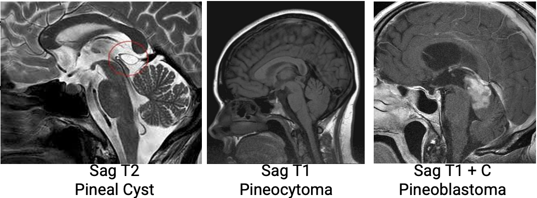
Pineal Gland Tumors
Pineal parenchymal tumors
Often incidental finding
cystic lesions most common
GRADE I: Pineocytoma
GRADE II/III: Papillary Pineal Tumor
GRADE IV: malignant Pineoblastoma
Germinoma
most common malignant germ cell tumor in the CNS
80-90% of all germinomas are located in the pineal region

Metastatic Brain Cancer
Secondary CNS neoplasms spreading via blood or lymph from distant primary sites
lung (20%)*
Breast
melanoma
colon
kidney
thyroid
typically found at gray-white matter junctions

MR Appearance
varies widely, difficult to diagnose
T1: iso - hypo
hemorrhagic = high signal
non hemorrhagic melanoma = high signal due to paramagnetic properties of melanin
T1+gad: can be intense uniform, punctate, or ring-enhancing
T2: hyperintense (can depend on hemorrhage/melanin)
mucinous mets - LOW
FLAIR: hyperintense (including edema)
DWI/ADC: edema is dark on DWI, bright on ADC. tumor is opposite


Autoimmune: Multiple Sclerosis
CNS inflammation that results in damage to the myelin sheath around the axons with the brain and spinal cord
most common in females (2:1 female:male)
Diagnosed during adolescence up to age 50
Symptoms:
Optic Neuritis (optic nerve lesion)
Ataxia (cerebellar lesion)
Parasthesia (cerebral lesion)
characteristically produce oval, periventricular white matter plaques
on FLAIR MRI (gold standard sequence)
Post contrast evals acuity of plaques (acute, subacute, or chronic)

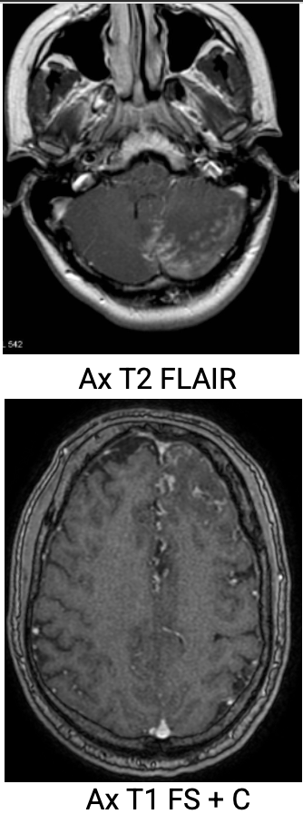
Autoimmune: Neurosarcoidosis
A CNS manifestation of systemic sarcoidosis where non-caseating granulomas form in brain tissue or meninges (~20% of sarcoidosis cases/1-% symptomatic)
Sarcoidosis: condition where immune system overreacts, creates granulomas (mass of granulation tissue)
Symptoms:
optic or facial palsies, hormone disorders (pituitary granulomas), seizure, weakness, AMS, dysphagia
MR Appearance:
T1: iso or hypo to gray matter
T2: hyper to surrounding tissue
T1+GAD: homogenous, parenchymal (top left) or leptomeningeal (bottom rt) enhancement

Autoimmune: Neurovasculitis
Inflammatory process affecting the lining of blood vessels in the brain and meninges. more common in males.
Primary: confined to CNS only
Secondary: inflammatory process or infection*
Further characteriation of Secondary Vasculitis determined by affected vessels
MR Appearance:
T2/FLAIR: high signal in white matter
GRE/SWI: detects microbleeds
MRA: vascular narrowing
3D T1: vessel wall imaging

Infection: Meningitis
Inflammation of the protective meningeal coverings of the brain and spinal cord. Can be viral, bacterial (pyogenic)*, or fungal. DX by LP
Symptoms:
Headache, fever, photophobia, stiff neck, N/V
left untreated can lead to abscess, hydrocephalus, cerebritis, thrombosis, infarcts, ventriculitis, empyema
Best evaluated on T1 post-contrast by observing leptomeningeal enhancement

Infection: Cerebral Abscess
Necrotic area of brain tissue surrounded by an inflamed vascular capsule resulting from cerebritis
Risk Factors: IVDU, CHD, lung infections, Sinus infections, dental abscesses, meningitis, sepsis
MR Appearance:
T1:
central/peripheral - low intensity (hyper to CSF)
ring enhancement post contrast
T2/FLAIR:
central/peripheral - high intensity (hypo to CSF, no FLAIR attenuation)
abscess capsule may be visible as thin rim of intermideiate - low signal
DWI: central high signal
SWI: low intensity rim

4 Stages of Brain Infection
Early Cerebritis (infections w/o capsule or pus)
Late Cerebritis
Early Abscess (encapsulation 10 days post onset)
Late Abscess (14 days or longer after encapsulation)
Neurocysticercosis
Parasitic CNS infection from ingesting tapeworm eggs from undercooked pork, leading to cyst formation in the brain parenchyma
Common in central/south american countries
Can take years for eggs to manifest in the brain tissue
MR Appearance:
T1: hyperintense
T2/FLAIR: hyperintense
T1+GAD: cyst wall enhancement

Leukodystrophies
A group of inherited genetic disorders causing abnormal glial cell function and defective myelin sheath development, causing abnormal white matter growth
all genetic but can originate from autoimmune, infectious, or metabolic conditions
effects newborns to adults
Symptoms:
abnormal movement, difficulty walking, dysphagia, learning/behavioral changes, hearing/vision changes
MR Appearance:
symmetric T2/FLAIR white matter hyperintensities

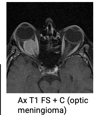
Optic Meningioma
Nerve sheath tumors
Rare
slow growing
benign
Choroidal Melanoma
Most common primary malignant intraocular tumor…tho still rare
Originate from melanin cells within the orbit
Aggressive, 50% met
