hematology exam 1 - globin chain synthesis, ODC, RBC metabolism
 Call Kai
 Learn
Learn Practice Test
 Spaced Repetition
 Match
Match Flashcards
 Knowt Play
1/21

There's no tags or description
Looks like no tags are added yet.
Name | Mastery | Learn | Test | Matching | Spaced | Call with Kai | Chat |
|---|
No analytics yet
Send a link to your students to track their progress
22 Terms
globin chain synthesis phases
embryonic
fetal
birth
1 year old
adult
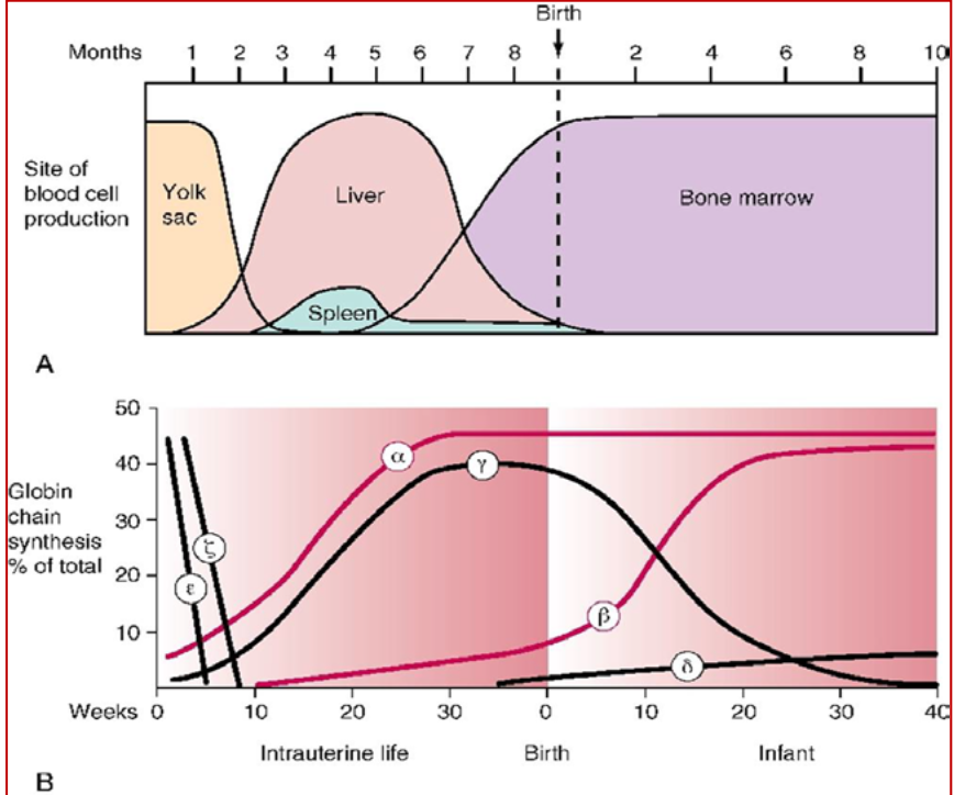
embyronic phase of globin chain synthesis
globin gene activation progresses from ζ to α gene and from the ε gene to the γ, δ, and β genes
ζ and ε chains are only present in the embryonic stage of development and are produced for 3 months after conception
type of Hb:
Gower 1 + 2, Portland
Gower 1: ζ2, ε2 subunits
Gower 2: α2 subunits, ε2 subunits
Portland: ζ2 subunits, γ2 subunits
DO NOT TRANSPORT OXYGEN
fetal phase of globin chain subunits
type of Hb:
fetal Hb (Hgb F or Hgb Portland) → α2, γ2
γ chain production occurs actively from the 3rd month of development to one year after birth
90-95% of total Hb levels before birth
Hgb F makes up 60% of Hb in newborns
Hgb F can carry oxygen, and levels decrease after 1-yr post birth
Hgb F is the main type of Hb produced by the liver and spleen
birth
type of Hb present
Hgb F (α2, γ2) → around 50-85% of total Hb
Hgb A (adult Hb) → α2, β2 → around 10-40% of total Hb
HbA2 → α2, δ2 → <1% of total hb
α is always present
γ chain production occurs actively from the 3rd month of development to one year after birth
1 year old globin chain synthesis phase
type of Hb present:
HbF → α2, γ2 = <2%
HbA → α2, β2 = >95%
HbA2 → α2, δ2 = 3.5%
adult globin chain synthesis phase
α is always present and the constant component in adult Hb
3-6 months after birth, γ chain production declines and β chain production increases to make Hgb A or adult HgB
Hgb A → 80-85% of Hb one year after birth
the majority of adult hemoglobin is HbA (α2β2), representing 95-98% of all adult Hb
Hgb A2 (HbA2, α2δ2) is 3-5% of adult Hb, produced around 3-5 wks of pregnancy
Hgb F represents <2% of adult Hb
globin chain synthesis graph
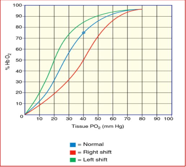
hemoglobin-oxygen dissociation curve
measurement of Hb’s affinity for oxygen
oxygen affinity is expressed as p50, where 50% of Hb is saturated with oxygen
in an avg human, p50 = 26 mmHg
measure the Hb-O2 saturation vs the partial pressure of oxygen = oxygen dissociation curve (ODC)
beginning of the curve
fully staurated Hb molecule containing full oxygen with a high oxygen lvl tension
as it travels, it releases oxygen to the surrounding tissues that need oxygen
partial pressure of oxygen O2
factors that can affect oxygen affinity
increases → oxygen affinity increases
pH
factors that can affect oxygen affinity
decrease (increase in H+) = Bohr effect, conformational change in the amino acids of Hb causing it to enter the T state (tensed) where oxygen binds less easily
increase (decrease in H+) = oxygen affinity increases
partial pressure of carbon dioxide (PCO2)
factors that can affect oxygen affinity
increase = oxygen affinity decreases for the same reason as pH, tissues are more acidic
2,3-BPG
factors that can affect oxygen affinity
molecule in RBCs that binds to Hb
decreases oxygen affinity
increased concentration of BPG in situations where oxygen is less available, such as high altitude and chronic lung diseases (cuz im assuming that it wants to deliver the oxygen to tissues and doesn’t want to keep it with RBC)
temperature
factor that can affect oxygen affinity
increase = oxygen affinity reduces
Hb tends to release oxygen more efficiently in warmer temps
ODC right and left shifts
right shift
Hb has less affinity for oxygen and releases the oxygen more compared to normal curve
cells are 50% saturated at 40 mmHG
can be caused by
anemia
rise in body temp
increase in 2,3-DPG
acidosis
left shift
Hb has a higher affinity for oxygen and doesn’t like to release oxygen to the tissues
cells are 75% saturated at 40 mmHg and will release about 12% of oxygen to the tissues
caused by
decrease in body temp
lower lvls of 2,3-DPG
alkalosis
higher lvls of methemoglobin and carboxyhemoglobin (by forcing hemoglobin into its relaxed R state)
blood transfusions (decrease 2,3-DPG)
higher hemoglobin F levels
carboxyhemoglobin
abnormal hemoglobin
iron is in the 2+ state
no oxygen will be delivered to the tissues because Hb has a higher affinity for carbon monoxide compared to oxygen (200x stronger binding)
methemoglobin
abnormal Hb
iron state is Fe3+ and has been oxidized
iron in this state can no longer bind to oxygen
sulfhemoglobin
abnormal Hb
iron is in the 2+ state
iron is not able to carry oxygen and can’t be reversed unless the RBCs are taken out of circulation
100x higher affinity for oxygen and competes with hemoglobin for oxygen
glycolytic pathway
90-95% of cell’s glucose consumption is metabolized by this pathway
glucose metabolized by this pathway to lactate or pyruvate = net gain of 2 moles of ATP per mole of glucose
if NADH is available in the cell, pyruvate is reduced to lactate
lactate or pyruvate formed is transported from cell to plasma and metabolized elsewhere in body
ATP necessary to maintain erythrocyte shape, flexibility, and membrane integrity and to regulate intracellular cation concentration
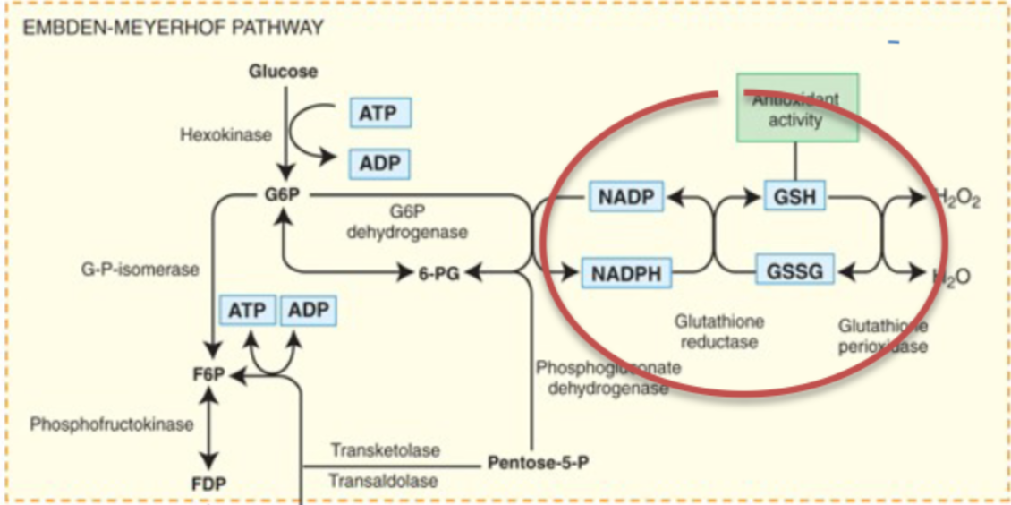
glycolysis generates NADPH, reduces glutathione and protects cell from oxidative injury
keep iron in Hb in the oxidated state (Fe 2+) and prevent hemoglobin iron from reducing (Fe3+) because in reduced state, iron CANNOT carry oxygen
what happensif no glucose for RBC
ATP for cation pumps is no longer available
cells can’t maintain normal intracellular cation concentration
cells become sodium and calcium loaded and potassium depleted
even though Na+ should be higher outside, K+ should be higher inside
cell accumulates water and changes from bioconcave disc to a sphere and is removed from circulation
Rapoport-Luebering pathway
controls the amount of 2,3-BPG produced
affects the oxygen affinity of Hb
key enzyme: BPG-synthase
part of the glycolytic pathway, bypasses the formation of 3-phosphoglycerate and ATP from 1,3-biphosphoglycerate (1,3-BPG)
instead, 1,3-BPG forms 2,3 BPG
erythrocyte sacrifices one of its two ATP producing steps in order to form 2,3-BPG
methemoglobin reductase pathway
EMP shunt
essential to maintain heme iron in the reduced state (ferrous) Fe++
methemoglobin (Hb with iron in oxidized ferric state, Fe3+) is generated simultaneously with the oxidative compounds discussed as O2 dissociates from heme iron
cannot bind oxygen
methemoglobin reductase w/ NADH produced by glycolytic pathway fxns to reduce the ferric iron in methemoglobin, converting it back to ferrous hemoglobin
W/O this sys: 2% of methemoglobin formed daily eventually builds up to 20-40%
certain oxidant drugs can interfere w/ this process and cause even higher levels of methemoglobin = cyanosis
hexose-monophosphate shunt
provides NADPH and glutathione to reduce oxidants that would shift the balance of oxyhemoglobin to methemoglobin
glutathione reductase, G6PD