Divisions of the Nervous System
 Call Kai
 Learn
Learn Practice Test
 Spaced Repetition
 Match
Match Flashcards
 Knowt Play
1/191

There's no tags or description
Looks like no tags are added yet.
Name | Mastery | Learn | Test | Matching | Spaced | Call with Kai | Chat |
|---|
No analytics yet
Send a link to your students to track their progress
192 Terms
Describe the structure, function and location of sensory (afferent) neurons
Structure: Short axon which branches off in 2 directions (pseudounipolar)
Function: Receives external stimuli and converts them to electrical impulses
Location: Sensory organs and sensory ganglia in the PNS
Describe the structure, function and location of motor (efferent) neurons
Structure: Large cell body, highly branched dendrites and a single long axon (multipolar)
Function: Receives signals from the CNS and transmits them to effectors
Location: Cell bodies are in the CNS but axons reach into the PNS
Describe the structure, function and location of inter (association) neurons
Structure: Multiple short dendrites and short axons (multipolar)
Function: Pass signals from sensory neurons to motor neurons
Location: Exclusively in the CNS
Give 5 examples of sensory (afferent) neurons
Photoreceptors (vision)
Mechanoreceptors (touch and hearing)
Thermoreceptors (temperature)
Chemoreceptors (taste and smell)
Nociceptors (pain)
Describe the stages from a stimulus to the generation of a nerve impulse
Stimulus detection: an environmental stimulus interacts with the dendrites/receptor cells of the sensory neuron
Receptor potential: the stimulus physically/chemically alters the cell membrane, causing ion channels to open. This changes the cell membrane voltage- creating a local electrical signal
Threshold reached: if the stimulus is strong enough, the receptor potential reaches the threshold
Action potential is generated: voltage-gated sodium channels open and changes the internal charge from negative to positive
Propagation and transmission: the action potential travels as a wave down the axon and triggers the release of neurotransmitters across the synapse
Describe the two types of motor (efferent) neurons
UMNs (upper motor neurons)
Originate in the cerebral cortex and the brainstem
Responsible for initiating voluntary movement
Do not directly contact the target muscles
Key neurotransmitter: glutamate
LMNs (lower motor neurons)
Originate in the brainstem and the spinal cord
Extend out of the CNS to directly innervate effector muscles and glands
Key neurotransmitter: acetylcholine
Describe how neurons are classified based on their level of myelination
Neurons are classified based on their nerve impulse velocity, using the Erlanger-Gasser classification system- which categorises fibres into types A, B and C
The velocity of the impulse depends on the axon diameter and the presence of a myelin sheath
Group A: heavily myelinated
Group B: moderately myelinated
Group C: unmyelinated (e.g in the tongue)
What are the 2 key mechanisms involved in myelination
Saltatory conduction between nodes of ranvier
Highly concentrated ion channels at the nodes of ranvier
Describe the impact of myelination on nerve cells
The myelin sheath drastically increases nerve impulse velocity, by forcing the electrical signal to ‘jump’ between gaps in the sheath- this is known as saltatory conduction
This prevents the electrical signal from degrading, allowing impulses to travel much faster
Describe the types of neuroglia
Schwann cells- wrap tightly around the axons of neurons, to form the myelin sheath, which acts as an electrical insulator. Schwann cells also release essential growth factors, which help repair and regenerate damaged axons
Satellite cells- surround the cell bodies of neurons located in the peripheral ganglia. They serve as physical and nutritional support, and release factors which protect neurons from degeneration
Names:
Astrocytes: star shapes cells which provide physical and nutritional support to neurons
Oligodendrocytes: specialised cells which form the myelin sheath around axons
Microglial cells: resident immune cells of the CNS, monitoring for pathogens
Ependymal cells: ciliated cells lining the CNS, which produce cerebrospinal fluid
What are NSAIDs? Include their target of action and an example
Non-steroidal anti-inflammatory drugs
Target of action: cyclooxygenase (COX)
Example: carprofen, meloxicam
What are corticosteroids? Include their target of action and an example
Steroid medications that mimic cortisol to suppress the immune system
Target of action: intracellular receptors
Example: prednisolone
Why should you not combine NSAIDs and corticosteroids?
Combining NSAIDs and corticosteroids is contraindicated, because it drastically increases the risk of severe, life-threatening GI ulceration, bleeding, and perforation
List 5 factors affecting drug mechanisms
Age
Gender
Diet
Environment
Body weight
Genetics
Organ functions
Neurological problems
Drug-food interactions
List the advantages and disadvantages of oral medication administration
Advantages:
Most convenient for owners
Cost-effective
Disadvantages:
Unreliable if vomiting/diarrhoea
Palatability
List the advantages and disadvantages of topical medication administration
Advantages:
Painless
Good for localised skin/eye conditions
Disadvantages:
Slow/erratic absorption
May be licked/rubbed off
List the advantages and disadvantages of rectal medication administration
Advantages:
Bypasses stomach acid
Good for vomiting/unconscious animals
Disadvantages:
Uncomfortable
Faeces can reduce absorption
List the advantages and disadvantages of subcutaneous medication administration
Advantages:
Simple to perform
Quick, predictable absorption
Disadvantages:
Reduced absorption in dehydrated/cold patients
Granulomas may form
List the advantages and disadvantages of intramuscular medication administration
Advantages:
Fast absorption
Useful for patients who are gardening to restrain
Disadvantages:
Painful for the patient
Higher risk of hitting nerves/blood vessels and causing muscle damage
List the advantages and disadvantages of intravenous medication administration
Advantages:
100% bioavailability instantly
Immediate therapeutic effect
Disadvantages:
High risk of anaphylaxis and adverse reactions
List the advantages and disadvantages of intranasal medication administration
Advantages:
Good for localised respiratory issues
Avoids gut enzymes
Disadvantages:
Variable absorption
Can cause mucosal irritation
Define the term ‘resting membrane potential’
The electrical charge difference across a cell’s plasma membrane when it is not actively sending signals (typically around -70mV)
Define the term ‘action potential’
A rapid, temporary spike in electrical voltage which travels along the cell membrane of a neuron/muscle cell
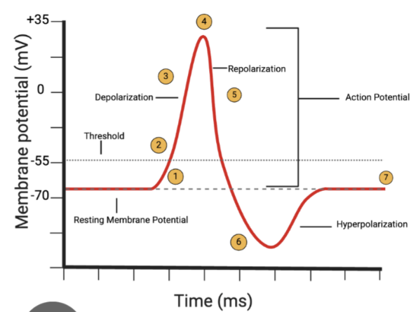
Describe the stages of neurotransmission along the axon, including a labelled graph
The voltage of the local membrane becomes less positive- resulting in the membrane reaching the ‘threshold’
Once the threshold is reached, voltage-gated sodium ion channels open, allowing sodium ions to flood into the cell: further depolarising the membrane
More voltage-gated sodium ion channels open- causing rapid depolarisation along the length of the membrane until the channels are inactivated and close
Voltage peaks at around +35mV- at which point, the membrane begins to repolarise
Repolarisation occurs due to the slow opening of potassium channels, and the outflow of potassium ions
Potassium ion channels stay open for a time, so that the membrane is briefly hyperpolarised
Resting membrane potential is restored as sodium ions leak in, and extracellular potassium ions are removed by astrocytes
Describe the mechanism of the sodium/potassium pump
An active transport protein
Uses ATP to constantly pump 3 sodium ions out of the cell, whilst pumping 2 potassium ions into the cell
This creates a slight negative charge inside
Describe the mechanism of the potassium ion leak channels
These are always open
Because potassium ions are always being pumped into the cell, potassium naturally leaks out down the concentration gradient
This establishes the slightly negative resting membrane potential
Describe the mechanism of the sodium ion voltage-gated channels
Normally closed at rest, but open in response to a stimulus/voltage change
Once open, sodium ions rush into the cell down the steep concentration gradient- depolarising the membrane and creating an action potential
Describe the mechanism of the potassium ion voltage-gated channels
These open after the cell has depolarised
They allow potassium ions to rapidly leave the cell
This causes repolarisation (and brief hyperpolarisation)
What is propagation?
Describe what it is affected by
This means the travel of an electrical signal down the axon of a neuron
It is affected by myelination:
Instead of moving continuously down the axon, myelination allows the signal to jump between nodes of Ranvier
The voltage-gated ion channels are dense at the nodes of Ranvier
Myelinated segments prevent ion leakage and conserve electrical charge
This increases speed and energy efficiency
Describe the action of lignocaine (lidocaine)
Temporarily halts nerve transmission in localised areas of the body, by blocking voltage-gated sodium ion channels to prevent the influx of sodium ions
When applied/injected, a neural portion of the drug is allowed to pass through the cell membrane
Once inside the cell, the drug becomes charged by combining with hydrogen ions
This charged molecule binds directly to the inner portion of the voltage-gated sodium ion channel
By binding to the channel, the drug locks it in an inactive state- physically blocking sodium ions from entering the cell
Because APs rely on sodium ion movement to travel, the nerve becomes temporarily ‘numb’
Describe the two types of sensory pathway
Those which register conscious perception
Those which control unconscious coordination
Describe the action of the spinocerebellar pathway
Convey unconscious proprioception from the limbs and trunk, directly into the cerebellum for motor refinement and posture
Dorsal and cuneocerebellar tracts: simple 2-neuron pathways
Ventral and rostral spinocerebellar tracts: serve as a feedback loop regarding spinal motor activity
Describe the function of the cuneate and gracile tracts
Manage conscious proprioception and discriminative touch
Uses a 3-neuron chain: first-order neurons enter the spinal cord and ascend without synapsing
Fasciculus gracilis: transmits sensory information from the pelvic limbs and caudal trunk
Fasciculus cuneatus: transmits sensory information from the thoracic limbs and cranial trunk
Decussation: both fasciculi synapse in their respective nuclei in the medulla oblongata
What is proprioception? Give a brief overview of how it works and the components involved
Proprioception is the subconscious ability to sense the body’s position, movement and balance in space without having to look
Sensory receptors constantly detect stretching, contracting and pressure
These receptors constantly send signals to the CNS- allowing the brain to calculate the exact orientation of the limbs
How is the white matter of the spinal cord divided?
The white matter of the spinal cord is divided into 3 bilaterally symmetrical pairs of columns (funiculi)
Dorsal funiculus (contains primarily ascending sensory tracts)
Lateral funiculus (contains a mix of both ascending and descending tracts)
Ventral funiculus (contains primarily descending motor tracts)
Describe the action and components of the ascending (sensory) tracts and descending (motor) tracts
Ascending (sensory) tracts
Relay afferent information up to the brainstem, cerebellum and cerebral cortex- typically following a sequence of three neurons
Spinothalamic tract
Spinocerebellar tract
Spinocervicothalamic tract
Descending (motor) tracts
Initiate voluntary movement, regulate muscle tone and maintain posture
Corticospinal tract (part of the pyramidal system)
Vestibulospinal, reticulospinal, rubrospinal tracts (part of the extrapyramidal system)
How are spinal cord pathways organised?
Spinal cord pathways are organised into bilaterally symmetrical tracts within the white matter
These tracts are primarily divided into ascending pathways (sensory) and descending pathways (motor)
What is the function of the thalamus?
The thalamus filters raw sensory inputs, and controls what information reaches conscious awareness
Describe the following fibre types, including their conduction times:
A-alpha (type I) myelinated
A-beta (type II) myelinated
A-delta (type III) thinly myelinated
C (type IV) non myelinated
A-alpha myelinated
80-120m/s
Can be type Ia (sense rate of change of muscle length) or type Ib (golgi tendon organs, sense muscle tension)
A-beta (type II) myelinated
33-75m/s
Muscle spindles and skin sensation
A-delta (type III) thinly myelinated
3-30m/s
Free endings, pain, cold
C (type IV) non myelinated
0.5-2 m/s
Pain, warmth
Describe the:
First order neuron
Second order neuron
Third order neuron
Involved in pain and temperature
First order neuron
Cell bodies reside in the dorsal root ganglion
They receive stimuli via free nerve endings (nociceptors and thermoreceptors) and enter the spinal cord dorsal horn
Second order neuron
Synapses in the dorsal horn
Axons decussate
They ascend via the lateral spinothalamic tract, in the ventrolateral funiculus of the spinal cord
Third order neuron
Axons terminate in the thalamus, which then projects to the somatosensory cortex for conscious localisation and perception
Describe the:
First order neuron
Second order neuron
Third order neuron
Involved in crude touch and pressure
First order neuron
Cell bodies are in the DRG
Second order neuron
Synapses in the dorsal horn
Ascend contralaterally via the anterior spinothalamic tract in the ventral funiculus
Third order neuron
Relays from the thalamus to the somatosensory cortex
Describe the:
First order neuron
Second order neuron
Third order neuron
Involved in fine touch and conscious proprioception
First order neuron
Cell bodies are in the DRG
Receptors detect fine touch, vibration and joint position
Second order neuron
Synapses in the medulla oblongata
The axons decussate at the medulla, and ascend via the medial lemniscus to the thalamus
Third order neuron
Projects from the thalamus to the somatosensory cortex
Describe the:
First order neuron
Second order neuron
Third order neuron
Involved in facial sensation
First order neuron
Housed in the sensory ganglion
Second order neuron
Reside in the sensory nuclei of the trigeminal nerve
Axons cross the midline and ascend to the thalamus
Third order neuron
Project to the face region of the somatosensory cortex
Describe the composition of the vagus nerve
It is made up of 80% sensory fibres and 20% motor fibres
Describe the origin and exit of the vagus nerve in the CNS
Origin
10th cranial nerve (CN X)
Between CN IX and XI
Exits as a series of rootlets from the lateral surface of the medulla oblongata
Exit
From the cranial cavity- the jugular foramen with CNs IX and XI
The vagus nerve also has fibres which are destined to be in the recurrent laryngeal nerve
Describe the distribution and branches of the vagus nerve in the head and neck
Below the base of the skull, the vagus nerves gives off:
Auricular branch (to external acoustic meatus : sensory)
A pharyngeal branch (to pharynx : sensory and motor to pharyngeal muscles)
The cranial laryngeal nerve (to larynx : sensory as far as the glottis, motor to cricothyroid m.)
Continuing caudally, the vagus nerve:
Joints the sympathetic trunk at the cranial cervical ganglion
This forms the vagosympathetic trunk
Describe the vagosympathetic trunk and the fibres that run within it
The vagosympathetic trunk is a structure that contains several components
Sympathetic fibres running cranially: to the head/neck
Parasympathetic fibres running cranially: from receptors and viscera
Parasympathetic fibres running caudally: to the heart and viscera
Describe the distribution and branches of the vagus nerve in the thorax
The vagus nerve:
Enters the thorax at the thoracic inlet
Separates from the sympathetic tract at the middle cervical ganglion
It does not synapse at the middle cervical ganglion, but just keeps on going
In the thorax, the vagus gives off:
Recurrent laryngeal nerve (axons from CN XI)
Cardiac branches: to cardiac plexus supplying the heart
Bronchial branches: to the pulmonary plexus supplying lungs
In the thorax:
The vagus on each side divides caudal to the heart, into a dorsal and ventral branch
May become a fine plexus
Dorsal branches unite on the oesophagus, to form the dorsal vagal trunk
Ventral branches unite to form the ventral vagal trunk
Dorsal and ventral vagal trunks pass through the oesophagus hiatus of the diaphragm
Describe the distribution of the recurrent laryngeal nerve
Fibres leave the vagal trunk in the thorax
Right RLN hooks around the right subclavian artery
Left RLN hooks around the aortic arch (longer route)
Describe the distribution and branches of the vagus nerve in the abdomen
Dorsal and ventral vagal trunk:
Provide preganglionic parasympathetics to abdominal plexuses
Supply preganglionic parasympathetics to the gut through to the transverse colon
Dorsal vagal trunk: fibres to coeliac and cranial mesenteric plexuses
Ventral vagal trunk: fibres to gastric and hepatic plexuses
Describe the distribution and branches of the vagus nerve in the abdomen of ruminants
The ruminant stomach is classically described as having dorsal and ventral vagal supplies, but there is crossover- so resecting one trunk does not denervate a section of the rumen
Dorsal vagal trunk: rumen (ruminal motor function)
Ventral vagal trunk:
Reticulum, omasum, abomasum
Gastric plexus, branches to duodenum, pancreas, liver, hepatic plexus
Describe Horner’s Syndrome
Damage to the sympathetic nerves within the vagosympathetic trunk (brain and spinal cord damage can also be a cause)
Usually one-sided
Loss of supply to:
Smooth muscle in the periorbital tunic
Smooth muscle of iris dilators
Results in:
Enophthalmos: globe retracts into orbit, due to tone in retractor bulbi mm., allowing the 3rd eyelid to move into view
Miosis: constricted pupil
Describe vagal indigestion
Occurs in ruminants, mainly cattle
Usually due to traumatic reticuloperitonitis (wire/hardware disease)
This is a chronic disease
Syndrome of ruminoreticular distension
Four types (I-IV)
Not acute bloat- it is chronic paralysis with poor motility
Surgery may be required, depending on the cause
Describe where these parts of the brain develop from:
Forebrain
Midbrain
Hindbrain
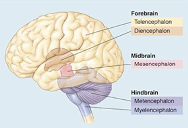
As the embryo develops, vesicles are formed- which form the basis of the brain and spinal cord
The forebrain develops from the telencephalon and diencephalon
The midbrain develops from the mesencephalon
The hindbrain develops from the metencephalon and myelencephalon
Which structures make up:
The forebrain
The hindbrain
The brainstem
Within the forebrain: cerebrum (made up of cerebral cortex), thalamus, hypothalamus, pituitary gland
Within the hindbrain: pons, medulla oblongata, cerebellum
The brainstem is made up of the medulla oblongata, pons and the midbrain
Draw a diagram of the developed brain and the areas of:
The forebrain
The midbrain
The hindbrain
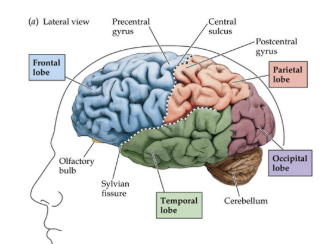
Describe the lobes which make up the cerebral cortex. Include a labelled diagram
Frontal lobe: at the front of the brain- involved in executive functioning, formulating plans and strategies
Parietal lobe: involved in somatosensation, receiving sensory information from around the body and the environment
Temporal lobe: incorporates the primary auditory cortex, which receives auditory information
Occipital lobe: receives visual information
Why do mice and sheep have significantly larger olfactory bulbs and rhiencephalons?
Mice and sheep have significantly larger olfactory bulbs and rhinencephalon, because they are macrosmatic animals (highly developed sense of smell)
In sheep, what is the rhiencephalon associated with?
In sheep, the rhinencephalon (nose brain) is associated with olfaction and emotion
Draw a complete labelled diagram of the brain
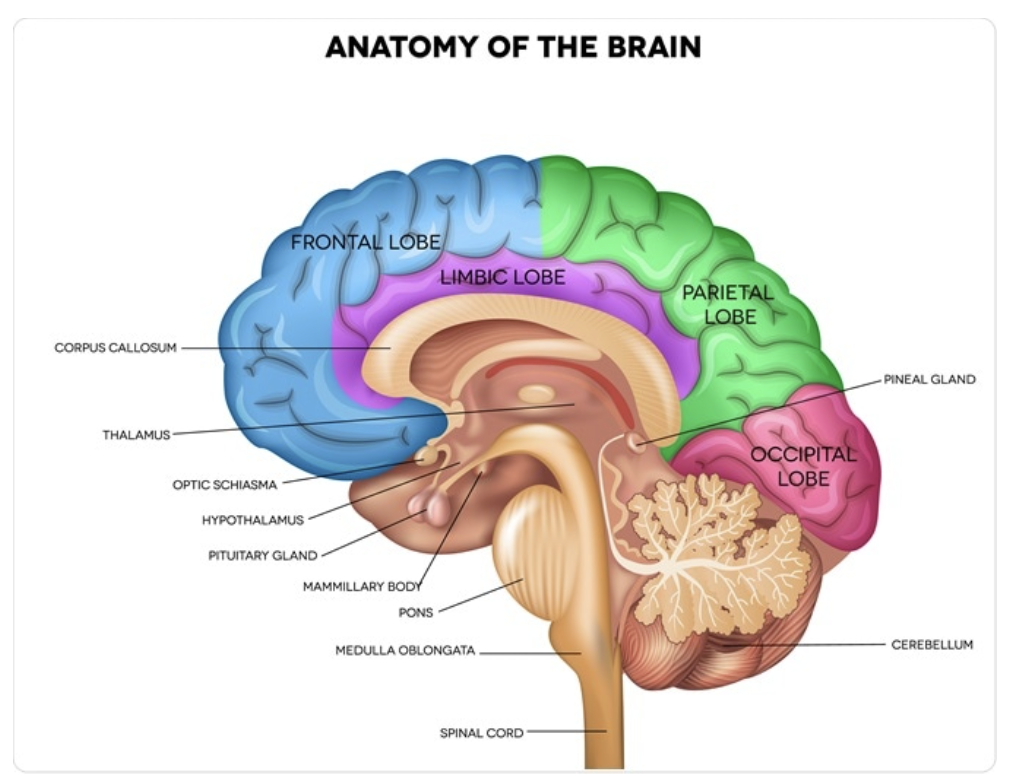
Describe the function of the cerebral hemisphere
The left brain is primarily responsible for logical, sequential and analytical tasks (language, logic, maths, detail)
The right brain specialises in holistic, spatial and intuitive processing (creativity, emotion, spatial awareness)
Both sides control the opposite half of the body, and communicate constantly via the corpus callosum
Describe the function of the basal ganglia
Regulate voluntary movement by filtering out unwanted actions, and selecting the right ones: motor control, cognition, habit formation, motivation
Describe the function of the hippocampus
Memory consolidation, spatial navigation, emotion and context- working closely with the limbic system
Describe the function of the corpus callosum
Integrating and transferring sensory/motor/cognitive signals- allowing both sides of the brain to communicate and coordinate essential daily functions
Describe the function of the limbic system
Made up of different structures within the brain
Regulating emotion, behaviour, memory, and motivation
Describe the function of the thalamus
Major relay station for sensory information
Integration of motor information from the cerebellum and basal ganglia, before passing on to the motor regions of the cortex
Describe the function of the medulla oblongata
Regulating heart rate, breathing, blood pressure and essential reflex actions
Describe the function of the pons
Relaying sensory-motor signals, generating REM sleep, regulating breathing patterns and controlling facial movements
Describe the function of the cerebellum
Integration and control of posture/movement
Describe the function of the cerebral cortex
Planning of movement, perception and learning
What is meant by the term ‘rhythm‘?
A function which oscillates or cycles at a regular frequency
Biological rhythms are the overt, measurable activities generated by some internal oscillator (or clock)
What is meant by the term ‘chronobiology‘?
A field of science that examines periodic (cyclic) phenomena in living organisms, and their adaptation to external (environmental) rhythms
What is meant by the term ‘circadian‘?
A daily rhythmical change in behaviour or in a physiological proces
What is meant by the term ‘infradian‘?
A rhythm with a period longer than the period of a circadian rhythm
What is meant by the term ‘ultradian‘?
A rhythm with a period shorter than the period of a circadian rhythm (e.g REM cycle in sleep)
What is meant by the term ‘zeitgeber‘?
Any external cue that entrains the internal timekeeping system of organisms
Describe the function of biological rhythms
Biological clocks are internal physical systems that enable organisms to live in harmony with the rhythms of nature (e.g cycles of day and night or the seasons). This is known as external synchronisation
Circadian rhythms also maintain temporal organisation of endogenous processes. This is known as internal synchronisation
The strongest zeitgeber (for both plants and animals) is light. Other zeitgebers include temperature, social interaction, pharmacological manipulation and eating/drinking patterns
Describe the biological clocks in:
The retina
The SCN
The pineal gland
Retina
A photo pigment (melanopsin) present in ganglion cells in the retina, whose axons transmit information to the SCN, the thalamus and the olivary pretectal nucleus
Suprachiasmatic nucleus (SCN)
A nucleus situated atop the optic chiasm
It contains a biological clock, that is responsible for organising many of the body’s circadian rhythms
Pineal gland
A gland attached to the dorsal tectum
It produces melatonin in response to signals from the SCN, and plays a role in circadian and seasonal rhythms
What are the inputs/outputs of the SCN in regards to biological rhythms?
Input
Light
Output
Sleep/wakefulness
Feeding and energy expenditure
Glucose homeostasis
Hormonal regulation
Body temperature regulation
Blood pressure and respiratory changes
What are the inputs/outputs of the peripheral clocks in regards to biological rhythms?
Input
Feeding/fasting
Signals from SCN
Output
Lipogenesis
Insulin secretion
Glucose clearance
Food absorption
Fat accumulation
What are the consequences if the biological rhythm is disrupted?
Sleep: poor sleep, depression, bipolar disorder, Alzheimers
Metabolism: obesity, type II diabetes, fatty liver disease
Microbiota: increased body weight
Cardiovascular: atherosclerosis, cardiovascular risk
Aging: life span, age-related markers
Cancer: enhanced cellular proliferation
Draw a labelled diagram of a synapse
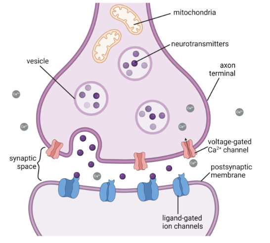
Describe the stages of neurotransmission across a synapse
Step 1 (Release)
When an AP reaches the end of the presynaptic terminal, it triggers the opening of calcium channels
The influx of calcium causes tiny sacs (called synaptic vesicles) to fuse with the cell membrane
The neurotransmitters inside are released into the synaptic cleft through exocytosis
Step 2 (Diffusion)
The neurotransmitters float across the synaptic cleft
They move down their concentration gradient
Step 3 (Binding)
On the postsynaptic neuron, the neurotransmitters lock into specialised receptor proteins
This binding opens ion channels in the receiving cell- allowing charged particles to flow in or out- which dictates whether the signal is passed on or halted
Step 4 (Activation)
To prevent continuous stimulation, the neurotransmitters must be cleared from the cleft
This is done via reuptake, enzymatic degradation or simple diffusion away from the synapse
Give the ionotropic and metabotropic receptor for:
Acetylcholine
Glutamate
GABA
Serotonin (5-HT)
Acetylcholine
Ionotropic: nicotinic receptors
Metabotropic: muscarinic receptors
Glutamate
Ionotropic: AMPA, NMDA, Kainate
Metabotropic: mGlu(1) to mGlu(8)
GABA
Ionotropic: GABA(A)
Metabotropic: GABA(B)
Serotonin (5-HT)
Ionotropic: 5-HT(3)
Metabotropic: 5-HT(1a) to 5-HT(7)
Describe the deactivation methods in a synapse
Reuptake
The recycling process by which a neurotransmitter is actively pumped back into the presynaptic neuron (or neighbouring glial cells) to terminate the signal
Specialised transporter proteins bind to the neurotransmitter and drive it back across the cell membrane, using an electrochemical gradient
Once inside the presynaptic cell, the neurotransmitter is either immediately repackaged into synaptic vesicles for future reuse, or metabolised by enzymes within the cell
Enzymatic deactivation
Enzyme deactivation terminates neurotransmitter signals by destroying the messenger in the synaptic cleft, using specialised catalytic proteins
An enzyme binds to the specific neurotransmitter, and breaks its molecular bonds (meaning it can no longer connect to postsynaptic receptors)
e.g acetylcholine: acetylcholinesterase breaks acetylcholine into choline and acetate
Describe the difference between ionotropic and metabotropic receptors
Ionotropic receptors
These receptors combine a receptor site and an ion channel into a single protein complex
When a neurotransmitter binds, the receptor changes shape: opening a pore that allows specific ions to flow across the membrane
These mediate fast synaptic transmission
Metabotropic receptors
These receptors do not contain an ion channel, and are instead coupled to intracellular G-proteins
When a neurotransmitter binds, the receptor activates the G-protein, initiating a cascade of biochemical reactions
These mediate slow synaptic transmission
Describe the blood supply to the brain
Blood supply to the brain is supplied by 5 main arteries:
Rostral cerebral arteries- supply the medial aspect of the cerebral hemispheres
Middle cerebral arteries- supply the lateral and ventrolateral aspects of the cerebral hemispheres
Caudal cerebral arteries- supply the occipital lobes
Rostral cerebellar arteries- supply the rostral aspects of the cerebellum
Caudal cerebellar arteries- supply the caudal and lateral aspects of the cerebellum
Draw a labelled diagram of the Arterial circle and the arteries that branch off/to it
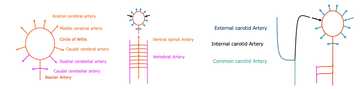
What is the rete mirabile?
The rete mirabile is a network of vessels located within the cavernous sinus. This is postulated to cool the blood, or reduce fluctuations from pulsatile flow.
Describe the blood supply to the spinal cord
Segmental arteries:
Cervical- vertebral artery
Thoracic- intercostal arteries
Lumbar- aorta
Describe the venous drainage of the brain and spinal cord
The brain and spinal cord are drained by a communicating system of sinuses.
Segmental ventral spinal vein draining:
Cervical- vertebral vein
Thoracic- azygous vein
Lumbar- azygous/caudal vena cava
What is the blood brain barrier?
This is a selective barrier between the circulating blood, and the parenchyma of the nervous system
Capillaries in the brain are surrounded by:
Closed, overlapping endothelium
Dense basement membrane
Processes of astrocytes
Selective barrier for exchange of substances
The blood brain barrier acts as a selective block to large molecules. Molecules can move across mainly by active transport, unless they are lipid soluble.
What is syringohydromyelia?
Syringohydromyelia is a condition characterised by a fluid-filled cyst (syrinx) within the spinal cord. It combines syringomyelia (a cavity in the spinal cord tissue) and hydromyelia (an expansion of the central canal). It disrupts normal cerebrospinal fluid (CSF) flow and requires medical monitoring or surgery.
What are the clinical uses of the meninges?
The meninges allow for epidural anaesthesia, collection of CSF and provides for the blood brain barrier
What are the denticulate ligaments and boundaries in relation to the meninges?
Denticulate ligaments are the focal firm attachment between the pia-arachnoid and the dura mater
Boundaries are continuous around the brain and spinal cord
Describe the layers of the meninges
Dura mater
Intracranially adherent to the periosteum of the skull
Spinal- separated from the periosteum by the epidural space, which is filled with fat
This is a tough layer, composed mainly of dense connective tissue
Arachnoid mater
This is a fine layer, pressed up against the dura mater with fine, wispy filaments which extend to and blend with the pia mater
Pia mater
This is a thin layer, which is adherent to the underlying brain/spinal cord
It blends with the fibres of the arachnoid
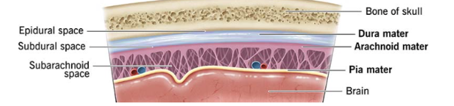
What is the function of the meninges?
The meninges function as protection, CSF containment, support and maintenance of the blood brain barrier
What is the function of the arachnoid villi in the meninges?
Arachnoid villi act as one-way valves to regulate pressure within the CSF. This drains into sagittal and other venous sinuses
Describe the formation and function of CSF
CSF is produced by the choroid plexus (a vascular proliferation of the ependymal layer) in lateral and third and fourth ventricles.
CSF is formed by dialysis of blood in the choroid plexus (ultrafiltration of plasma, and active transport). There is further production from ependymal linings and vessels within the pia mater.
The CSF functions as protection, support and nutrition.
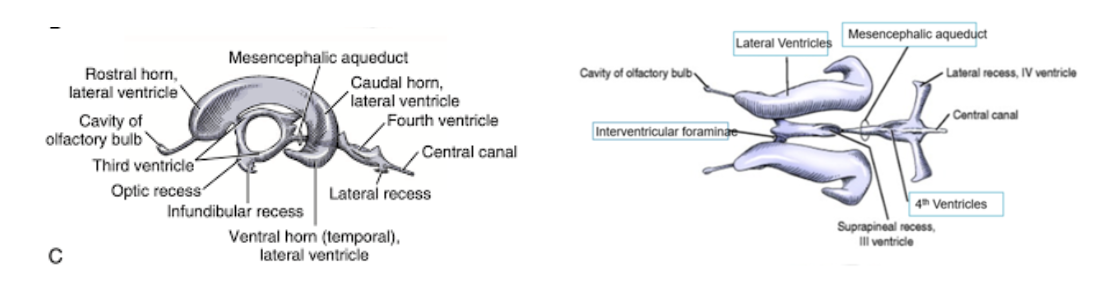
Draw out the ventricular system of the brain
From above
From the side