immunology test 1 JMU
 Call Kai
 Learn
Learn Practice Test
 Spaced Repetition
 Match
Match Flashcards
 Knowt Play
1/132

There's no tags or description
Looks like no tags are added yet.
Name | Mastery | Learn | Test | Matching | Spaced | Call with Kai | Chat |
|---|
No analytics yet
Send a link to your students to track their progress
133 Terms
2 major functions of immunity
1. protect against infectious diseases by recognizing and defending us against foreign substances
2. detect and kill mutant cells
also immunity
-self tolerance
-immunological memory
host defenses
barrier defenses, innate immunity, adaptive immunity
innate immunity
defenses against any pathogen
defense mechanisms that act immediately at the beginning of an infection.
-not pathogen specific
-overall effect is to induce a state of inflammation!!
-inflammation
-TLR4
-recognition and destruction of pathogens
-complement system
adaptive immune response
The response of antigen-specific B and T lymphocytes to antigen, including the development of immunological memory.
-clonal expansion
-dendritic cells
barrier host defenses
mucosal surfaces of the GI, respiratory, and urogenital tracts -where pathogens normally enter the body
the skin is the body's first line of defense
barrier defenses mechanical
skin- epithelial cells joined by tight junctions; longitudinal flow of air or fluid (peeing)
gut-epithelial cells joined by tight junctions; longitudinal flow of air or fluid (peeing)
lungs-epithelial cells joined by tight junctions; movement of mucus by cilia
eyes/nose/oral-epithelial cells joined by tight junctions; tears, nasal cilia
barrier defenses chemical
skin- fatty acids; antimicrobial peptides
gut- low pH, antimicrobial enzymes; antimicrobial peptides
lungs- pulmonary surfactant; antimicrobial peptides
eyes/nose/oral- antimicrobial enzymes in tear and saliva; antimicrobial peptides
antimicrobial peptides- all epithelial cells produce
barrier defenses microbiological
skin- normal microbiota
gut-normal microbiota
lungs-normal microbiota
eyes/nose/oral- normal microbiota
microbiota- the community of microbiobes that inhabit a particular niche
physical barriers colonized by commensal bacteria protect against infection by pathogen
commensal bacteria
compete with pathogens for nutrients and space and they secrete antibacterial proteins that inhibit pathogenic species
antibiotic treatment can disrupt colon microbiota by
1. colon is colonized by large numbers of commensal bacteria
2. antibiotics kill many of these commensal bacteria
3. pathogenic bacteria gain a foothold and produce toxins that cause mucosal injury
4. red and white blood cells leak into gut between injured epithelial cells
Steps of innate immunity
1. recognition of pathogen by cell surface receptors(they can bind to more than one pathogen)
2. recruitment of destructive "effector mechanisms" that kill and eliminate pathogen
-effector cells such as pathogens
-serum proteins called complement which flag pathogens for phagocyte destruction or attack pathogens directly

inflammation
heat, redness, swelling, pain, loss of function
-involves endothelium

endothelium
blood and lymphatic capillaries are composed endothelial cells

inflammation steps
-cytokines are released by host cells in response to pathogens and this leads to a state on inflammation
1. wound introduces bacteria which activate effector cells to secrete cytokines
2. vasoDILATION and inc vascular permeability in endothelial cells allow protein and inflammatory cells to leave blood and enter tissue
3. the tissue becomes inflamed ; edema formation (swelling)
adaptive immunity
-immune response specific to the pathogen
-involves WBCs and B and T cells
-leads to immunological memory
-innate often only slows the spread of infect until adaptive response can respond
innate vs adaptive immunity
innate - fast (hours), fixed, limited # of specificities, constant during response
Adaptive Immunity- slow response (days to weeks), variable, has numerous highly selective specificities, and improves during the response.
clonal selection and expansion
1. lymphocytes are exposed to pathogens
2. replicate cells that have adapter to pathogen (all specific for the same pathogen)
3. create effector cells and memory cells- responsible for immunological memory
lymphocytes- subtypes of white blood cell in immune system. include natural killer cells (which function in cell-mediated, cytotoxic innate immunity), T cells (for cell-mediated, cytotoxic adaptive immunity), and B cells (for humoral, antibody-driven adaptive immunity).
Primary adaptive immune response
the 1st time one is exposed to a specific pathogen
Secondary adaptive immune response
subsequent exposure to the same pathogen- creates a stronger, fast acting response because of high numbers of memory cells specific for pathogen
*without innate, adaptive doesnt occur
Hematopoiesis
blood cell formation
hematopoetic stem cells
1. self renewing
2. gives rise to all other blood cells
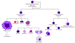
granulocytes
wbc's containing granules in the cytoplasm with multilobed nuclei
neutrophil: excellent of phagocytosis, most common, 1st to arrive
eosinophil: granules stain orange with dyes
basophil: rarest, granules stain purple
what is pus?
dead neutrophils after they engulf and kill bacteria
what is the stage before matured into a macrophages
monocytes
monocytes
Circulate in the blood and then migrate to tissues where they differentiate into macrophages
macrophages
located in tissues, good at phagocytosis, provide early warning to other cells and orchestrate local response to infection
typically phagocyte to sense infection- pathogen phagocytosis, cytokine secretion
dendritic cells (myeloid lineage)
located in tissues
pick up pathogen and bring it to lymph node
adaptive immune.
mast cells (myeloid lineage)
located in tissues
granules contain histamine
responsible for allergic diseases
natural killer cells (NK cells)
in blood
pursue diseased cells (such as those infected by viruses or cancer)
first to react to viral infections
B and T cells
-two types of lymphocytes
-in blood and lymphatic tissues.
-responsible for adaptive immunity
-B cells can mature into a plasma cell
plasma cells
a fully differentiated B cell that secrete antibodies
leukocytes
make up blood WBCs
Proportion of Leukocytes
neutrophil: 40-75%
eosinophil: 1-6%
basophil:
layers of white blood cells

antigens (ags)
any molecule or molecular fragment that can be recognized and bound by a BCR, TCR, or Ab
usually not born with it
usually a protein

effector cells
on encountering their specific antigen, B and T cells differentiate into effector cells by clonal expansion
a terminally differentiated activated lymphocyte that can kill pathogens or remove them from the body without the need for further differentiation
B cell ->
encounters antigen and turns to Ab-secreting plasma cell
Tcell ->
encounters antigen and turns to a Cytotoxic Tcell (kills cells infected with virus or certain bacteria
or
Helper Tcell (secretes cytokines that help other immune cells become fully activates
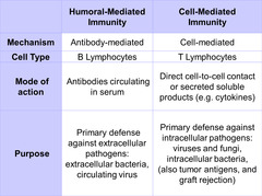
humoral immunity
immunity due to antibodies and their actions by which they combat infection
1. neutralization- antibody binds to pathogen and either inhibits growth or prevent replication, make it not able to bind to us
2. opsonization- coating surface of extracellular pathogen and makes it easier to be ingested
primary lymphoid tissues
-where lymphocytes develop
-bone marrow (Bcells) and thymus (Tcells)
secondary lymphoid tissues
-where lymphocytes are stimulated and respond to pathogens (Ags) & where clonal selection & expansion occurs
-lymph nodes, spleen, adenoids, tonsils, appendix, peyers patch of small intestine
no thymus=
no Tcells and few Abs (Tcells are required for Bcell ab production)
high risk for infection
lymphatics
plasma that leaks from blood capillaries can pick up pathogens or their components.
this fluid is collected by open ended lymphatic capillaries & is carried to the nearest lymph node
ultimately returns the lymph to the blood
lymph and lymphocyte recirculation
driven slowly by body movement. one way valves keeps lymph moving in one direction
after time, they leave via efferent lymphatics and return to the blood
circulated lymphocytes meet pathogens in draining lymph node. this is where adaptive immunity is initiated.
theyre taken to the closest lymph node
lymphocytes divide and differentiate into effector cells (clonal expansion)
lymph node
divided into, cortex(mostely Bcells), paracortex(mostly Tcells), medulla
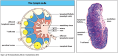
blood circulation through lymph node
during infection, pathogen specific B cells proliferate to form dense areas called germinal centers
inc lymphocyte proliferation is why an infection causes lymph nodes to swell
infection generally causes enlarged nodes to be tender
germinal center
when B cells proliferate to form dense areas
lymphadenopathy
swollen lymph nodes
not filled with fluid , they have a lot of cells from clonal expansion
Activation of Adaptive Immunity
inflammatory reaction in tissues leads to recruitment of WBCs to site
pathogens and their components then travel to the draining lymph node via lymphatics
T cells cant make B cells without antibodies
**
spleen
provides adaptive immunity to BLOOD infections
red pulp- major function is to remove old and damaged RBCs (lots of macrophages)
white pulp- secondary lymphoid tissue, organization similar to lymph node, *no lymphatic drainage, antigens must enter via the central artery, lymphocytes enter/leave via venous blood
congential asplenia
born w/o a spleen, more susceptible to bacteria infections, genetic immunodeficiency
Mucosa-associated lymphoid tissue (MALT)
Concentrations of lymphatic tissue without a connective tissue capsule
most secondary lymphoid tissue is associated with the gut
1. gut associated lymphoid tissue (GALT)
2. Bronchial associated lymphoid tissue (BALT)-lines respiratory tract
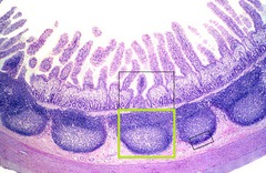
peyers patches
large collections of lymphoid tissue found in the submucosa of the small intestine
pathogens are transported from gut lumen across mucosa by specialized cells called M cells
extracellular pathogens
susceptible only to soluble, secreted immune molecules
ex. Abs (cant pass plasma membrane), complement
intracellular pathogens
only susceptible to soluble molecules when microbes first enter the body and when released the host cell dies
the complement system
proteins become covalently attached or fixed to pathogen surface (complement fixation)
made in liver; circulate in blood as inactive enzymes
complement protein 3 (C3) is most imp

Fixation of complement
key event: cleavage of C3 and the covalent attachment of C3b to the surface of a pathogen
C3b marks the pathogen for destruction by phagocytes and can organize formation of proteins complexes that damage the pathogens membrane

3 pathways of complement system
alternative, lectin, classical
all are part of innate immunity, but classical is kind of innate and adaptive
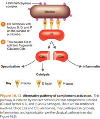
alternative pathway
pathogen surface creates local environment conducive to complement activation
1st to act
1st step is the spontaneous hydrolysis of a thioester bond forming iC3
-allows factor B to bind and be cleaved by factor D
-leads to form soluble C3 convertase called iC3Bb
-soluble iC3Bb cleaves C3 and then C3b binds covalently to the pathogen surface
leads to positive feedback; one of the progressive amplification of C3 cleavage, more C3b is created
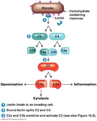
lectin pathway
mannose-binding lectin binds to pathogen surface
2nd to act
classical pathway
C-reactive protein or antibody binds to specific antigen on pathogen surface
3rd to act

regulatory proteins
determine the extent and site of C3b deposition
complement control proteins function to inc complement binding to pathogens and to limit the binding to human cells
Decay-accelerating factor (DAF)
membrane bound complement regulatory protein on human cells that inactivates alternative C3 convertase that has bound
opsonization
coating antigen with antibody enhances phagocytosis
C3b is an opsonin

complement receptor 1
on cells like macrophages trigger the uptake and breakdown of C3b coated pathogens
lyctic or terminal pathway
requires a C5 convertase
-a second C3b binds to the alt. C3 convertase(C3bBb) and forms the alt C5 convertase (C3b2Bb)
-C5 is cleaved from C3b2Bb to form fragments C5a and C5b
-C5b functions to initiate the formation of the membrane attack complex which makes hole in membrane
formation of a membrane attack complex
is NOT a direct function of antibodies
C6 and C7 bind to C5b and assembly on the pathogen membrane then C8 binds and induces polymerization of multiple C9 proteins that form a pore
leads to death of the cell

Anaphylatoxins (C3a, C5a)
small peptides released during complement activation that induce local inflammation
-act to induce inflammation, recruiting fluid and inflammatory cells to sites of infection
they bind to neutrophils/monocytes inc their adherence to vessel walls, promote migration toward site of comp. fixation and inc phag.
endothelial cells, phagocytes, mast cells

phagocytic receptors
recognize bacterial carbs and lipids and triggers macrophage phagocytosis
macrophage phagocytosis
type of receptor mediated endocytosis and degradation within phagolysosomes
Toll-like receptors (TLRs)
10 signaling receptors that sense infection and are present on many types of innate cells, including macrophages, dendritic cells and neurophils
expressed on the cell surface and within cells
each type of TLR is specific for a diff pathogen component
signaling through TLRs usually leads to the production of inflammatory cytokines

TLR4
recognizes LPS & leads to production of cytokines, adhesion molecules and other proteins necessary for inflammation
signaling involves adapter proteins and protein kinases that lead to activation of transcription factor called Nuclear Factor kB

TLR4
1. a complex of TLR4, MD2, CD14, and LP5 is assembled at the macrophage surface
2. myD88 bindes TLR4 and activates IRAK4 to phosphorylate TRAF6 which leads to the phosphorylation and activation of IKK (inhibitor if kB kinase)
3. IKK phosphorylates IkB leading to its degradation and the release of NFkB which enters nucleus
4. NFkB activates transcription of genes for inflammatory cytokines which are synthesized in the cytoplasm and secreted in the ER
infants without IKK
lack IKK
TLR4 is not activated, there would be no cytokine gene transcription
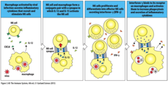
activated macrophages
release cytokines that promote inflammation
release CXCL8, which recruits NK cells from the blood
-macrophages secrete IL-12 which activates and stimulates proliferation of NK cells
-activated NK cells in turn release INF-gamma that further activate macrophages
cytokines
small soluble proteins secreted by cells that influence other cells by binding to a specific surface receptor
IL-1beta and TNF-alpha
induces blood vessels to become more permeable, enabling effector cells and fluid containing soluble effector molecules to enter the infected tissue
IL-6
induces fat and muscle cells to metabolize, make heat and raise the temperature in the infected tissue
CXCL8
recruits neutrophils from the blood and guides them to the infected tissue
IL-12
recruits and activates NK cells that in turn secrete cytokines that strengthen the macrophages' response to infection
septic shock (sepsis)
gram negative bac. is in the blood which express LPS
lotssss of macrophages in spleen and liver become activated via TLR-4 and secretes significant amt of TNK-alpha systematically into blood stream
primary site of hematopoiesis
above 1 yr
bone marrow
why is adaptive immune response slow to respond
clonal selection and expansion
major goal of inflammatory response
recruit inflammation cells to site of infection
neutrophils
the most common type of WBC
recruited by macrophages from blood into the tissues in large numbers during infection.
1st cell recruited
elevations in their numbers in blood is a sign of infection
major function: phagocytosis and killing; die within hours after phagocytosis (pus)
inflammatory cytokines
recruit neutrophils from the blood to inflamed tissues
extravastion
overall movement of cells from within capillaries into tissues in 4 steps
1. Rolling Adhesion
in presence of infection and inflammatory cytokines (IL-1b, TNF-a,CXCL8) the endothelium expresses an adhesion molecule that bind to specific sugars on neutrophils
only adhere to vein surfaces

2. tight binding
cytokines induces endothelium to express ICAM adhesion molecules which bind integrin adhesion molecules on neutrophils
neutrophil now becomes immobilized on vascular endothelium near the sire of infection
3. Diapedesis
neutrophils flatten and squeeze out of capillaries and enter connective tissue

4. migration
by using their chemokine receptors for CXCL8 neutrophils migrate up the concentration gradient toward source of chemokine, which is macrophages in the infected tissue
Mannose receptor
cell-surface receptor on dendritic cells, macrophages, and other leukocytes that binds to mannose residues on the surfaces of pathogens
the binding of bacteria to neutrophil innate receptors induce phagocytosis and microbial killing
N-formyl-Met receptor: is the starting amino acid in prokaryotic protein synthesis
killing of bacteria by neutrophils
involves fusion of 2 types of cytoplasmic granules and lysosomes with the phagosome
1. bacterium is phagocytosed
2. phagosome fuses with azurophilic and specific granules
3. pH of phagosome rises, antimicrobial response is activated and bacterium is killed (NADPH oxidase raises pH)
4. pH of phag. dec, fusion with lysosome allows acid hydrolases to degrade the bacterium completely
5. neutrophil dies by apoptosis and is phagocytosed

acute phase response
a response to an acute illness that produces specific blood proteins called acute phase proteins
IL-1B, IL-6,TNF-a are endogenous pyrogens that induce fever
at higher temps bacteria and viruses grow more slowly
C-reactive protein
elevations in this is used to diagnose infection, inflammation, and tissue damage
concentrations rise within 2 hours of inflammation and peak at 48 hours
triggers the classical pathway
-requires C1-composed of C1q and 2 proteases C1r and C1s
- Creactive proteins bind to C1q, results in activation of C1s
-C1s cleaves both C4 and C2 forming the classical C3 convertase (C4bC2a)

Mannose-binding lectin & C-reactive protein
-both bind to structures unique to bacteria and serve as opsonins
-mannose binding can activate the complement pathway via the lectin pathway
-c reactive can activate complement pathway via classical
Lectin complement pathway
initiated by mannose binding lectin
-MASPs and MBL -associated protease
-complement C2 and C4 are cleaved
-C4a is another anaphylatoxin
-end result is formation of classical C3 convertase (C4bC2a)that cleaves C3 to C3b and a
- C3 b is primarily used to form alternate C3 convertase (C3bBb)

Type 1 interferons
IFN-alpha and IFN-beta
-essentially all humans cells that become virally infected make these cytokines
-uninfected cells do not synthesize INFs but INF receptors are always present on the surface of all cells
-once secreted, can act in an autocrine or paracrine fashion
every cell except RBCs can make cytokines