MSK TBL 16/ILS 10: Knee Joint Anatomic Imaging & Functional Anatomy
 Call Kai
 Learn
Learn Practice Test
 Spaced Repetition
 Match
Match Flashcards
 Knowt Play
1/60

There's no tags or description
Looks like no tags are added yet.
Name | Mastery | Learn | Test | Matching | Spaced | Call with Kai | Chat |
|---|
No analytics yet
Send a link to your students to track their progress
61 Terms
knee joint
formed by femur, tibia, & patella
femoral & tibial condyles articulate
patella articulates w/ femoral trochlear [patellar] surface
fibula NOT involved
patella
largest sesamoid bone
located within tendon of quadriceps femoris
articulates w/ femur but NOT tibia
attaches to tibial tuberosity by continuation of quadriceps = patellar ligament
function
prevent wear & tear on quadriceps tendon
increase angle of pull of quads, magnifiying its power
tibia
weight-bearing, medial bone
has tibial tuberosity where patellar ligament inserts
has medial malleolus w/ malleolar groove for tendons of tibialis posterior & flexor digitorum longus muscles
another groove, posterolateral to malleolus groove, for tendon of flexor hallucis longus muscle
provides attchmnt for deltoid ligament of ankle joint
fibula
little to no weight bearing function
provides attchment for muscles
head [apex] provides attchment for fibular collateral ligament of knee joint
knee joint btwn femur & tibia
condylar type of synovial joint
knee joint btwn femur & patella
saddle joint
knee ligaments are taut in
full extension, accompanied by medial rotation of femur on tibia
knee joint stability depends on which muscle
quadriceps
knee joint stabilized laterally by
biceps & gastrocnemius [lateral head] tendons
IT tract
fibular collateral ligaments
knee joint stabilized medially by
sartorius
gracilis
gastrocnemius [medial head]
semitendinosus
semimembranosus
tibial collateral ligament
fibrous joint capsule of knee
capsule attched to margin of articular surfaces
has opening posterior to lateral tibial condyle
passage of popliteus tendon
median infra-patellar synovial fold
infra-patellar fat pad that divides articular cavity of knee into R. & L. compartments
extracapsular ligaments of knee
tibial collateral ligament [medial collateral ligament, TCL]
fibular collateral ligament [lateral collateral ligament, FCL]
patellar ligament
oblique popliteal ligament
arcuate popliteal ligament
TCL
from medial femoral epicondyle to medial tibial condyle
injury to this ligament → concomitant damage to medial meniscus
prevents medial displacement
taut on extension
FCL
separated from lateral meniscus & capsule of joint by tendon of popliteus muscle
from lateral femoral epicondyle to head of fibula
taut on extension
oblique popliteal ligament
recurrent expansion of tendon of semimembranosus
posteriorly attched btwn medial tibial condyle & lateral tibial condyle
resists hyperextension & lateral rotation
arcuate popliteal ligament
strengthens joint capsule posterolaterally
intracapsular extra-synovial ligaments
anterior cruciate ligament [ACL]
posterior cruciate ligament [PCL]
ACL
from anterior intercondylar region [superior, posterior, & lateral]
attach to lateral femoral femoral condyle [posteromedially]
prevents posterior displacement of femur on tibia
prevents hyperextension
weaker than PCL
taut during extension
often injured in conjuction w/ tibial collateral ligament & medial meniscus
unhappy triad
![<ul><li><p>from anterior intercondylar region [superior, posterior, & lateral] </p><ul><li><p>attach to lateral femoral femoral condyle [posteromedially]</p></li></ul></li><li><p>prevents posterior displacement of femur on tibia</p><ul><li><p>prevents hyperextension</p></li></ul></li><li><p>weaker than PCL</p></li><li><p>taut during extension</p></li><li><p>often injured in conjuction w/ tibial collateral ligament & medial meniscus </p><ul><li><p><strong>unhappy triad</strong></p></li></ul></li></ul><p></p>](https://assets.knowt.com/user-attachments/a3f4e30b-275f-4642-8796-b2c8897c7a31.png)
PCL
from posterior intercondylar area of tibia & passes upward, forward, & medially to insert into lateral surface of medial femoral condyle
stronger than ACL
prevents backward sliding of tibia on femur
limits hyperflexion of knee
taut during flexion

menisci of knee
fibrocartilage
shock absorbers
attched along their external margins to tibia & fibrous joint capsule [coronary ligaments]
attached at their ends to intercondylar eminence
only peripheral 1/3 [red zone] is vascularized
inner 2/3 avascular
central tears heal poorly
medial meniscus
C-shaped [semicircle]
![<p>C-shaped [semicircle]</p><p></p>](https://assets.knowt.com/user-attachments/f32c1a6a-1cf8-483d-b241-7bbfdf485de6.png)
lateral meniscus
nearly circular

transverse ligament of knee
binds anterior horns [ends] of lateral & medial semilunar cartliages [menisci]
![<p>binds anterior horns [ends] of lateral & medial semilunar cartliages [menisci]</p>](https://assets.knowt.com/user-attachments/8380a8d6-d815-41bf-9711-a5de03c91166.png)
anterior bursae of knee joint
suprapatellar [quadriceps] bursa
prepatellar bursa
deep & subcutaneous infrapatellar bursae
posterior bursae of knee
popliteus bursa
anserine bursa
gastrocnemius bursa
semimembranosus bursa
articularis genus
small muscle that pulls bursa away from joint during extension of knee
bursae that DON’T communicate w/ articular cavity of knee
prepatellar bursa
subcutaneous & anterior to patella
deep & subcutaneous infrapatellar bursae
deep could be aggravated in runners
semimembranosus bursa
4 bursae that communicate w/ synovial cavity of knee joint
GAPS
Suprapatellar [quadriceps] bursa
popliteus bursa
anserine bursa
gastrocnemius bursa
all deep to corresponding tendons of area
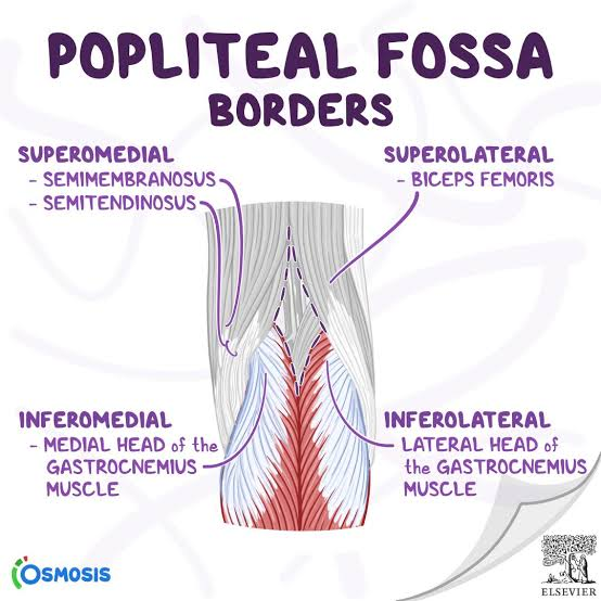
Popliteal Region/Fossa Boundaries
fat-filled, diamond-shaped depression posterior to knee joint
superolateral: biceps femoris long head
superomedial: semimembranosus
inferolateral & inferomedial: lateral & medial heads of gastrocnemius
floor: popliteal surface of femur, posterior knee joint capsule
reinforced by oblique popliteal ligament & popliteus muscle
roof: skin & popliteal fascia
Popliteal region/fossa contents
popliteal artery & vein
tibial & common fibular nerves
small saphenous vein
genicular branch of obturator nerve
popliteal lymph nodes

popliteal artery
begins at adductor hiatus [adductor magnus muscle] as continuation of femoral artery
terminates at inferior border of popliteus muscle by dividing into anterior & posterior tibial arteries
deepest structure in popliteal fossa
popliteal vein is posterior & slightly lateral to artery
tibial nerve is posterior & slightly lateral to vein
gives genicular branches to knee
peripheral artery disease [PAD]
narrowing of arteries
popliteal pulse evaluates blood flow to leg
popliteal artery entrapment [PAE]
muscle hypertrophy or knee trauma
assess w/ popliteal pulse
blood supply to knee
genicular branches
superior
medial
lateral
Inferior
medial
lateral
middle
descending genicular artery
anterior tibial recurrent artery
popliteal vein
formed by union of anterior & posterior tibial veins at inferior border of popliteus muscle
terminates at adductur hiatus, where it becomes femoral vein
tributaries: small saphenous vein
nerve supply of knee joint
femoral
saphenous nerve & nerve to vastus
obturator
via genicular branch
common fibular [deep & superficial]
lateral sural cutaneous nerve arises here
tibial
medial sural cutaneous nerve arises here
popliteal [baker] cyst
swelling behind knee
caused by arthritis, meniscus injury, or herniation or tear of joint capsule
impairs flexion & extenion of knee
pain worse when knee extended
“housemaid’s knee”
prepatellar bursitis
“clergyman’s knee”
infrapatellar [superficial] bursitis
fractured distal femur or dislocated knee may injure
popliteal artery
transverse patellar fracture
from blow to knee or sudden contraction of quadriceps muscle
proximal fragment of patella pulled superiorly w/ quadriceps tendon
distal fragment remains w/ patellar ligament
tibial plateau / bumper fracture
fracture of lateral tibial condyle caused by an automobile bumper
usually associated w/ common fibular [peroneal] nerve injury
patellofemoral syndrome
“runner’s knee”
patella tracks improperly [usually laterall] in femoral trochlear groove
vastus medialis atrophy
presents as anterior knee pain
chondromalacia patella
pain in front of knee
erosion of articular cartilage on posterior surface of patella → bone-on-bone friction
seen in prior knee trauma, runners, or pple that exercise a lot
genu varum
“bowleg”
femur abnormally vertical
small Q angle
excess weight place on medial aspect of knee joint
→ arthrosis [destruction of knee cartilages] & fibular collateral ligament overstressed
genu valgum
“knock knee/ K legs”
lateral angulation of leg
large Q angle
weight bearing line falls lateral to center of knee
tibial collateral ligament overstretched
Unhappy triad / O’Donoghue’s triad / terrible triad
ACL
MCL
Medial meniscus
anterior drawer test
tests ACL
Lachman test
knee slightly bent, examiner stabilizes thigh while pulling shin forward
tests ACL
posterior drawer test
tests PCL
valgus stress test
pt supine w/ knees flexed to 30 degrees
clinician applies valgus stress to knee
checks for MCL injury
Varus stress test
pt supine w/ knees flexed to 30 degrees
clinician applies varus stress to knee
checks for LCL injury
menisci can get torn during
forceful rotation or twisting
vertical meniscal tear
perpendicular to tibial plateau
horizontal meniscal tear
parallel to tibial plateau, splitting meniscus into upper & lower halves
bucket handle meniscal tear
longitudinal tear where torn portion of meniscus forms handle-shaped fragment
arthroscopy
performed to repair meniscal tear
McMurray test
test for meniscal tears
pt supine, clinician flexes pts knee & externally rotates foot
apply valgus stress to knee & extend knee
tests medial meniscus
(+) test = reproduction of pain & locking
lateral meniscus tested similarly, except foot rotated internally & varus force applied while extending knee
“LIME”
to name direction of displacement
use orientation of DISTAL bone
what rules out meniscus injury?
if theres no joint line tenderness or effusion, likely not meniscal