BIOS242 Exam 1 Review Live Lecture Flashcards
 Call Kai
 Learn
Learn Practice Test
 Spaced Repetition
 Match
Match Flashcards
 Knowt Play
1/94

There's no tags or description
Looks like no tags are added yet.
Name | Mastery | Learn | Test | Matching | Spaced | Call with Kai | Chat |
|---|
No analytics yet
Send a link to your students to track their progress
95 Terms
Medical Microbiology
Deals with microbes that cause disease in humans and animals
Agricultural Microbiology
Relationships between microbes and farm animals and crops
Public Health Microbiology and Epidemiology
Monitor and control the spread of diseases in communities
Immunology
Investigates protective substances and cells produced in response to infection
Industrial Microbiology
Safeguards our food and water
Biotechnology
Environmental Microbiology
Effect of microbes on Earth’s diverse habitats (aquatic, soil)
Medical Microbiology Example
Unfortunately, the strawberries in MAs PIIE were contaminated with Salmonella enterica, causing gastrointestinal distress in people who ate the pie
Agricultural Microbiology Example
MA is having trouble finding other ingredients to make her PIIE since local blueberries have developed fungal fruit rot
Public Health Microbiology and Epidemiology Example
MA sold her PIIEs at a local farmer’s market and there is now a community wide outbreak of Salmonella enterica
Immunology Example
To test people with gastrointestinal distress for Salmonella enterica, the local clinic performs blood tests to look at the immune response to the microorganism
Industrial Microbiology Example
MA is really upset over making so many people sick with her PIIEs, so she indulges in her favorite meal- cheese, bread, and a good glass of wine
Environmental Microbiology Example
MA decides to grow all her own PIIE fruit from now on, so she takes her soil to be sampled to make sure it has a good balance of microbes for “fruitful” growth
Pasteur’s Experiment
Louis Pasteur used a swan-neck flask to demonstrate that microbial contamination comes from the air, not from spontaneous generation. He showed that sterile broth in the flask remained free of microbes until exposed to air, proving that microorganisms are present in the environment.
Louie Pasteur
Invented pasteurization
Developed the germ theory of disease
Eventually develops a vaccine for rabies
Robert Hooke
First to observe microbes
Antonie van Leeuwenhoek
Made small, crude microscopes to examine fabric
Drew “animalcules” from rainwater and scrapes from his teeth
Robert Koch
Developed postulates to link a specific organism to a specific disease
John Needham
Attempted to disprove spontaneous generation but failed
Did not realize that boiling did not kill all organisms
Koch’s Postulates
Find evidence of a particular microbe in every case of a disease
Isolate that microbe from an infected subject and cultivate it in pure culture in the laboratory; perform full microscopic and biological characterization
Inoculate a susceptible healthy subject with the laboratory isolate and observe the same resultant disease
Reisolate the same agent from this subject
John Tyndall
Demonstrated that some microbes in the dust and air have very high heat resistance
Something stronger than boiling was needed to destroy these microbes
Joseph Lister
First to utilize handwashing and misting operating rooms with antiseptic chemicals
Credited with developing ASEPTIC technique
Oliver Wendell Holmes
Observed that mothers who gave birth at home experienced fewer infections than mothers who gave birth at the hospital
Dr. Ignaz Semmelweis
Showed that women became infected in the maternity ward after examinations by physicians who had been working in the autopsy rooms without washing their hands
Carl von Linne
Also known as Linnaeus
Established rules for classification
Binomial Naming System
Scientific name is Genus name followed by species name
EXAMPLE: Staphylococcus aureus
Light Microscopes: Bright Field
Most widely used
Light Microscope: Interference
Well defined images that appear 3D
Electron Microscopes: Transmission
Detailed views of cell organelles and viruses; metal coat
Electron Microscopes: Scanning
Most dramatic and realistic 3D images
Simple Stains
Stains that give color to organisms
Gives contrast and color
Allows size, shape, and arrangement to be seen
Does not tell anything about the organism
Differential Stains
Tells us about the microorganism by staining specific structures or components, highlighting differences between cellular types and allowing for identification.
DNA Replication
Happens in a semiconservative manner
Original parent strand (template strand) opens→ Enzymes come in with new DNA
Each daughter molecule is identical to the parent in composition
The template strand is an original parental DNA strand
The newly synthesized DNA is made in a discontinuous fashion
Transcription
DNA is transcribed into mRNA (before translation)
Translation
mRNA is translated into protein (after transcription) and the translation happens via a ribosome
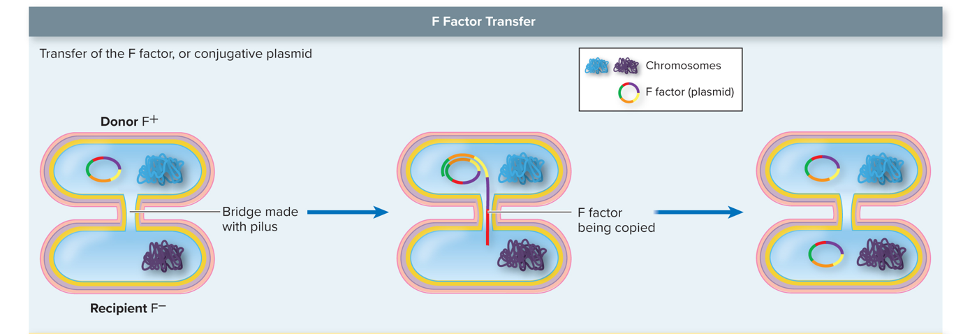
Conjugation
Involving a plasmid that is replicated and is sent from Bacteria A to Bacteria B through pili (pilus) that come together to allow for the transfer of the plasmid.
THINK PLASMID
Transformation
Involves a bacterial cell dying and releasing all of its genetic material into the environment, and other bacteria can take up this DNA and integrate it into their own genome.
If the DNA is not beneficial, they do not keep it, and they later get rid of it. If they are beneficial, they keep them and use them
THINK FREE DNA
Transduction
Viruses (bacteriophages) that specifically infect bacteria and transfer genetic material from one bacterium to another during the infection process.
THINK BACTERIOPHAGE
Transposons
Can jump from chromosome to chromosome, chromosome to plasmid, and plasmid to chromosome.
Very widespread among cells and viruses.
THINK JUMPING GENE
Spontaneous Mutation
A random change in the DNA arising from errors in replication that occur randomly
Induced mutations
Result from exposure to known physical or chemical agents that damage DNA (known as mutagens)
Ionizing Radiation (y rays, X rays)
Causes breaks in the DNA strands, leading to mutations or cell death. It is a type of induced mutation.
Ultraviolet Light Radiation
Causes cross-links between adjacent pyrimidines
Point Mutations
Small Mutations that affect only a single base in a gene
Involve addition, deletion, or substitution of single bases
Missense Mutation
Any change in the code that leads to the placement of a different amino acid
EXAMPLE: THE OLD CAT ATE THE FAT DOG → THE OLD RAT ATE THE FAT DOG
EXAMPLE 2: Met - Thr - Asp - Glu - Met - Lys → Met - Thr - Glu - Glu - Met - Lys
Nonsense Mutation
Changes a normal codon into a stop codon
Causes a very short protein that is usually nonfunctional
EXAMPLE: THE OLD CAT ATE.
EXAMPLE 2: Met - Thr - Asp - Glu - Met - Lys → Met - Thr - Asp
Silent Mutation
Alters a base but does not change the amino acid
EXAMPLE: THE OLD CAT ATE THE FAT DOG → THE OLD KAT ATE THE FAT DOG
EXAMPLE 2: Met - Thr - Asp - Glu - Met - Lys → Met - Thr - Asp - Glu - Met - Lys
Frameshift Mutations
Alter the reading frame of the mRNA
Due to INSERTIONS or DELETIONS
Nearly always result in a nonfunctional protein
Every amino acid after the mutation is different from what is coded for in the original DNA
EXAMPLE: THE LDC ATA TET HEF ATD OG THE GOL DCA TAT ETH EFA TDO G
EXAMPLE 2: Met - Thr - Asp - Glu - Met - Lys → Met - Thr - Thr - Asp - STOP
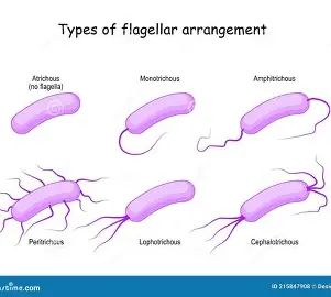
Flagella
Long, whip-like structures used for cell movement in prokaryotic and eukaryotic organisms. They enable mobility by propelling the organism through its environment.
THINK MOTILITY/MOBILITY
Pili
Short, hair-like structures on the surface of bacteria that facilitate attachment to surfaces and enable conjugation, allowing for the transfer of genetic material between cells.
THINK ADHESION AND CONJUGATION
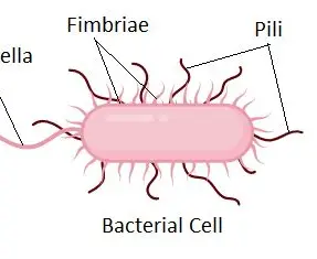
Frimbriae
Short, hair-like structures on bacteria that enhance adhesion to surfaces and can play a role in forming biofilms. They are distinct from pili and primarily involved in attachment.
THINK ATTACHMENT AND BIOFILM FORMATION
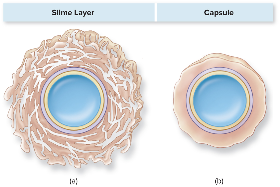
Slime Layer
A loose, unorganized layer of polysaccharides and proteins that surrounds some bacterial cells, providing protection and aiding in adhesion to surfaces.
THINK BIOFILM
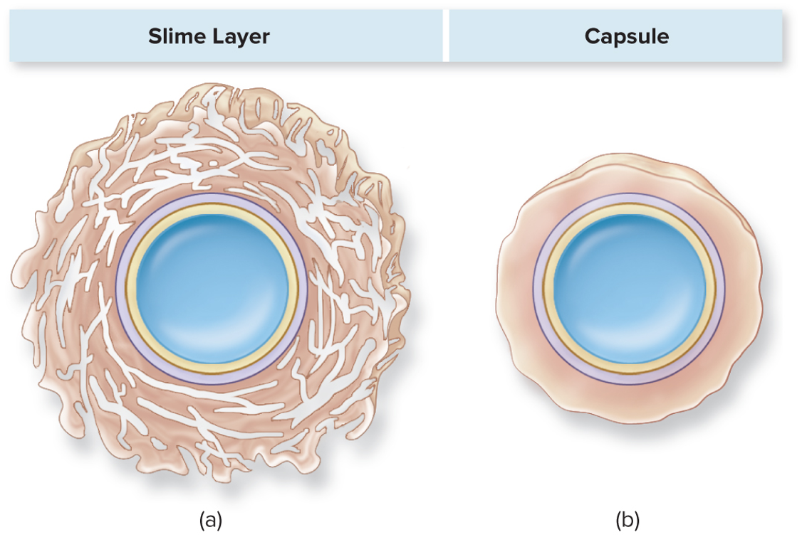
Capsule
A well-organized layer of polysaccharides or proteins that fully encases bacterial cells, providing protection against phagocytosis and aiding in adherence to surfaces.
THINK PHAGOCYTOSIS
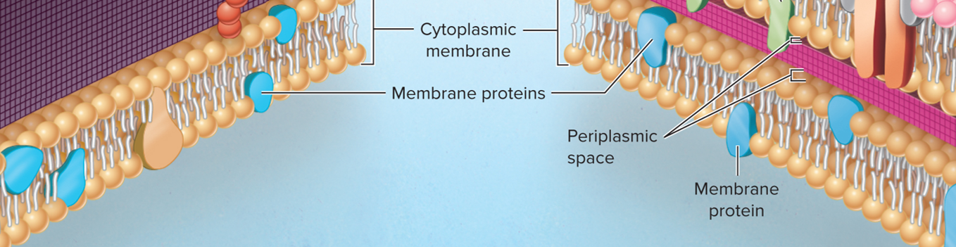
Cell Envelope (Cell Membrane)
A thin, flexible barrier around the cell that protects it and regulates the movement of substances into and out of the cell.
Gram Positive Organisms
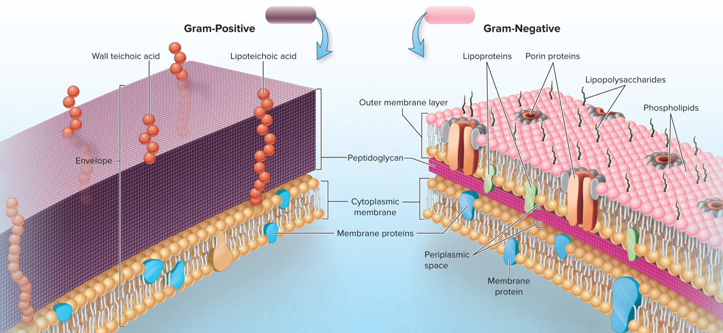
Bacteria that retain the crystal violet stain used in the Gram staining procedure, appearing purple under a microscope. They typically have a thick peptidoglycan layer in their cell wall.
Gram Negative Organisms
Bacteria that do not retain the crystal violet stain used in the Gram staining procedure appear pink under a microscope. They typically have a thin peptidoglycan layer and an outer membrane.
Cell Envelope (Cell Wall)
The cell envelope includes the cell wall, which provides structural support and protection to bacterial cells. It plays a crucial role in maintaining cell shape and can influence the interaction of the bacteria with its environment.
Cell Envelope (Gram Positive Cell Wall)
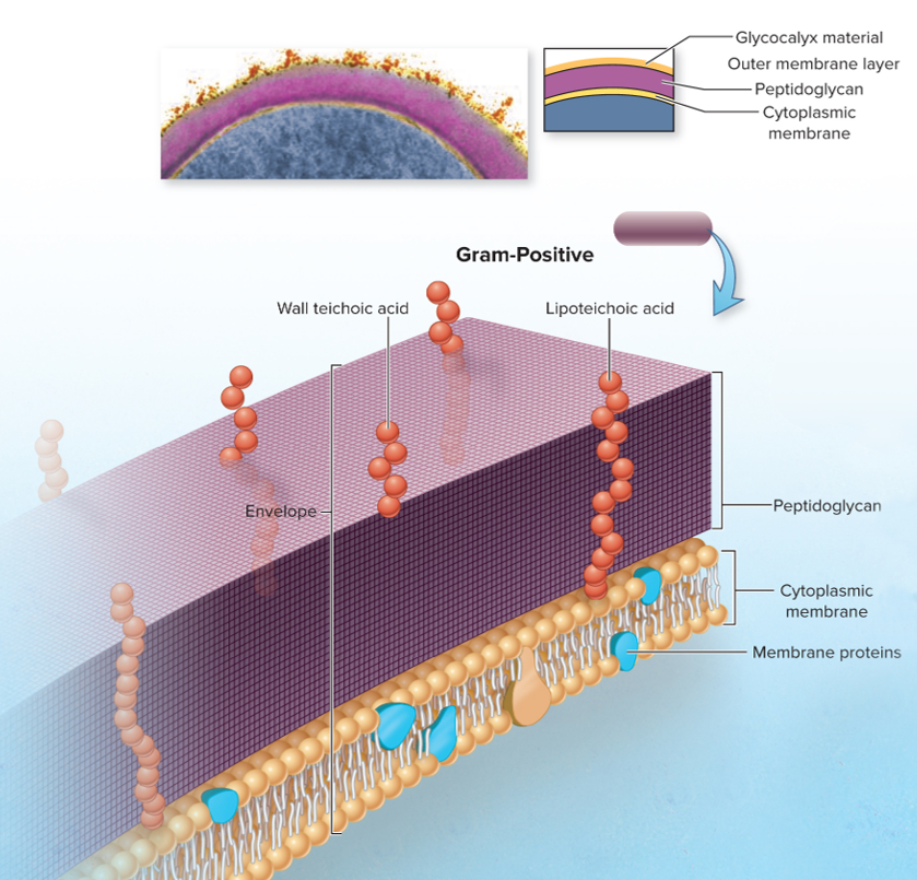
The part of the cell envelope, specifically in Gram-positive bacteria, is characterized by a thick peptidoglycan layer that provides structural integrity and retains the crystal violet stain during Gram staining, contributing to their purple appearance.
THINK THICK PEPTIDOGLYCAN LAYER, TECHOIC ACIDS, PURPLE GRAM STAIN FOR GRAM POSITIVE CELL WALLS
Techoic Acids
Polymers found in the cell walls of Gram-positive bacteria are composed of glycerol or ribitol phosphate, which play roles in cell wall maintenance and regulation of cell division.
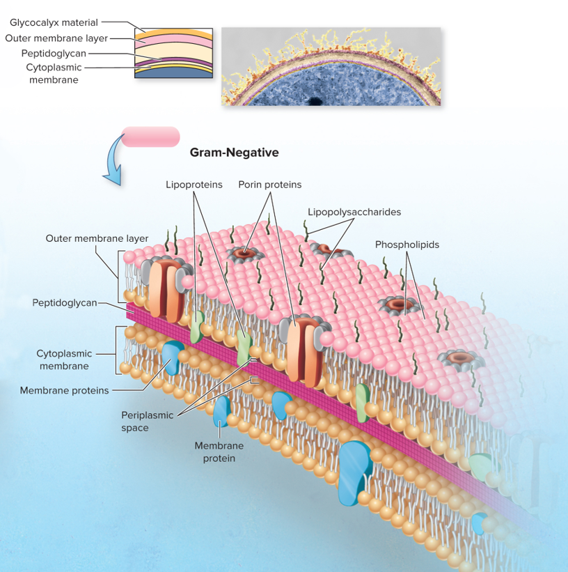
Cell Envelope (Gram Negative Cell Wall)
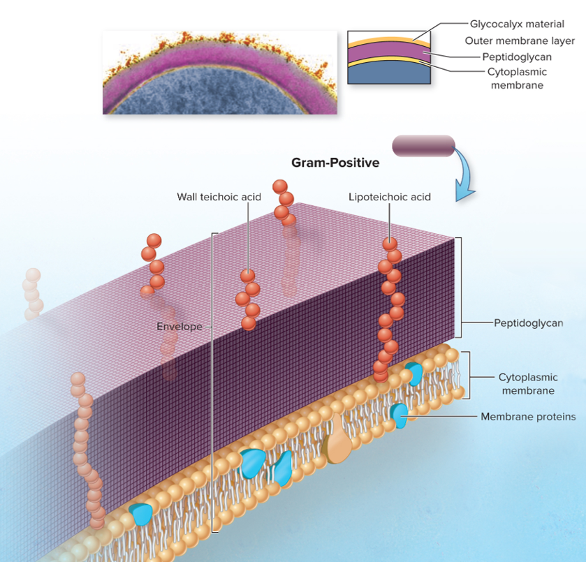
The part of the cell envelope, specifically in Gram-negative bacteria, characterized by a thin peptidoglycan layer located between the inner and outer membranes. This structure is less rigid than in Gram-positive bacteria and does not retain the crystal violet stain, appearing pink after Gram staining.
THINK THIN PEPTIDOGLYCAN LAYER, OUTER MEMBRANE CONTAINING LIPOPOLYSACCHARIDES, PINK GRAM STAIN FOR GRAM NEGATIVE CELL WALLS
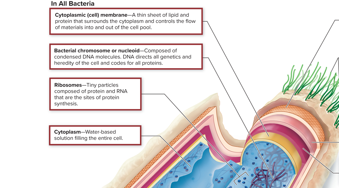
Nucleoid
The irregularly shaped region within prokaryotic cells where the genetic material, typically a single circular DNA molecule, is located. It is not surrounded by a membrane, distinguishing it from the nucleus in eukaryotic cells.
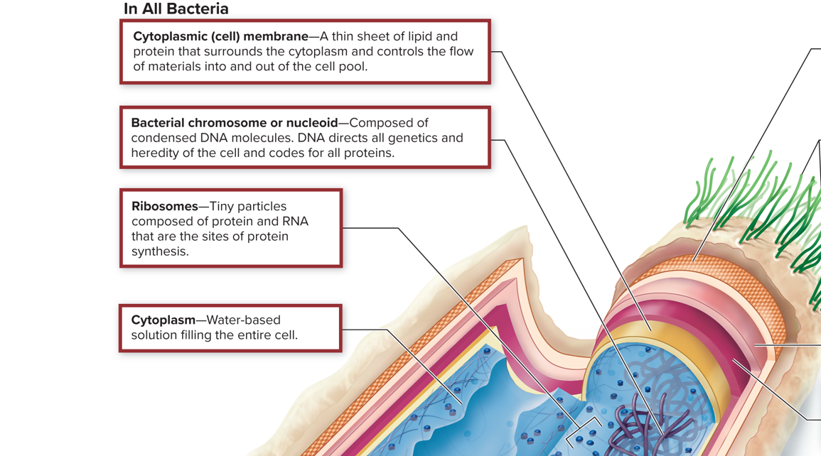
Ribosomes
Cellular structures responsible for protein synthesis, made up of ribosomal RNA and proteins, are found in both prokaryotic and eukaryotic cells.
In bacteria, there’s only a single circular one of these structures. This is so they can replicate faster.
Cytoplasm
The gel-like substance within the cell membrane, consisting of water, salts, and organic molecules. It houses organelles and is the site of numerous biochemical reactions.
Internal Bacterial Cell Structure: Plasmids
Small, circular DNA molecules separate from chromosomal DNA in prokaryotic cells. They can carry genes that provide bacteria with beneficial traits, such as antibiotic resistance.
THINK BENEFICIAL GENES
Internal Bacterial Cell Structure: Endospores
Highly resistant structures formed by certain bacteria to survive extreme conditions, containing a copy of the bacterial genome and essential proteins. They allow bacteria to endure harsh environments until conditions are favorable for growth.
THINK PROTECTION
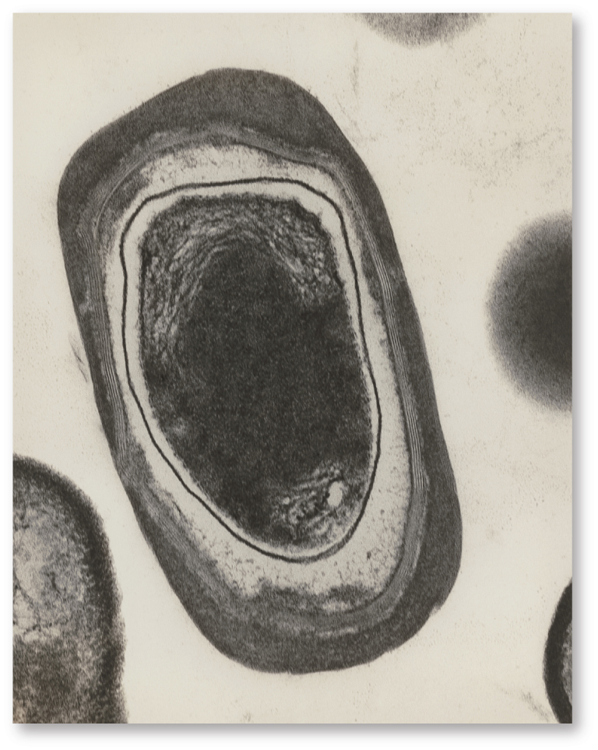
Prokaryote: Bacteria
Single-celled organisms without a nucleus or membrane-bound organelles, characterized by their simple structure and ability to thrive in diverse environments. They reproduce asexually through binary fission.
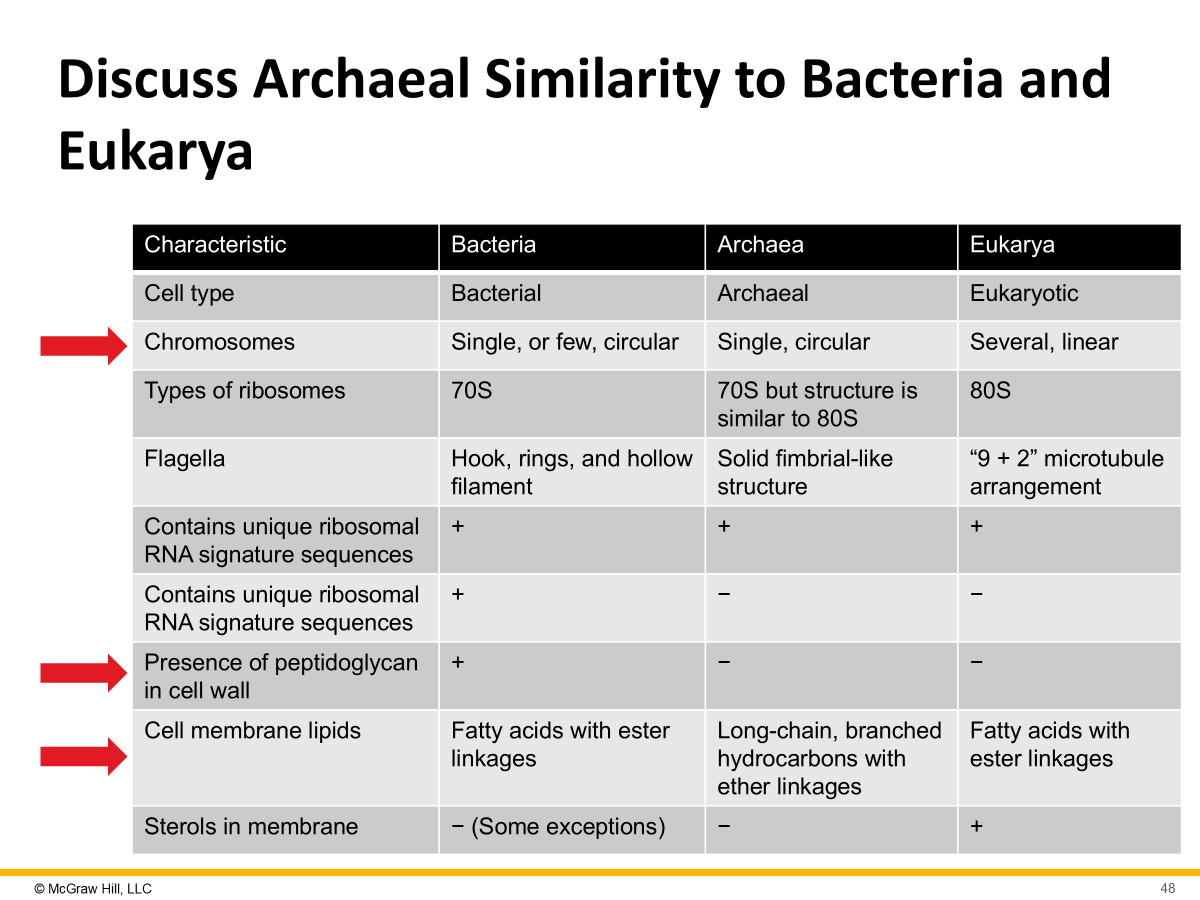
Prokaryote: Archaea
Single-celled microorganisms similar to bacteria but with distinct genetic and biochemical differences. Often thrive in extreme environments and can have unique metabolic pathways.
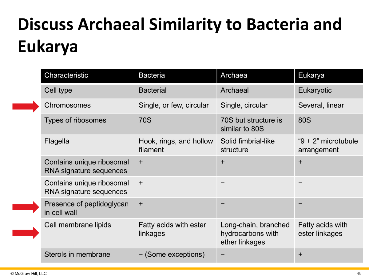
Eukaryote: Eukarya
Organisms with complex cells containing a nucleus and membrane-bound organelles. They include plants, animals, fungi, and protists, and reproduce sexually and asexually.
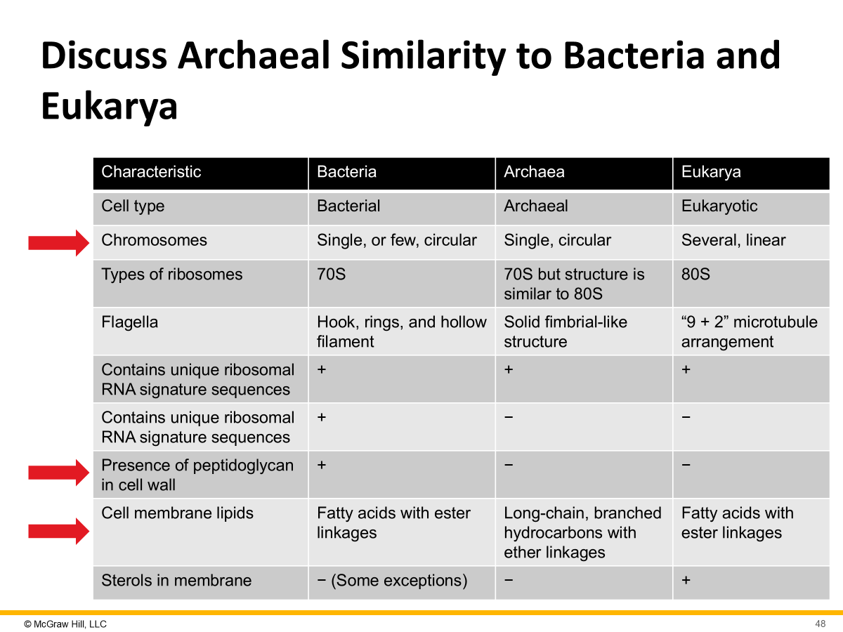
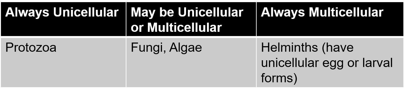
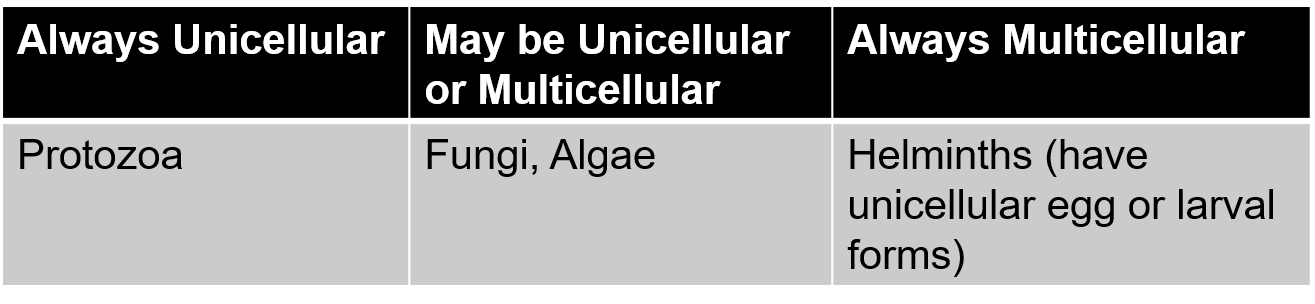
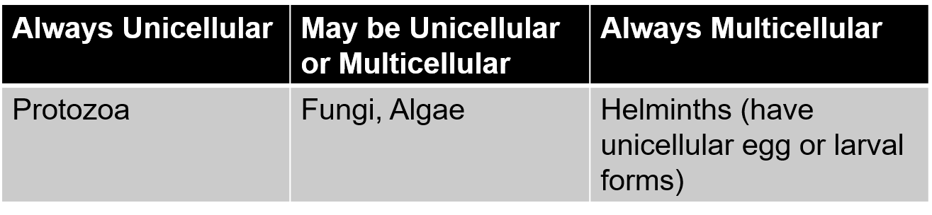
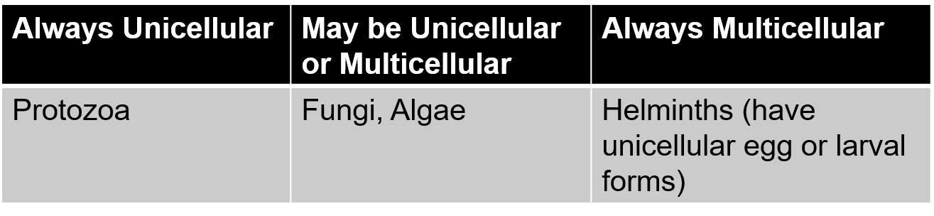
Protozoa
Single-celled eukaryotic organisms, often motile, that can be free-living or parasitic. They play various roles in ecosystems and can reproduce through asexual or sexual means.
Fungi
A diverse group of eukaryotic organisms, including yeasts, molds, and mushrooms, that obtain nutrients through absorption. They play crucial roles in decomposition and nutrient cycling.
Algae
Photosynthetic eukaryotic organisms that live in water or moist environments. They can be unicellular or multicellular and serve as a vital part of aquatic ecosystems, producing oxygen and serving as a food source.
Helminths
Multicellular parasitic worms, including nematodes and platyhelminths, that can infect various hosts. They often have complex life cycles and cause diseases in humans and animals.
Stages of Mitosis
The phases of cell division that result in two identical daughter cells are prophase, metaphase, anaphase, and telophase.
Each stage involves specific processes for chromosome alignment, separation, and cell division.
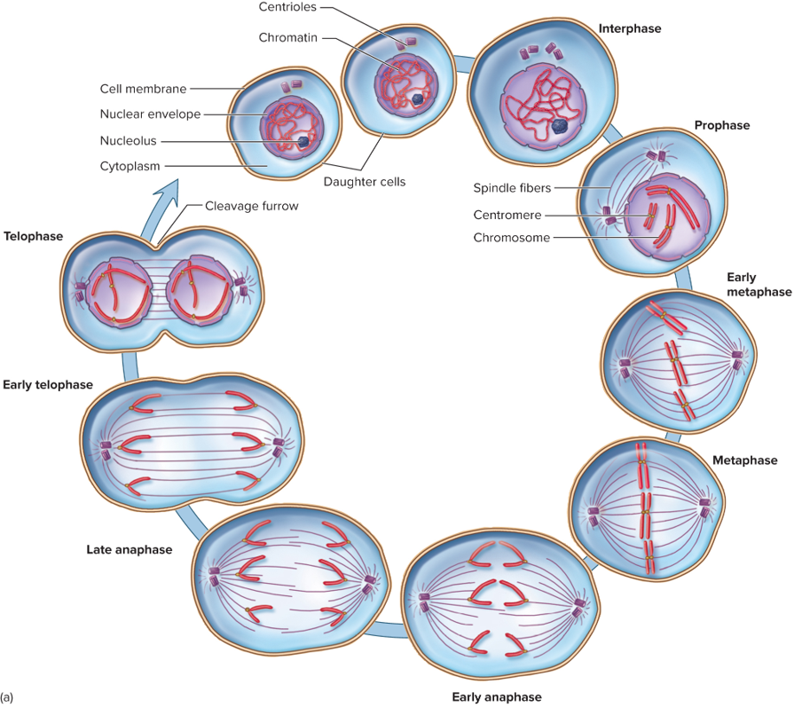
Interphase
The phase of the cell cycle that occurs before mitosis, where the cell grows, duplicates its DNA, and prepares for division. It consists of G1, S, and G2 phases.
THINK PREPARATION
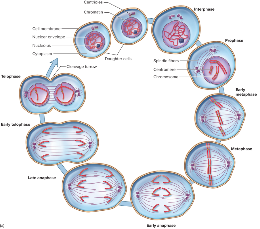
Prophase
The first stage of mitosis, where chromosomes condense and become visible, the nuclear envelope begins to break down, and spindle fibers start to form.
THINK CHROMOSOMES CONDENSE
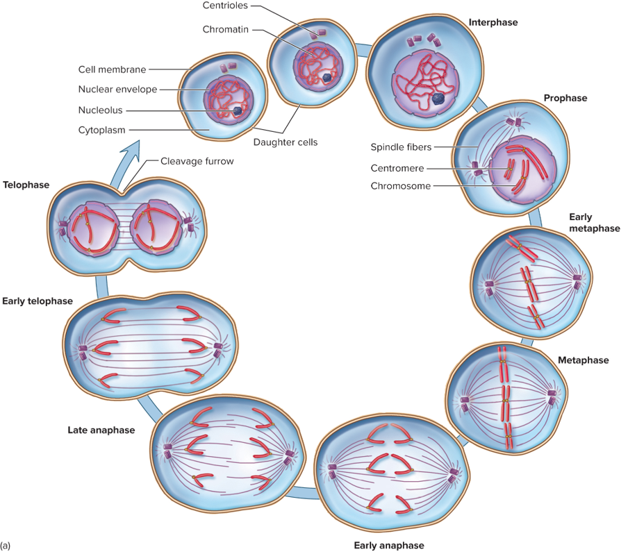
Metaphase
The second stage of mitosis, during which chromosomes align along the metaphase plate in the center of the cell, ensuring proper separation into daughter cells.
THINK MEET IN THE MIDDLE
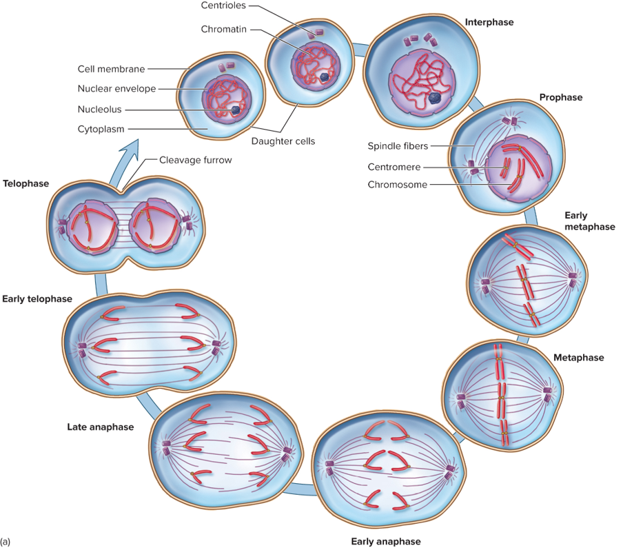
Anaphase
The stage of mitosis where sister chromatids are pulled apart towards opposite poles of the cell by spindle fibers, ensuring each daughter cell will receive an identical set of chromosomes.
THINK PULL APART
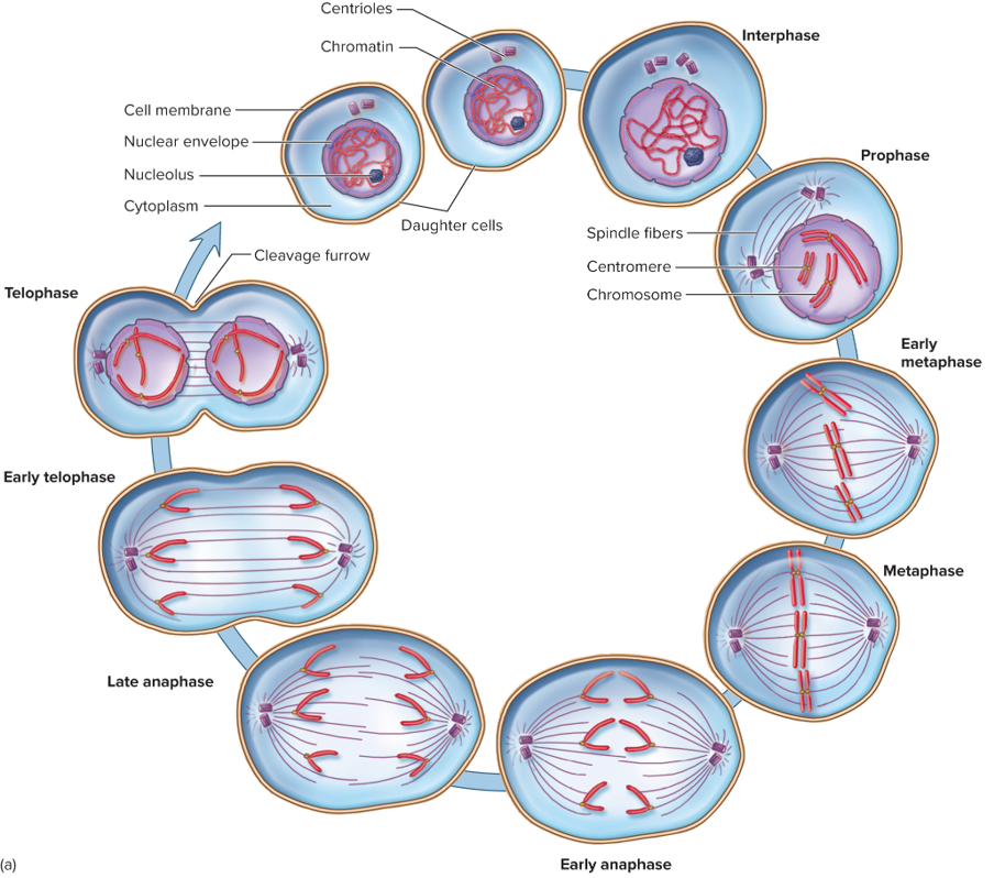
Telophase
The final stage of mitosis, where chromosomes begin to de-condense back into chromatin, the nuclear envelope re-forms around each set of chromosomes, and the cell prepares to divide into two daughter cells.
THINK CHROMOSOMES DE-CONDENSE
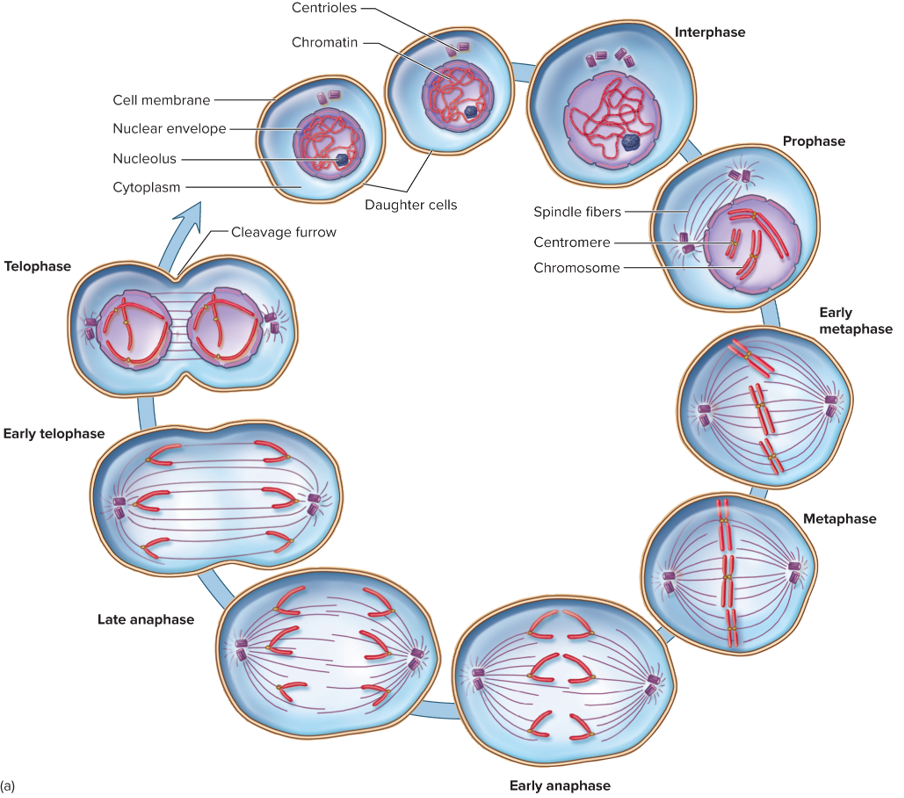
Cellular Organization, Cell Wall Composition, and Reproductive Strategy of Microbial Eukaryotes Chart
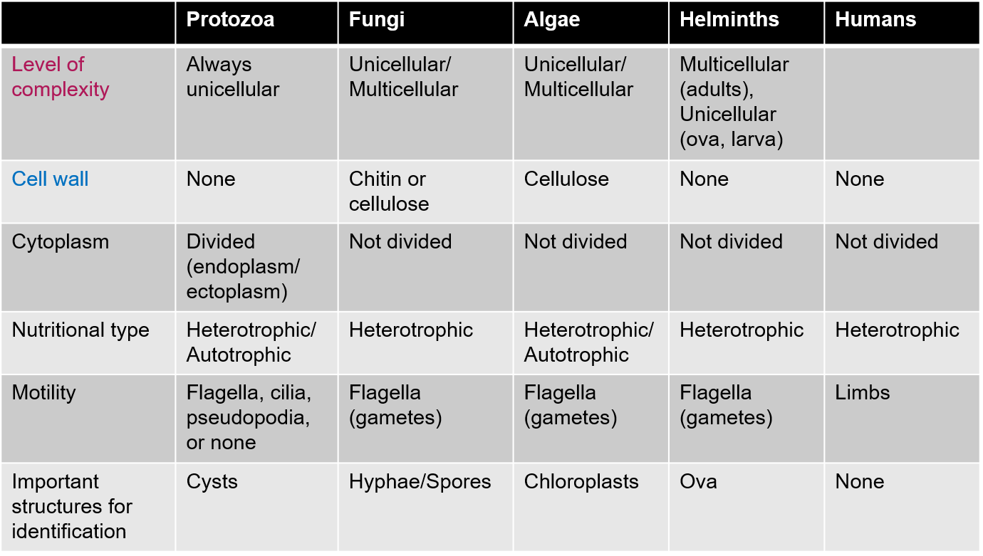
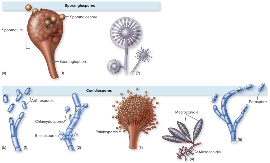
Sporangiospores
A type of asexual spore produced within a sporangium, specifically found in some fungi and plants. They play a key role in the reproduction and dispersal of these organisms.
Formed by successive cleavages within a sporangium
Stalk bursts open and spores are released
THINK SPORES INSIDE OF A SACK, ENCLOSED
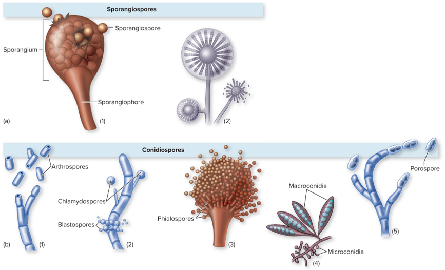
Conidiospores or Conidia
Asexual spores produced by filamentous fungi, formed at the ends of specialized stalks called conidiophores. These spores are released into the environment for dispersal and reproduction.
Pinch off from fungi
Free spores and not enclosed by a spore-bearing sac
THINK FREE SPORES, NOT ENCLOSED
Helminths Reproductive Strategy
Reproduce sexually, with complex life cycles involving multiple hosts. They often produce large numbers of eggs or larvae, which contribute to their transmission and persistence in the environment.
Can be male or female
Can also be hermaphroditic, with both male and female sex organs in the same worm
Virus Characteristics
Acellular infectious agents that require a host cell to replicate. They consist of genetic material (DNA or RNA) encased in a protein coat and can infect various organisms, including humans, animals, plants, and bacteria.
Lack cellular structure and metabolic processes
Too small to be seen with the naked eye; must be viewed under an electron microscope and metal staining
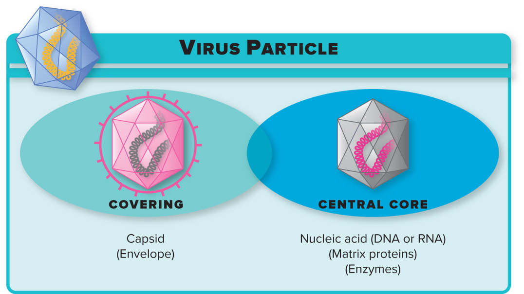
Virus: Capsid and Nucleic Acid
Genetic material in the central core made by one or more nucleic acid strands of either DNA or RNA
External protein coat covering the genetic material
Some viruses will have one or two enzymes
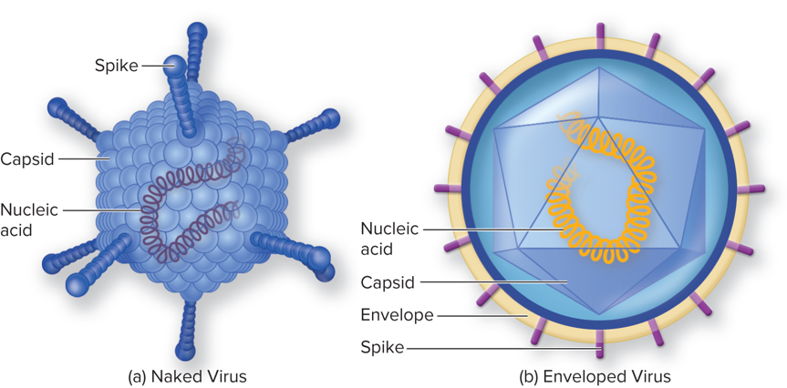
Viruses: Enveloped or Naked and Spikes
Viruses can be classified as enveloped or naked based on the presence or absence of a phospholipid bilayer surrounding the capsid.
Spikes are protein structures on the surface of enveloped viruses that are essential for attachment and entry into host cells.
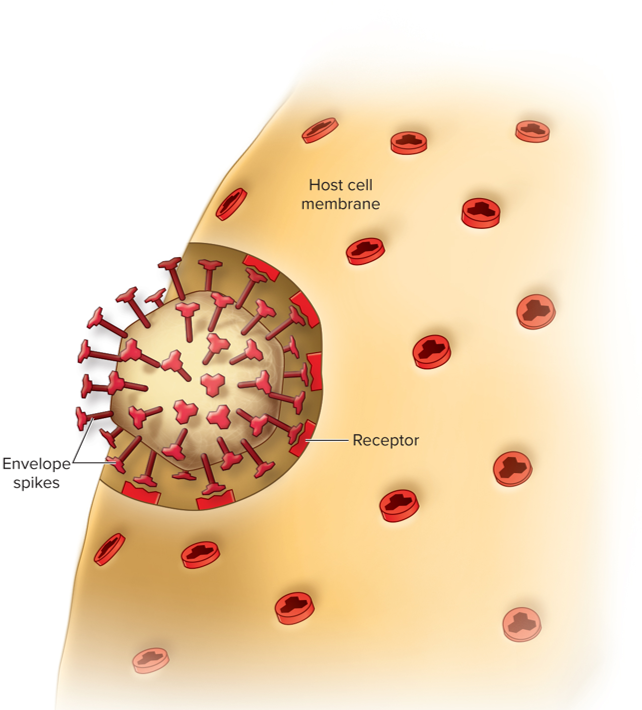
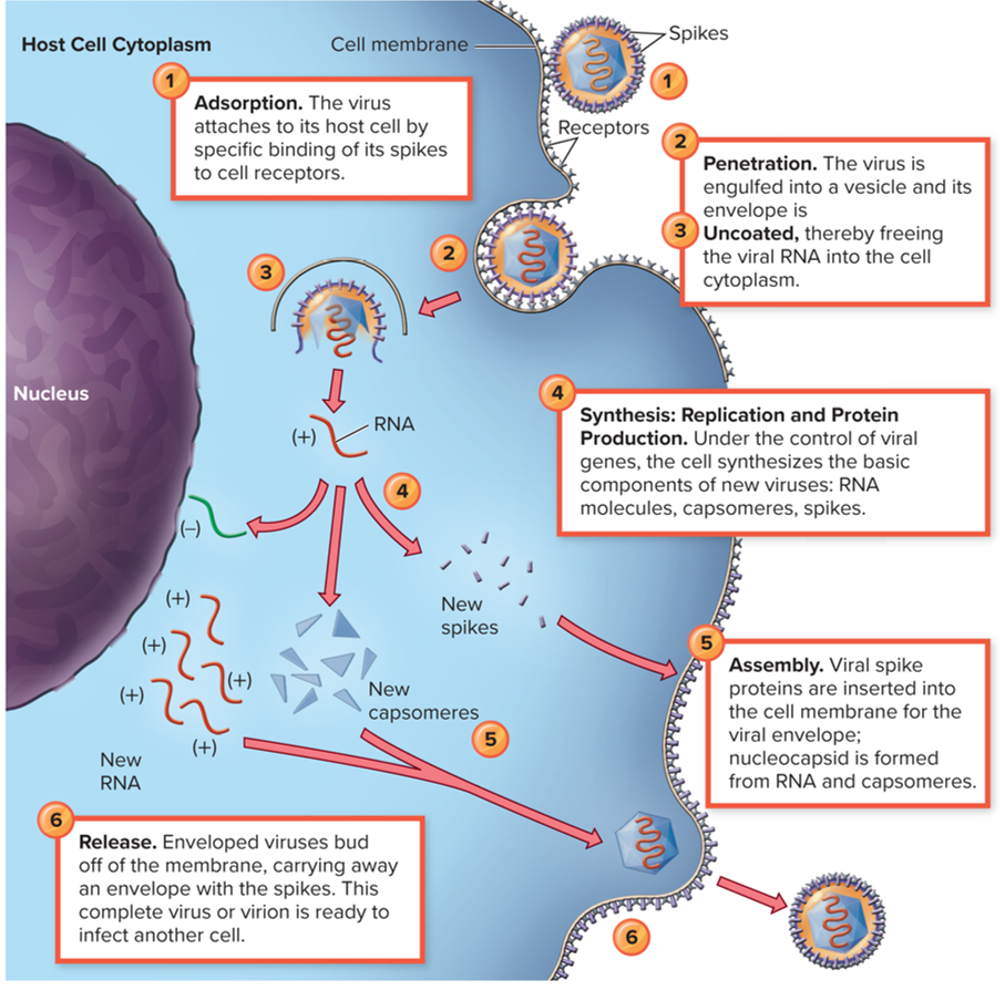
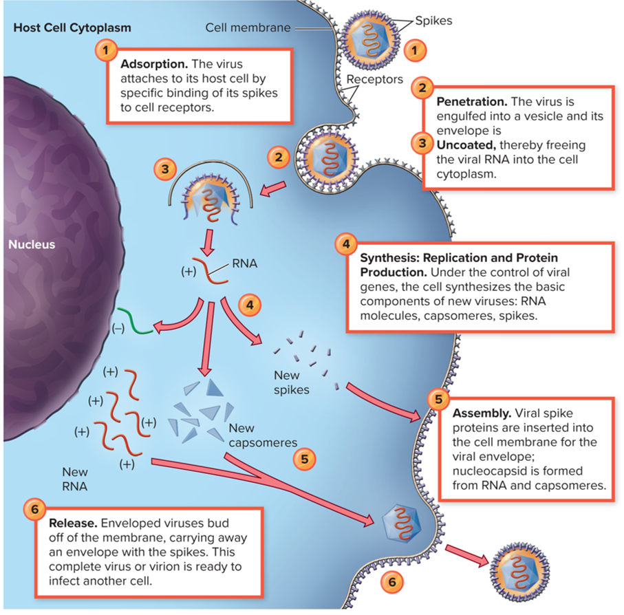
Adsorption/Attachment
The virus uses spikes to bind to the receptor.
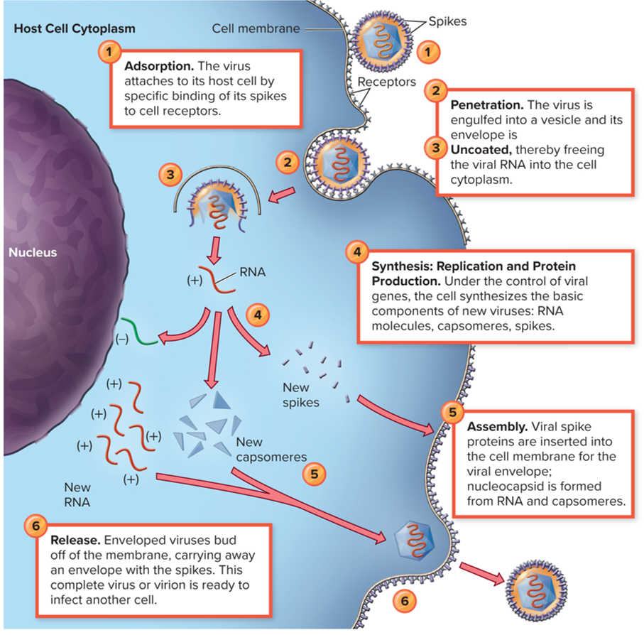
Penetration/Entry
The virus enters the host cell; the envelope can either fuse with the membrane or it can be taken in by endocytosis.
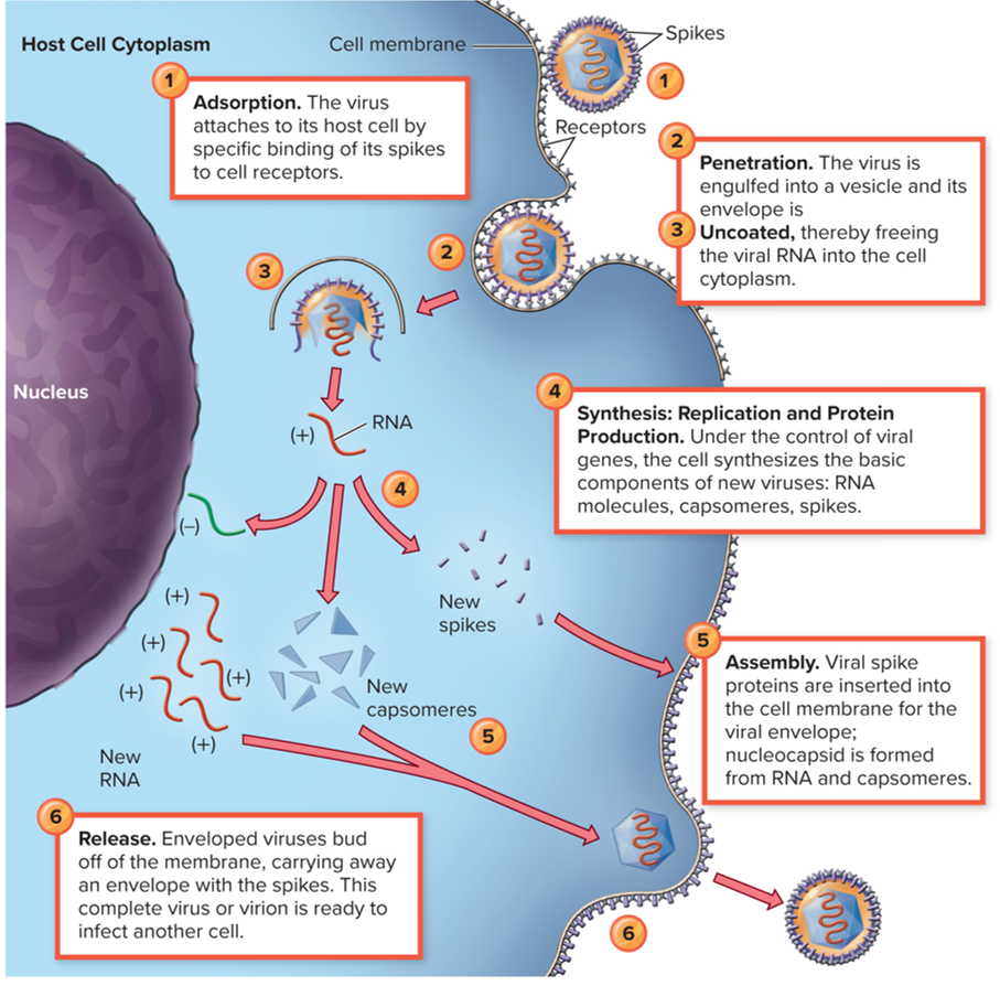
Uncoating
The protein coat is taken off, and the genetic material is exposed to the ribosomes and enzymes present in the host cell. Viruses don’t have any ribosomes or enzymes, so they must release their genetic material so they can use all of the structures that are in the host cell.
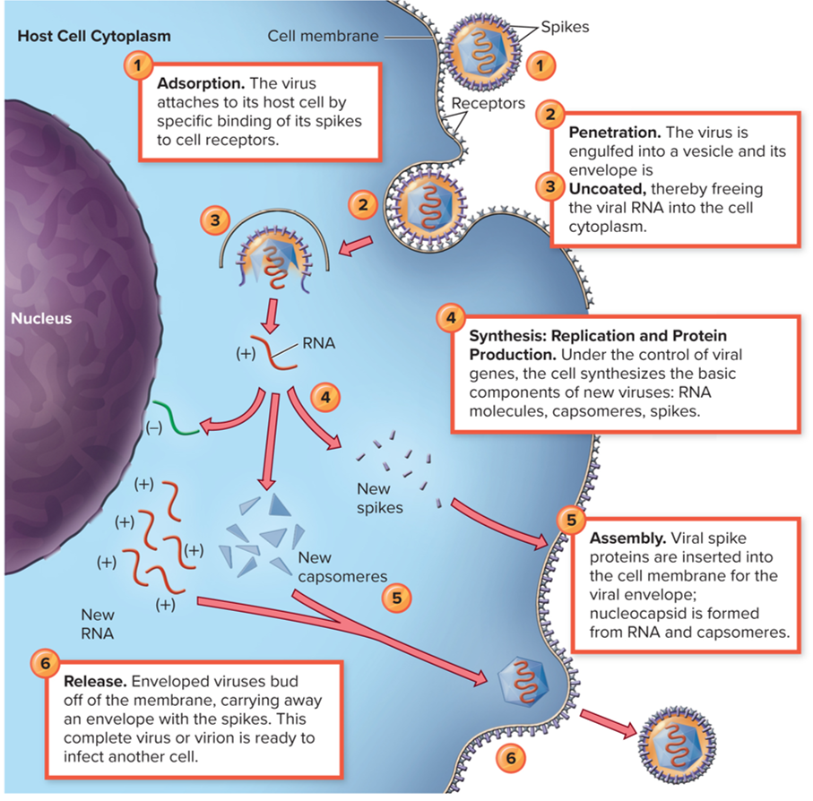
Synthesis
Replication of protein production: this is where viruses will copy their genome, whether it’s RNA to RNA or DNA to DNA. This is where the replication of the genome happens and also where protein synthesis happens.
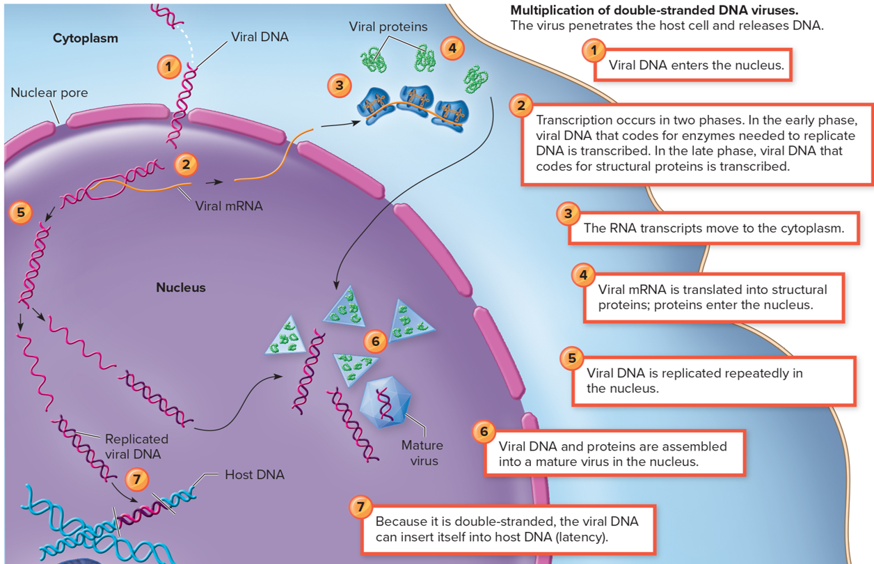
Synthesis: Multiplication of double-stranded DNA viruses
Viral DNA enters the nucleus
Transcription occurs in two phases. In the early phase, viral DNA that codes for enzymes needed to replicate DNA is transcribed. In the late phase, viral DNA that codes for structural proteins is transcribed.
The RNA transcripts move to the cytoplasm
Viral mRNA is translated into structural proteins; proteins enter the nucleus
Viral DNA and proteins are assembled into a mature virus in the nucleus
Because it is double-stranded, the viral DNA can insert itself into host DNA (latency)
Assembly
Viruses will put everything together, including proteins. If it’s going to be an enveloped virus, they’ll put their spikes in the envelope of the host and then exit the host.
Release
The virus will exit the host by budding out, in which the virus will take some of the membrane with it and become an enveloped virus.
If the virus is not an enveloped virus, it will usually be spit out in a vesicle, the same way it entered.
Another way of exit would be exploding the host cell.
Persistent Infections
Infections that last for weeks, months, years, or a lifetime, allowing the virus to replicate without causing immediate cell death. These infections can result in viral latency or continuous presence in the host.
Can be latent in the cytoplasm or in the DNA
Once incorporated into the host DNA, the virus is called a provirus
Some viruses remain in a chronic latent state where they become periodically reactivated
Prions
Infectious proteins that cause neurodegenerative disease in animals
Proteins that bind together and form holes in the brain
Prion disease is always fatal (both animals and humans can get and always die from)
No cellular structure
Viroids
Infectious strands of naked RNA that infect plants
No cellular structure
Replicate differently than viruses because they don’t have both parts (protein and genetic material)
Virus, Prions, and Viroid Differences
Viruses are infectious agents made of genetic material and a protein coat; prions are misfolded proteins that cause disease, and viroids are infectious RNA molecules that lack a protein coat.
THINK VIRUSES: BOTH COMPONENTS (DNA/RNA AND PROTEINS), PRIONS ONLY PROTEIN; VIROIDS ONLY GENETIC MATERIAL (RNA)