A & P 314 Lab Exam 1 Quiz 2
1/49
There's no tags or description
Looks like no tags are added yet.
Name | Mastery | Learn | Test | Matching | Spaced | Call with Kai |
|---|
No analytics yet
Send a link to your students to track their progress
50 Terms
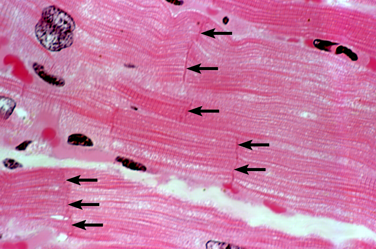
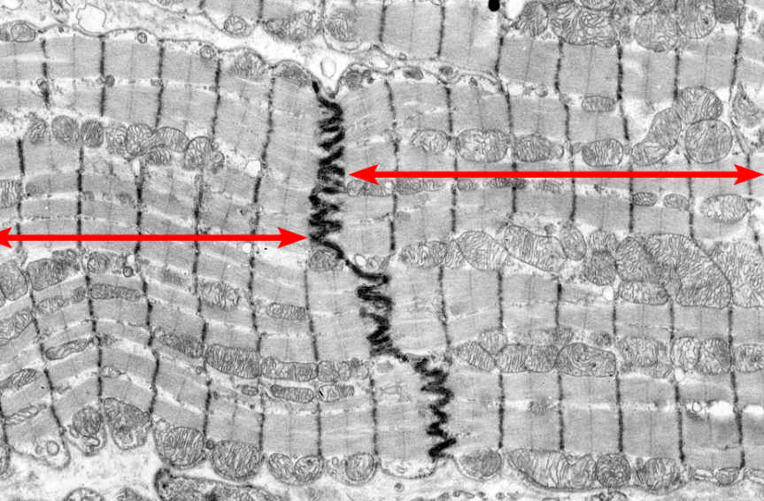
intercalated disc
structure
Gap junctions
Small cell to cell junctions, which allow for the cytoplasm of one cell to continuously flow into the cytoplasm of an adjacent cell
Cardiac muscle tissue
tissue type
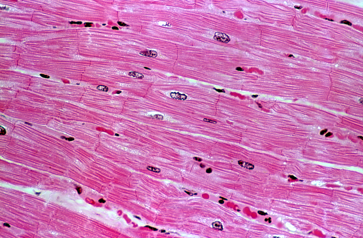
Cardiomyocyte
Cell type
parietal pericardium
outer layer of the pericardial sac, E
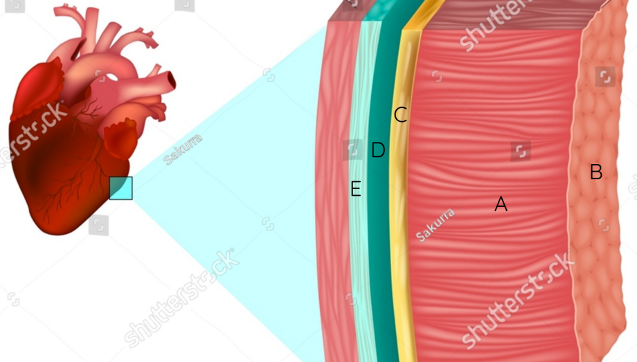
visceral pericardium
inner layer of the pericardial sac, epicardium, C

epicardium
outer layer of the heart, visceral pericardium, C

endocardium
slippery inner lining of the heart, B

pericardial space
space between the two layers of the pericardial sac, D

pericardial fluid
lubrication in the pericardial space
myocardium
muscular layer between epi- and endocardium, A

apex
inferior portion of the heart that comes to a point, blue
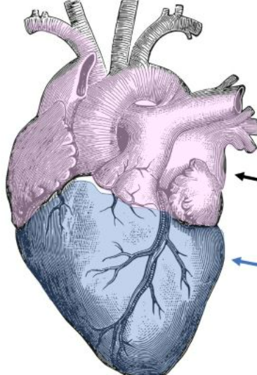
base
superior margin of the heart, line drawn across the large blood vessels, purple

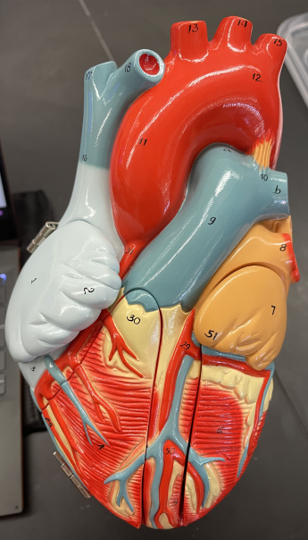
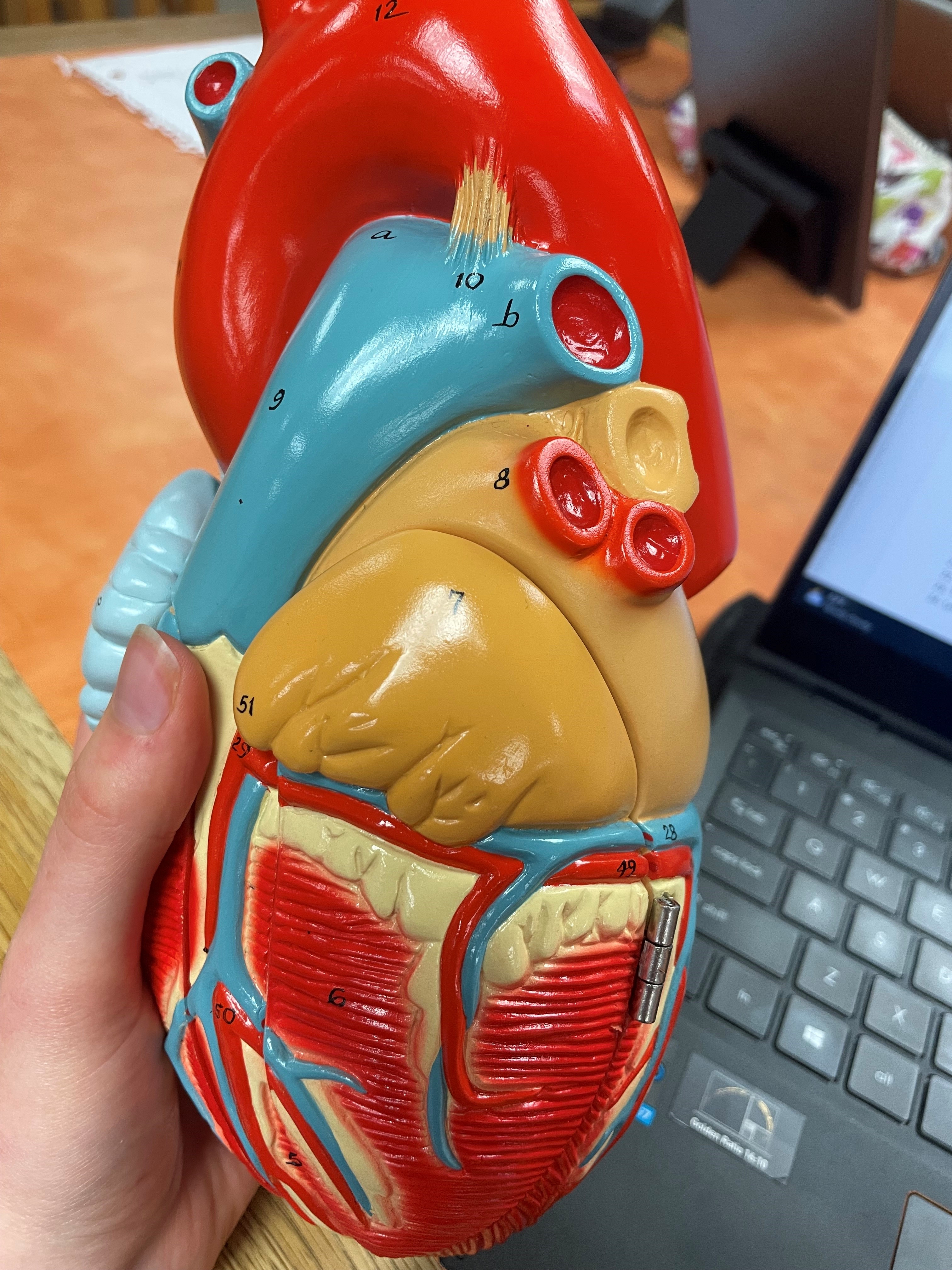
auricle
flap-like extensions of the atria that extend anteriorly and increase the amount of blood that the atria can hold, 2 and 51

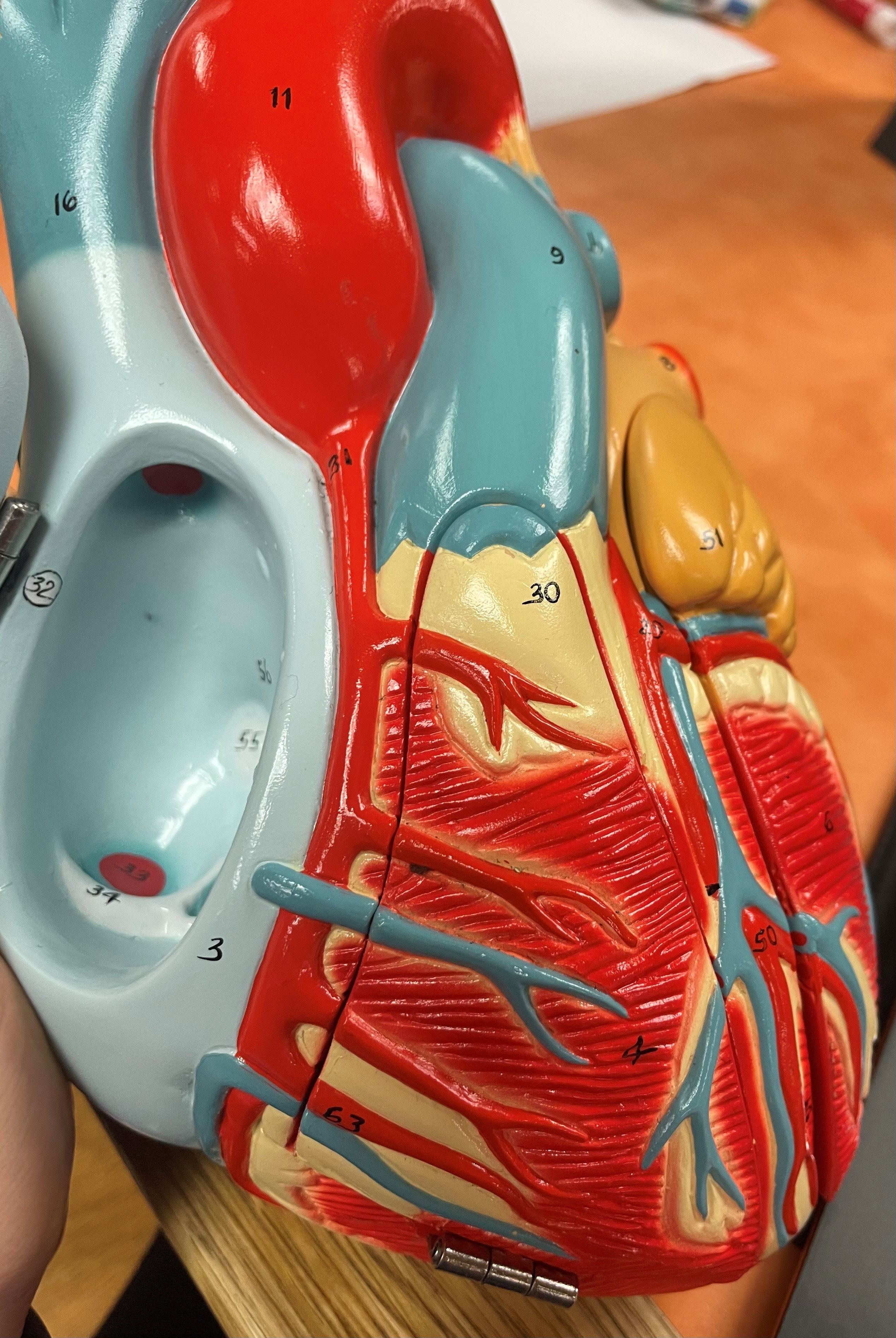
Anterior Interventricular Sulcus
shallow indentations in the front of the heart between ventricles that is filled with adipose tissue and coronary blood vessels, 5

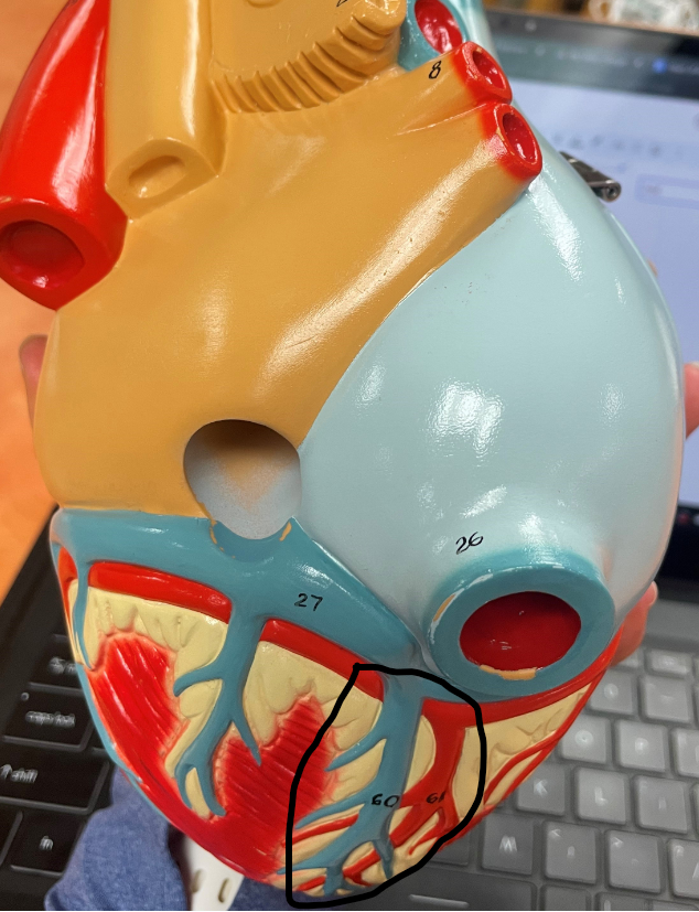
Posterior Interventricular Sulcus
shallow indentations in the back of the heart between ventricles that is filled with adipose tissue and coronary blood vessels
right atrium
first part of the heart that deoxygenated blood drains into from the Superior Vena Cava and Inferior Vena Cava, 1

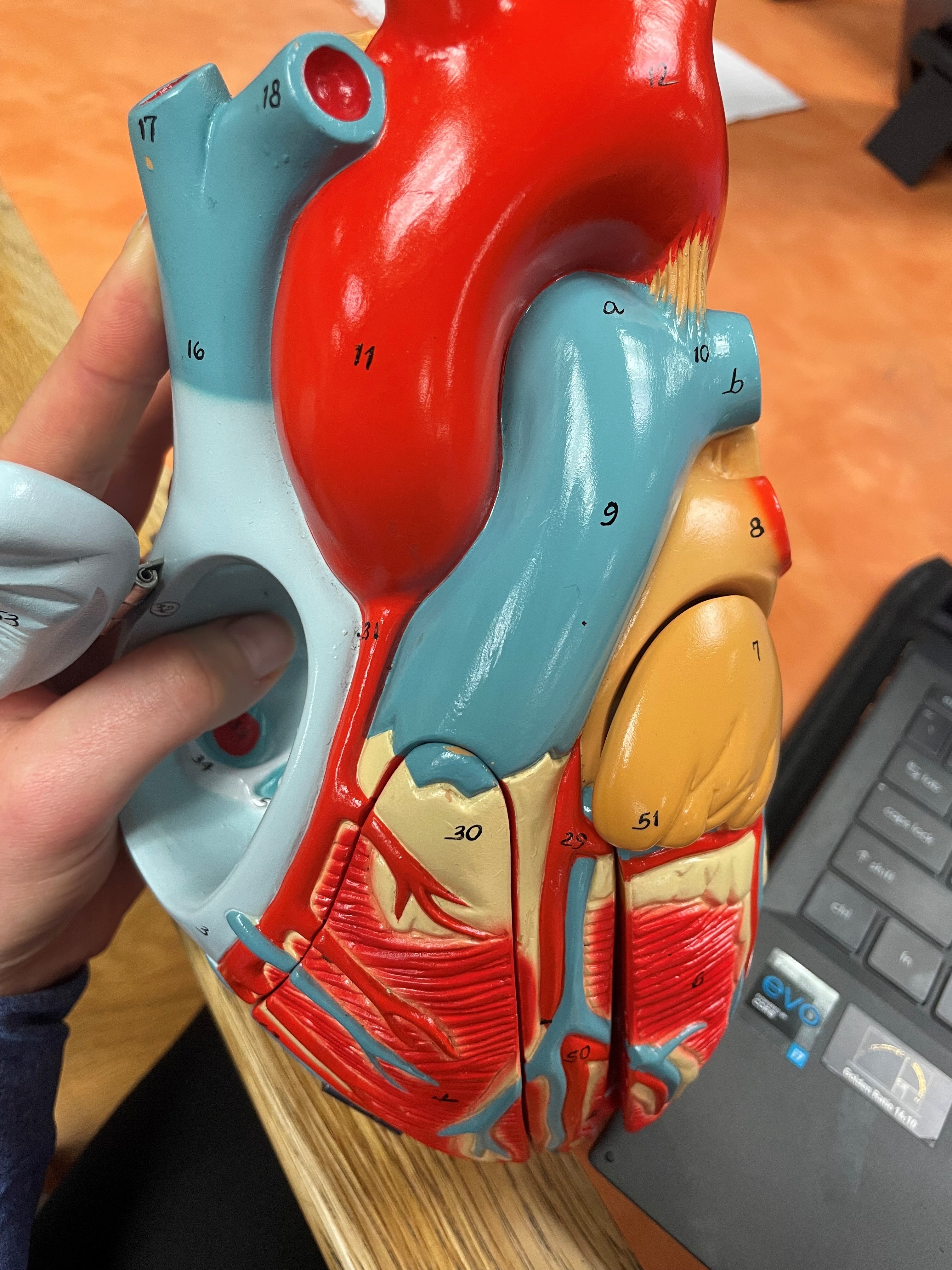
Superior Vena Cava
drains deoxygenated blood from the top of the body into the right atrium, 11
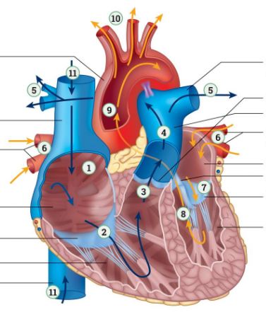
Inferior Vena Cava
drains deoxygenated blood from the bottom of the body into the right atrium, 11

right atrioventricular valve
valve that deoxygenated blood flows through to get from the right atrium to the right ventricle, 2

Left Pulmonary Vein
drains oxygenated blood from the left lung into the left atrium, 6

Right Pulmonary Vein
drains oxygenated blood from the right lung into the left atrium, 6

left atrium
receives oxygenated blood from the lungs via the Left and Right Pulmonary Veins, 7

left atrioventricular valve
valve that oxygenated blood moves through when going from the left atrium and enters left ventricle, 41
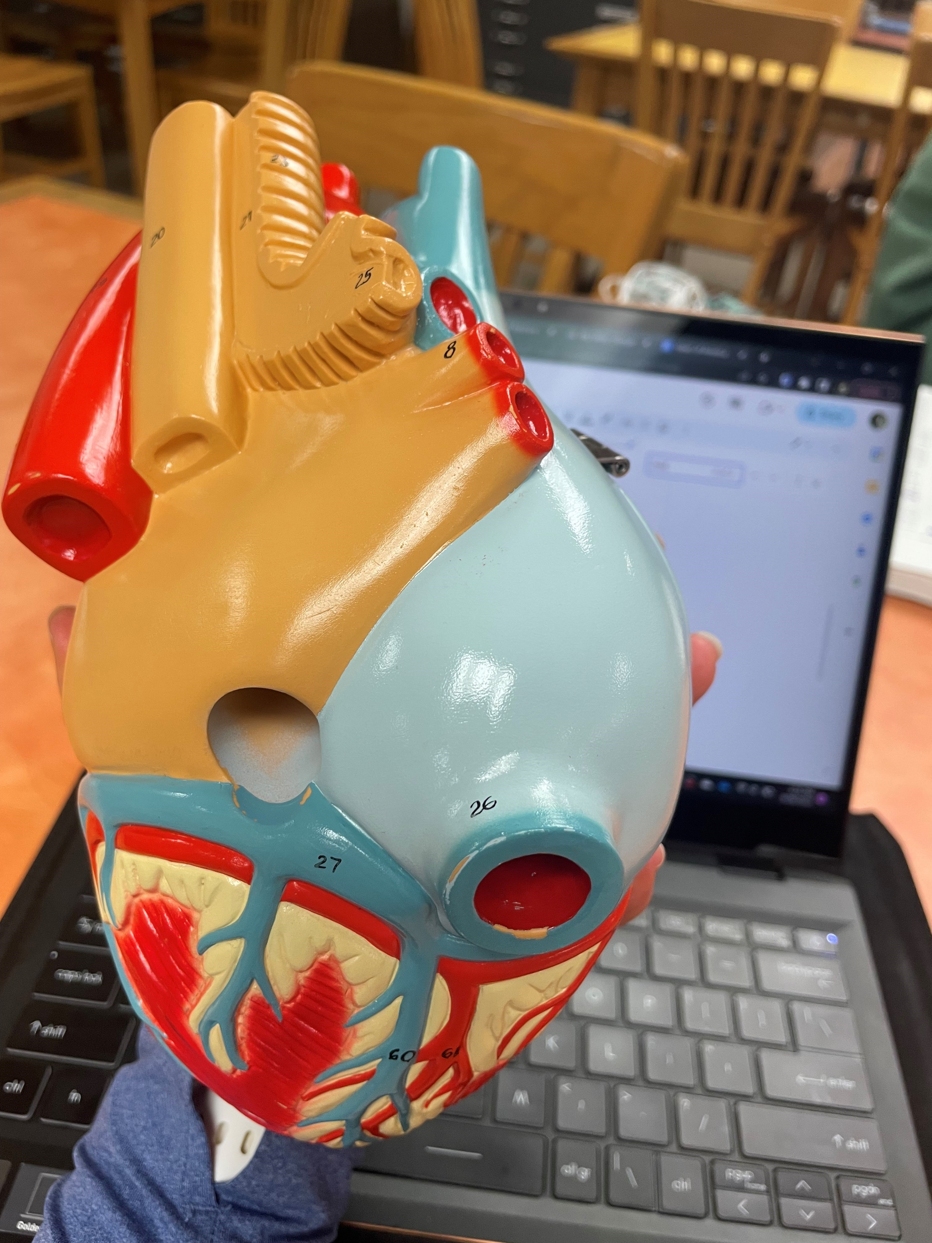
aortic valve
valve that oxygenated blood flows through on its way out of the left ventricle, 9

right ventricle
pumps deoxygenated blood to the lungs via the pulmonary trunk, 4

pulmonary valve
valve that deoxygenated blood goes through to enter into the pulmonary trunk from the right ventricle, 3

pulmonary trunk
artery that transports deoxygenated blood away from the right ventricle to the lungs, splits in the Left Pulmonary Artery and the Right Pulmonary Artery, 4

Left Pulmonary Artery
takes deoxygenated blood away from the right ventricle to the left lung, 5

Right Pulmonary Artery
takes deoxygenated blood away from the right ventricle to the right lung, 5

left ventricle
pumps oxygenated blood to the body via the aorta, 6

aorta
artery carrying oxygenated blood from the left ventricle, 10

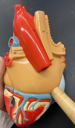
ascending aorta
transports oxygenated blood superiorly, 11
aortic arch
portion of the aorta that curves over the superior margin of the heart, 12

descending aorta
transports oxygenated blood inferiorly, 19

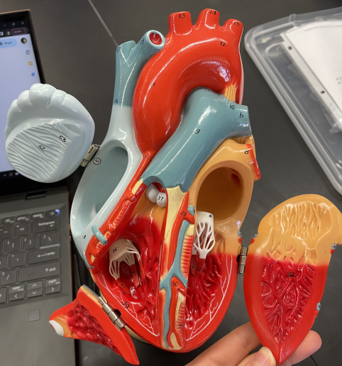
chordae tendineae
hold atrioventricular valves in place by extending downward into either ventricle and anchoring into papillary muscles
papillary muscles
small extensions of tissue that hold chordae tendinea into place, 40, 42, 62

interatrial septum
separates the atria
interventricular septum
separates the ventricles
right coronary artery
first branch off the aorta to the right to supply the heart with blood, splits into two branches, 31
right marginal artery
supplies the anterior of the right ventricle, branch off the right coronary artery, 63
posterior interventricular artery
supplies the posterior of the left and right ventricle, branch off the right coronary artery, 61

left coronary artery
first branch off the aorta to the left supply the heart with blood, splits into two branches, 29

anterior interventricular artery
mostly supplies the anterior of the left ventricle but does send some blood to the anterior of the right ventricle too, branch off the left coronary artery, 50

circumflex artery
wraps around the right margin of the heart, under the left auricle, and eventually merges with the posterior interventricular artery via small anastomoses, 49
Anastomoses
A small blood vessel, which allows blood to bypass a capillary bed
great cardiac vein
begins in the anterior interventricular sulcus and will follow the circumflex artery as it wraps around the left margin of the heart to merge with the coronary sinus, 28
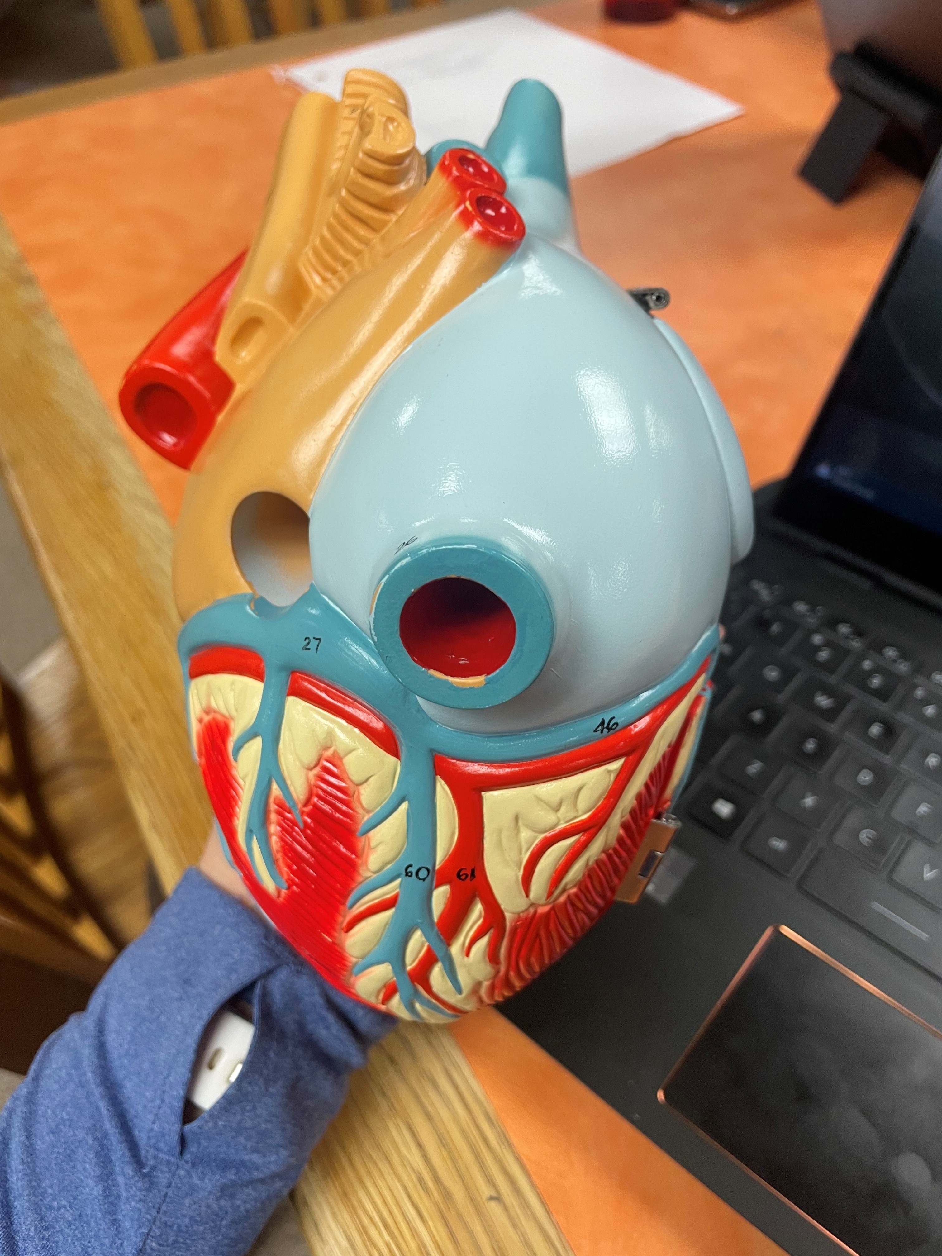
coronary sinus
the convergence of all the major veins of the heart, and it will drain blood directly into the right atrium, 27
small cardiac vein
forms on the right margin of the heart (alongside the marginal artery) and will wrap around the right side of the heart to drain into the coronary sinus, 46

middle cardiac vein
forms in the posterior interventricular sulcus and drains directly into the coronary sinus, 60