Brain and Behaviour Wks 1-5
 Call Kai
 Learn
Learn Practice Test
 Spaced Repetition
 Match
Match Flashcards
 Knowt Play
1/58

There's no tags or description
Looks like no tags are added yet.
Name | Mastery | Learn | Test | Matching | Spaced | Call with Kai | Chat |
|---|
No analytics yet
Send a link to your students to track their progress
59 Terms
What does somatic mean?
Relating to the physical body rather than the mind.
What is somatic intervention?
Brain substances are manipulated in a precise and controlled manner, and consequent effects on behavior are noted.
What are the 3Rs of using animals in research
Replace, reduce, refine
What did Franz Joseph Gall do?
Developed the practice of phrenology; claiming that the bumps on the skull measured a person’s character and mental abilities. It was a misguided method, but still influential.
Sensory neurons vs motor neurons
Sensory neurons: Pseudounipolar neurons and bipolar neurons
Motor neurons: Efferent neurons
What are the Brodmann Areas?
Identifying distinct functional areas based on their cellular organisation in the brain. Brodmann created maps of the brain based on detailed microscopic examinations of brain tissue.
How are the brain’s sensory maps organised?
Topographically: the neighbouring areas of the body or environment are represented by neighbouring groups of neurons in the brain.
Differentiate the CNS and the PNS
Central nervous system: The control centre
Peripheral Nervous system: The communication network
What is the PNS responsible for and what direction does it travel?
Information transfer and processing.
If somatic sensory input, TOWARDS the CNS, but if motor output then AWAY from the CNS.
If you touch something hot, the sensory nerves carry the heat signal and message up to the CNS. THEN, motor neurons carry the commands back down AWAY FROM the CNS, causing your hand to pull away from the hot surface.

What is the Somatic Nervous system
Controls conscious and voluntary body movements.
Divided between sensory input and motor output.
They comprise the nerves that connect the brain to the major muscles and sensory systems of the body.
The sensory output carries signals towards the CNS (in the brain), and the motor output carries signals away from the CNS towards the related muscles.
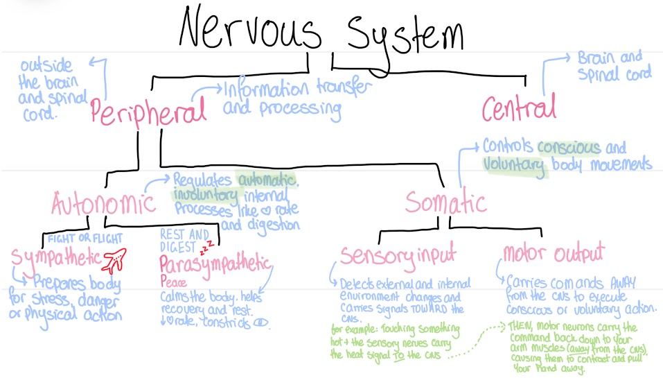
What is the Autonomic Nervous System
Regulates automatic, involuntary internal processes like heart rate and digestion.
Divided into the sympathetic nervous system and parasympathetic nervous system.
Sympathetic: S for stress. Fight or flight. Prepares the body for stress, danger or physical action.
Parasympathetic: P for peace. Rest and digest. Calms the body, helps recovery and rest, lowers heart rate and constricts pupils.
Where in the spine does information come from in the CNS?
Information travelling INTO the CNS (sensory input) always comes through the back of the spine.
Instructions to the body from the CNS (motor output) always come out the front.
What is the process of neuron’s attaining information
The information is received by the dendrites, and the neuron passes on the information through the axon terminals.

What are synapses?
The gaps between neurons.
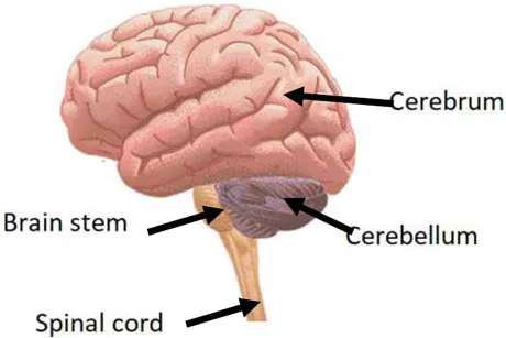
What are the four main divisions of the brain:
Cerebrum, brainstem, cerebellum and spinal chord
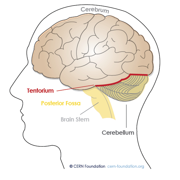
What separates the cerebellum from the cerebral hemisphere?
The tentorium cerebelli. It is a tough, sheet-like layer of the dura mater.
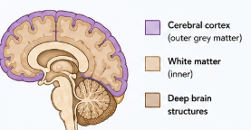
Is cerebral cortex white or grey matter?
The cerebral cortex is grey matter, which processes information.
Immediately beneath the cortex is white matter, which is responsible for information transfer. These cells are covered in myelin, which is a fatty layer around the neuron.
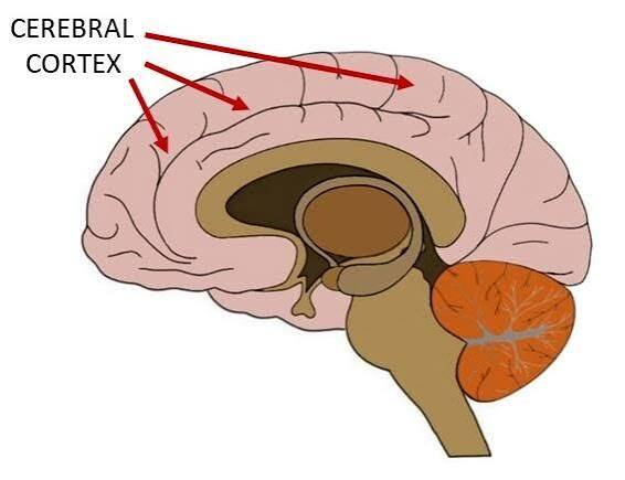
What is the cerebral cortex divided into?
4 major lobes
Frontal: Manages voluntary movement, reasoning, planning, emotion and problem-solving.
Parietal: Processes sensory information: touch, temperature, pain and spatial awareness.
Temporal: Handles memory formation, hearing and language comprehension.
Occipital: Acts as the primary visual processing centre for shapes, colour and recognition.
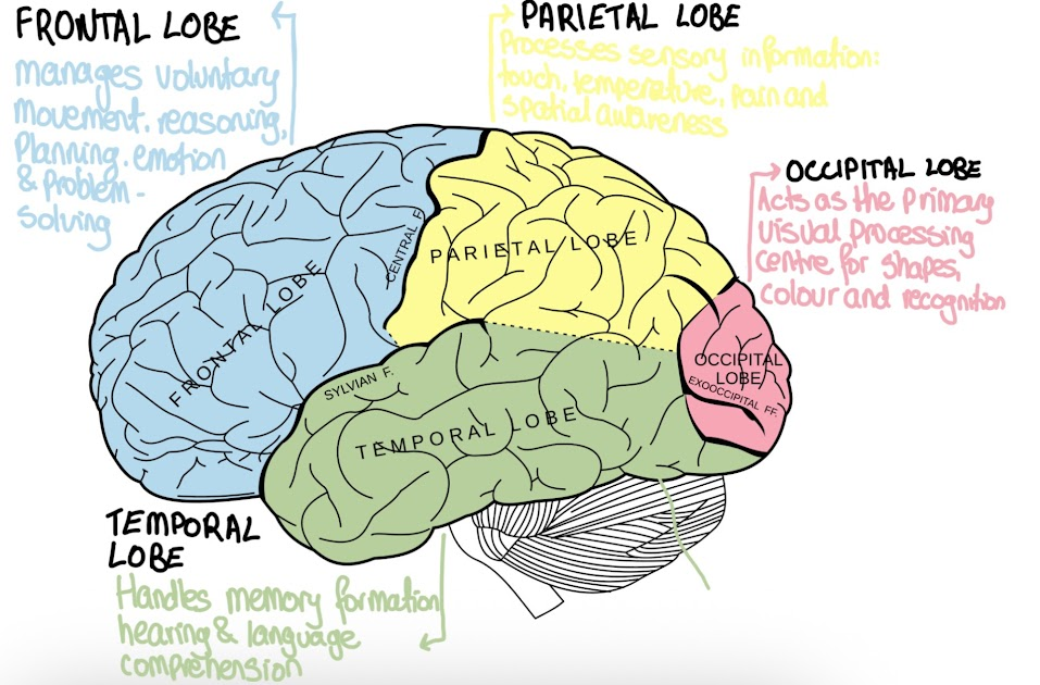
What are the frontal and parietal lobes separated by?
What are the gyri and sulci?
The frontal and parietal lobes are separated by the central sulcus.
The gyri are the hills, and sulci are the valleys. (Sulky = down or sad; sulci = down = valleys)
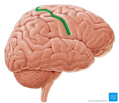

Label this diagram and briefly describe their responsibility
Longitudinal fissure: the deep groove that physically separates the left and right halves of the brain’s cerebrum.
Lateral fissure: Separates the temporal lobe from the frontal and parietal lobes.
Central sulcus: Separates the frontal and parietal lobes.
Cingulate sulcus: The deep groove on the inner surface of the brain that separates the cingulate gyrus from the frontal and parietal lobes.
Occipital and parietal lobes: Occipital: visual processing centre. Parietal: Sensory information.
Parieto-occipital sulcus: Separates the parietal lobe from the occipital lobe on the medial surface of the brain.

What is the difference between the cerebrum and cerebral cortex?
Cerebrum: Large, upper part of the brain, including the cerebral cortex AND the underlying white matter and deep structures.
Cerebral Cortex: The thin, outer layer of grey matter covering the surface of the cerebrum.
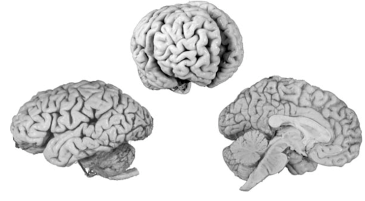
Describe these views
Left figure: lateral view
Middle figure: oblique view
Right figure: Medial surface or sagittal surface
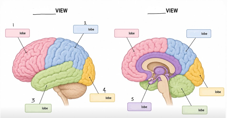
Label the parts of the brain and the views
1: Frontal Lobe
2: Parietal Lobe
3: Temporal Lobe
4: Occipital Lobe
5: Limbic Lobe

What are the functions of these 4 gyri, and where are they located?
Precentral gyrus
Postcentral gyrus
Superior Temporal gyrus
Cingulate gyrus
Precentral gyrus: The execution of voluntary physical movements. Located in the frontal lobe.
Postcentral gyrus: Processes touch and temperature. Located in the parietal lobe.
Superior Temporal gyrus: Processes sound and spoken language. Located in the temporal lobe.
Cingulate gyrus: Regulates emotion, behaviour and memory. Located in the limbic lobe.
The surface of all four of these gyri is made of grey matter.

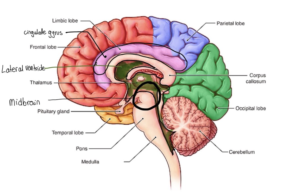
Label this diagram
A: Limbic Lobe
B: Cingulate Gyrus
C: Frontal Lobe
D: Lateral Ventricle
E: Thalamus
F: Midbrain
G: Pituitary Gland
H: Temporal Lobe
I: Pons
J: Medulla
K: Cerebellum
L: Occipital
M: Corpus callosum
N: Parietal Lobe
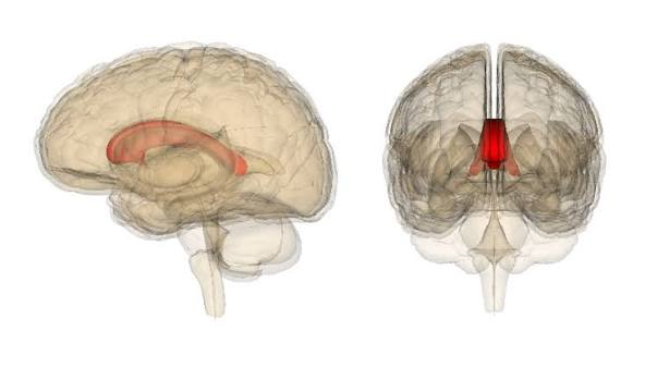
What is the corpus callosum
The massive white matter joining the two hemispheres.
What is part of the brain stem?
Midbrain, pons, medulla
Potassium and what is the resting potential of the cell?
Potassium (K+) can move into and out of the cell at will. They are kept in an equilibrium state that keeps the resting potential of the cell at -70mV.
Sodium and what is the resting potential of the cell?
Sodium (Na+) cannot move freely across the cell membrane. It is actively pumped out of the cell by the sodium-potassium pump.
At rest, sodium ions are kept outside the cell.
The resting potential is -70 mV.
The sodium-potassium pump
It exchanges three sodium for two potassium. The pump needs energy to function and it’s a large part of the energy required by the brain.
What is the order of an action potential?
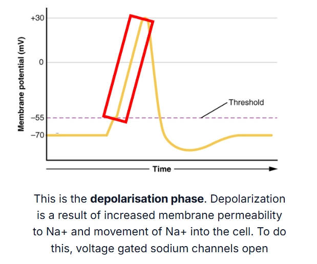
Resting membrane potential, graded potential, depolarisation phase, repolarisation and hyperpolarisation
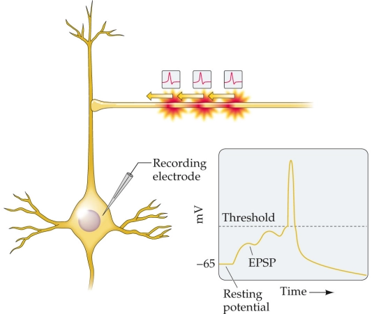
What is a graded potential (EPSP)?
An action potential was elicited when the graded potential reached the threshold (-55mV).
What is the depolarisation phase?
The inside of a cell membrane becomes less negative (and temporarily positive) as positively charged sodium ions rush inside as voltage gated sodium channels opened.
What is the repolarisation phase?
The stage where the cell’s membrane potential returns to a negative resting value after the peak of depolarisation.
The inside of the cell changes from positive charge back to a negative charge.
The voltage-gated sodium (Na+) channels close, stopping positive sodium ions from entering the cell.
Voltage-gated potassium (K+) channels open, allowing positive potassium ions to rush out of the cell.

What is the hyperpolarisation phase
The membrane potential becomes more negative than the normal resting state. The voltage-gated potassium channels close slowly after repolarisation, allowing positive potassium ions (K+) to keep leaving the cell for a short time.
The voltage drops (-90mV) because too many positive ions exit. This extra-negative charge creates a short refractory period.
How to measure how strong the signal is?
NOT a measure of how big or small the action potential was, but by how many there were.
How long do the voltage-gated sodium channels stay open for?
About a millisecond
At the peak of the action potential, what does the membrane potential reach?
+40mV
An action potential is generated in the axon __.
Hillock
What is the all-or-nothing law?
An individual nerve or muscle fiber will either respond completely to a stimulus or not at all.
What receives electrical signals from other neurons?
Dendrites
What are Schwann cells and where are they found?
In the peripheral nervous system: Schwann cells are glial cells that create the protective myelin insulation around the nerves outside the brain and spinal cord.
What are oligodendrocytes?
In the central nervous system: The glial cells that create myelin around axons in the brain and spinal cord.
What are the three main parts of a chemical synapse
The presynaptic terminal
The synaptic cleft
The postsynaptic ending
Ionotropic vs metabotropic neurotransmitters
Can be excitatory (making an action potential more likely). Or inhibitory (making an action potential less likely).
Ionotropic → fast
Metabotropic → slow (because the receptor doesn’t open an ion channel directly, but instead makes a g-protein inside the cell).
Agonist, antagonist, inverse agonists, neuromodulators meaning
AGONIST: If a drug has the same effect as the neurotransmitter.
ANTAGONIST: If a drug blocks the effect of a neurotransmitter.
INVERSE AGONISTS: Some neurotransmitters can bind to the same site as the neurotransmitter and have the exact opposite effect.
NEUROMODULATORS: Drugs can also bind to a site that is separate from the site that the neurotransmitter binds to, and can impact the likelihood that the neurotransmitter will bind, having an allosteric effect.
What is a neuromuscular junction
A specialised synapse that connects a motor neuron to a muscle fibre.
What is spatial summation?
Spatial and temporal summation are ways a neuron adds up signals. Spatial summation occurs when multiple sender cells fire simultaneously at different locations.
Both processes combine voltage changes to reach the threshold required to fire an action potential.

What is temporal summation?
Spatial and temporal summation are ways a neuron adds up signals.
Temporal summation happens when a single sender cell fires rapid signals back-to-back.
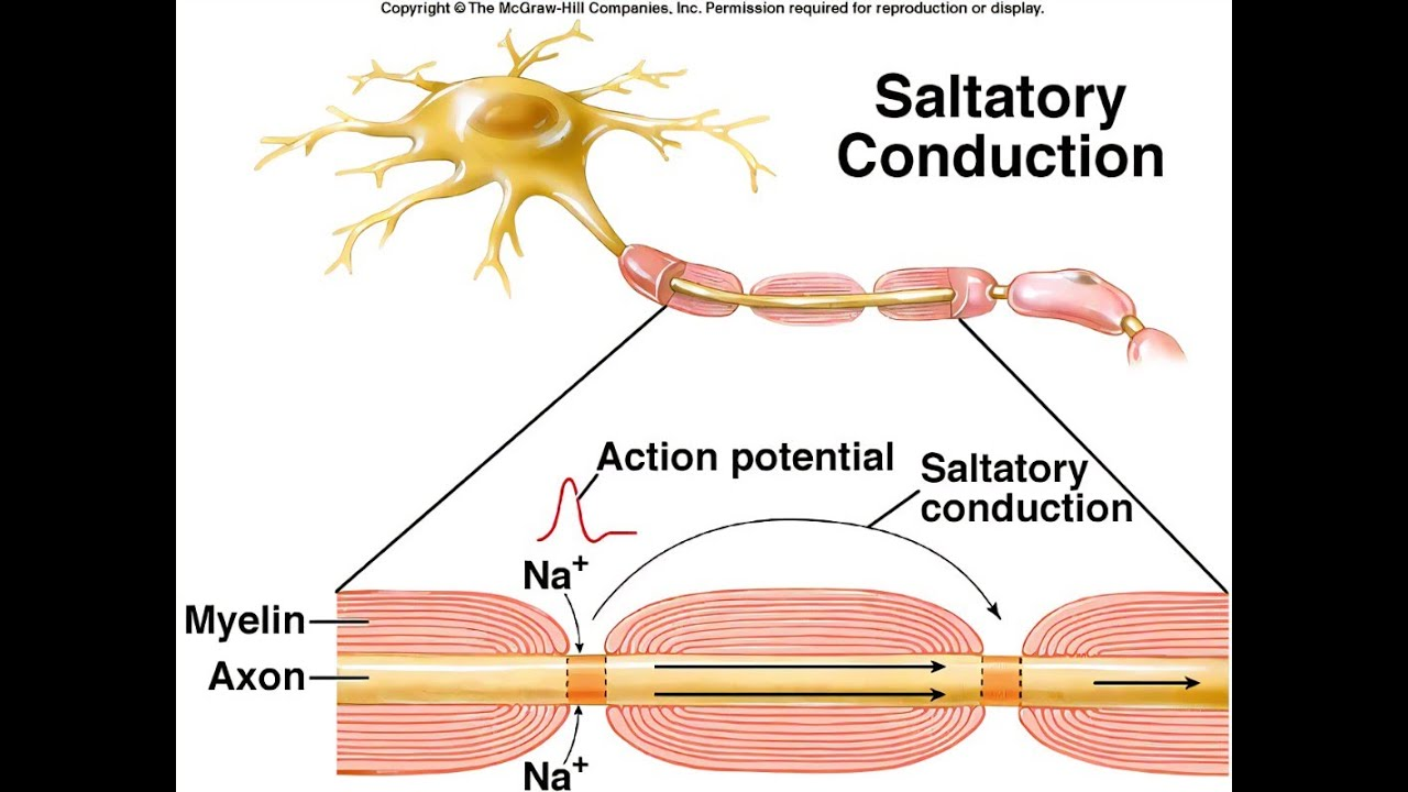
What is saltatory conduction?
The rapid way an action potential jumps along a myelinated nerve fibre from one gap to the next.
It makes nerve signal transmission much faster and more energy-efficient than continuous conduction.
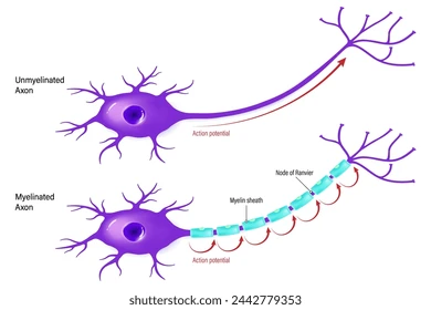
Does every action potential undergo saltatory conduction?
No. Saltatory conduction only happens in myelinated axons. In these axons, the action potential ‘jumps’ from one gap in the myelin, called a ‘node of Ranvier’ to the next.
What is the function of the glial cell microglia
Support blood vessels and nerves in the central nervous system
What is the function of ependymal glial cells
Produce, circulate and regulate cerebrospinal fluid (CSF)
What is the function of satellite glial cells
Provide physical, metabolic and chemical support to neurons in the peripheral nervous system.
IPSP vs EPSP
IPSP: When an incoming signal causes the inside of a receiving neuron to become more negative, moving its voltage further away from the threshold needed to fire an action potential.
EPSP: When a neurotransmitter binds to a receptor on a neuron’s postsynaptic membrane, causing positive ions to flow into the cell and make the inside less negative.
Glutamate
The main excitatory neurotransmitter in the nervous system
GAMA and glycerine
The main inhibitory inhibitory transmitter in the nervous system
What is dura matter?
The tough, thick outermost membrane that covers and protects your brain and spinal cord.
What is Broca’s area responsible for?
Left frontal lobe of the brain that is mainly responsible for producing speech and managing language grammar.